
Article Figures & Data
Figures
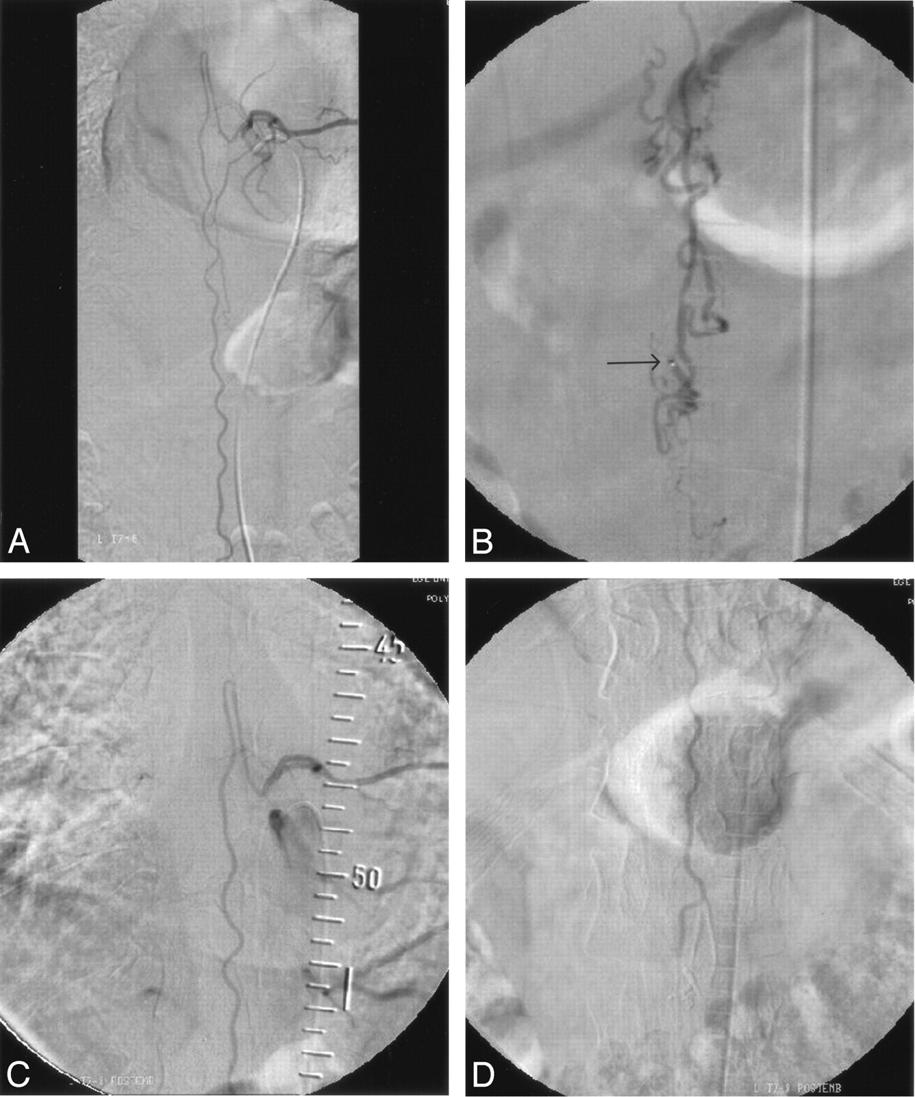
- Fig 1.
Case 2.
A, Sagittal T2-weighted MR image shows serpentine flow voids along the medulla spinalis with abnormal intramedullary signal intensity.
B, Selective angiogram of right L2 artery shows filling of the posterior spinal artery supplying the fistula at the level of conus medullaris.
C, Injection through the microcatheter (midway to fistula) depicts more clearly the fistula site located just distal to sharp bend, an ideal position for embolization. Please note that diameter of the feeder is slightly larger than the 1.5F (0.5 mm) microcatheter.
D, After embolization, selective right L2 artery angiogram reveals occlusion of the fistula with preservation of the posterior spinal artery.
E, Sagittal T2-weighted MR image 20 months after treatment shows that the signal intensity voids around the cord consistent with enlarged perimedullary veins and intramedullary abnormal signal intensity have disappeared.
- Fig 2.
Case 1.
A, Selective angiogram of left T8 intercostal artery shows slightly enlarged artery of Adamkiewicz and tortuous anterior spinal artery.
B, Injection through the microcatheter placed just inside the fistula site at the level of conus medullaris shows typical dilated perimedullary veins. Arrow indicates microcatheter tip.
C and D, After embolization, selective angiograms of left T8 intercostal artery (C) upper and (D) lower part. The fistula is occluded with patent anterior spinal artery up to the level of conus medullaris.
- Fig 3.
Case 4.
A and B, Selective angiograms of left T12 intercostal artery, early (A) and late (B) phases, show filling of the anterior spinal artery supplying the fistula located in the filum terminale. Arrows indicate 2 fistula sites. Note the serpentine perimedullary veins filling in ascending fashion.
C, Injection through the microcatheter placed at the level of conus medullaris shows first fistula (arrow) in the L3–L4 disk level. Please note triple-axial technique (guiding catheter within long vascular sheath) to improve pushability of the microcatheter. Despite the more caudal fistula located in the filum terminale, proximal occlusion of the feeder at the level of conus medullaris (current position) is safe.
D, After embolization, selective left T12 artery injection confirms occlusion of the fistula and preservation of the anterior spinal artery up to the level of conus medullaris.


Tables
Summary of five patients with type I perimedullary spinal arteriovenous fistula
Patient No./Age (y) Arteriovenous Fistula Treatment LEMF Sphincter Function Follow-up (mo) Location Segmental Artery Feeding Artery Pre- Post- Pre- Post- 1/46 Conus Left T8 ASA Emb. 2 4 Poor Good 43 2/43 Conus Right L2 PSA Emb. 2 4 Poor Good 46 3/34 Conus Left L2 ASA Emb. 2 4 Poor Poor 49 4/67 Filum terminale Left T12 ASA Emb. 1 3 Fair Good 30 5/67 Conus Left L4 Radiculopial artery Surgery 1 3 Poor Fair 37 Note.—LEMF indicates lower extremity motor function; ASA, anterior spinal artery; PSA, posteriolateral spinal artery; Emb., embolization.
In this issue


Jump to section
Related Articles
Cited By...
- Clinical features and outcomes of perimedullary arteriovenous fistulas: comparison between micro- and macro-type lesions
- Endovascular management of spinal arteriovenous malformations
- Endovascular Treatment of Spinal Arteriovenous Lesions: Beyond the Dural Fistula
- Segmental Artery Exchange Technique for Stable 4F Guiding-Catheter Positioning in Embolization of Spinal Vascular Malformations