Advertisement

Main menu

  • Home
  • Content
    • Current Issue
    • Accepted Manuscripts
    • Article Preview
    • Past Issue Archive
    • Video Articles
    • AJNR Case Collection
    • Case of the Week Archive
    • Case of the Month Archive
    • Classic Case Archive
  • Special Collections
    • AJNR Awards
    • Low-Field MRI
    • Alzheimer Disease
    • ASNR Foundation Special Collection
    • Photon-Counting CT
    • View All
  • Multimedia
    • AJNR Podcasts
    • AJNR SCANtastic
    • Trainee Corner
    • MRI Safety Corner
    • Imaging Protocols
  • For Authors
    • Submit a Manuscript
    • Submit a Video Article
    • Submit an eLetter to the Editor/Response
    • Manuscript Submission Guidelines
    • Statistical Tips
    • Fast Publishing of Accepted Manuscripts
    • Graphical Abstract Preparation
    • Imaging Protocol Submission
    • Author Policies
  • About Us
    • About AJNR
    • Editorial Board
    • Editorial Board Alumni
  • More
    • Become a Reviewer/Academy of Reviewers
    • Subscribers
    • Permissions
    • Alerts
    • Feedback
    • Advertisers
    • ASNR Home

User menu

  • Alerts
  • Log in

Search

  • Advanced search
American Journal of Neuroradiology

American Journal of Neuroradiology

ASHNR American Society of Functional Neuroradiology ASHNR American Society of Pediatric Neuroradiology ASSR
  • Alerts
  • Log in

Advanced Search

  • Home
  • Content
    • Current Issue
    • Accepted Manuscripts
    • Article Preview
    • Past Issue Archive
    • Video Articles
    • AJNR Case Collection
    • Case of the Week Archive
    • Case of the Month Archive
    • Classic Case Archive
  • Special Collections
    • AJNR Awards
    • Low-Field MRI
    • Alzheimer Disease
    • ASNR Foundation Special Collection
    • Photon-Counting CT
    • View All
  • Multimedia
    • AJNR Podcasts
    • AJNR SCANtastic
    • Trainee Corner
    • MRI Safety Corner
    • Imaging Protocols
  • For Authors
    • Submit a Manuscript
    • Submit a Video Article
    • Submit an eLetter to the Editor/Response
    • Manuscript Submission Guidelines
    • Statistical Tips
    • Fast Publishing of Accepted Manuscripts
    • Graphical Abstract Preparation
    • Imaging Protocol Submission
    • Author Policies
  • About Us
    • About AJNR
    • Editorial Board
    • Editorial Board Alumni
  • More
    • Become a Reviewer/Academy of Reviewers
    • Subscribers
    • Permissions
    • Alerts
    • Feedback
    • Advertisers
    • ASNR Home
  • Follow AJNR on Twitter
  • Visit AJNR on Facebook
  • Follow AJNR on Instagram
  • Join AJNR on LinkedIn
  • RSS Feeds

AJNR Awards, New Junior Editors, and more. Read the latest AJNR updates

Research ArticlePEDIATRICS

Posterior Fontanelle Sonography: An Acoustic Window into the Neonatal Brain

Flavia Correa, Goya Enríquez, José Rosselló, Javier Lucaya, Joaquim Piqueras, Celestino Aso, Elida Vázquez, Arantxa Ortega and Alfredo Gallart
American Journal of Neuroradiology August 2004, 25 (7) 1274-1282;
Flavia Correa
  • Find this author on Google Scholar
  • Find this author on PubMed
  • Search for this author on this site
Goya Enríquez
  • Find this author on Google Scholar
  • Find this author on PubMed
  • Search for this author on this site
José Rosselló
  • Find this author on Google Scholar
  • Find this author on PubMed
  • Search for this author on this site
Javier Lucaya
  • Find this author on Google Scholar
  • Find this author on PubMed
  • Search for this author on this site
Joaquim Piqueras
  • Find this author on Google Scholar
  • Find this author on PubMed
  • Search for this author on this site
Celestino Aso
  • Find this author on Google Scholar
  • Find this author on PubMed
  • Search for this author on this site
Elida Vázquez
  • Find this author on Google Scholar
  • Find this author on PubMed
  • Search for this author on this site
Arantxa Ortega
  • Find this author on Google Scholar
  • Find this author on PubMed
  • Search for this author on this site
Alfredo Gallart
  • Find this author on Google Scholar
  • Find this author on PubMed
  • Search for this author on this site
  • Article
  • Figures & Data
  • Info & Metrics
  • Responses
  • References
  • PDF
Loading

Article Figures & Data

Figures

  • Tables
  • Fig 1.
    • Download figure
    • Open in new tab
    • Download powerpoint
    Fig 1.

    Diagram shows the locations of the AF, PF, and mastoid fontanelle (MF).

  • Fig 2.
    • Download figure
    • Open in new tab
    • Download powerpoint
    Fig 2.

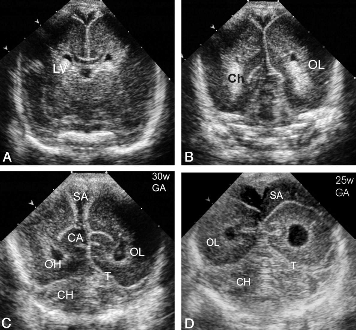
    Normal coronal sections of the PF (superior to inferior). CA indicates calcar avis; Ch, choroid plexus; CH, cerebellar hemispheres; LV, lateral ventricle trigone; OH, occipital horn; OL, occipital lobe; SA, subarachnoid space; and T, tentorium.

    A–C, Images in a neonate at 30 weeks’ gestational age.

    A, The most superior coronal view.

    B, The middle coronal view.

    C, The inferior view.

    D, Same section as in C in a neonate at 25 weeks’ gestational age shows a more rounded configuration of occipital horns and a wide (up to 15 mm) subarachnoid space with internal echogenic dots corresponding to vessels.
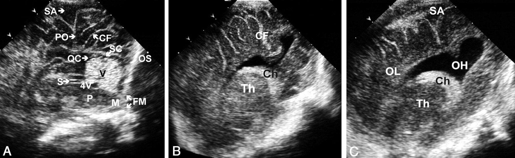
  • Fig 3.
    • Download figure
    • Open in new tab
    • Download powerpoint
    Fig 3.

    Normal sections of the PF. CF indicates calcarine fissure; Ch, choroid plexus; FM, foramen magnum; M, medulla; OH, occipital horn; OL, occipital lobe; OS, occipital squama; PO, parieto-occipital fissure; QC; quadrigeminal cistern; S, aqueduct of Sylvius; SA, subarachnoid space; SC, supracerebellar cistern; Th, thalamus; V, vermis; and 4V, fourth ventricle.

    A, Midsagittal view.

    B, Parasagittal view.

    C, More lateral parasagittal view.

  • Fig 4.
    • Download figure
    • Open in new tab
    • Download powerpoint
    Fig 4.

    Sonograms obtained in a neonate with a presumptive diagnosis of posterior fossa malformation at prenatal sonography.

    A, AF midline sagittal view shows a cystic image (asterisk) in the posterior fossa behind the cerebellum.

    B, PF midline sagittal view clearly demonstrates that the lesion corresponds to a megacisterna magna, depicting the normal-sized cerebellum vermis and fourth ventricle. Note that the cystic lesion (asterisk) extends around the cerebellum without compressing it, as often occurs with arachnoid cysts.

  • Fig 5.
    • Download figure
    • Open in new tab
    • Download powerpoint
    Fig 5.

    Images obtained in a premature neonate with systemic candidiasis treated with prolonged antibiotic therapy; disease was confirmed on the basis of blood culture findings.

    A, AF right parasagittal sonogram demonstrates multiple, rounded, hyperechoic nodules—some with central umbilication—affecting the basal nuclei (arrowheads).

    B, PF right parasagittal sonogram also shows multiple, rounded, hyperechoic lesions affecting the frontal and parietal lobes (arrowheads).

    C, Parasagittal T1-weighted MR image confirms the sonographic findings. (The child survived, and the lesions [arrowheads] calcified.)

  • Fig 6.
    • Download figure
    • Open in new tab
    • Download powerpoint
    Fig 6.

    Images of cerebellar abscesses in a 45-day-old neonate presenting with AF bulging and lethargy. A culture of fine-needle aspiration material isolated S aureus.

    A, PF coronal sonogram reveals bilateral occipital horn dilatation and enlarged left cerebellar hemisphere with two hypoechoic lesions (arrowheads).

    B, PF left parasagittal sonogram shows lateral ventricle dilatation and the cerebellar abscesses (arrowheads).

    C, Contrast-enhanced T2-weighted MR image confirms the sonographic findings (arrowheads).
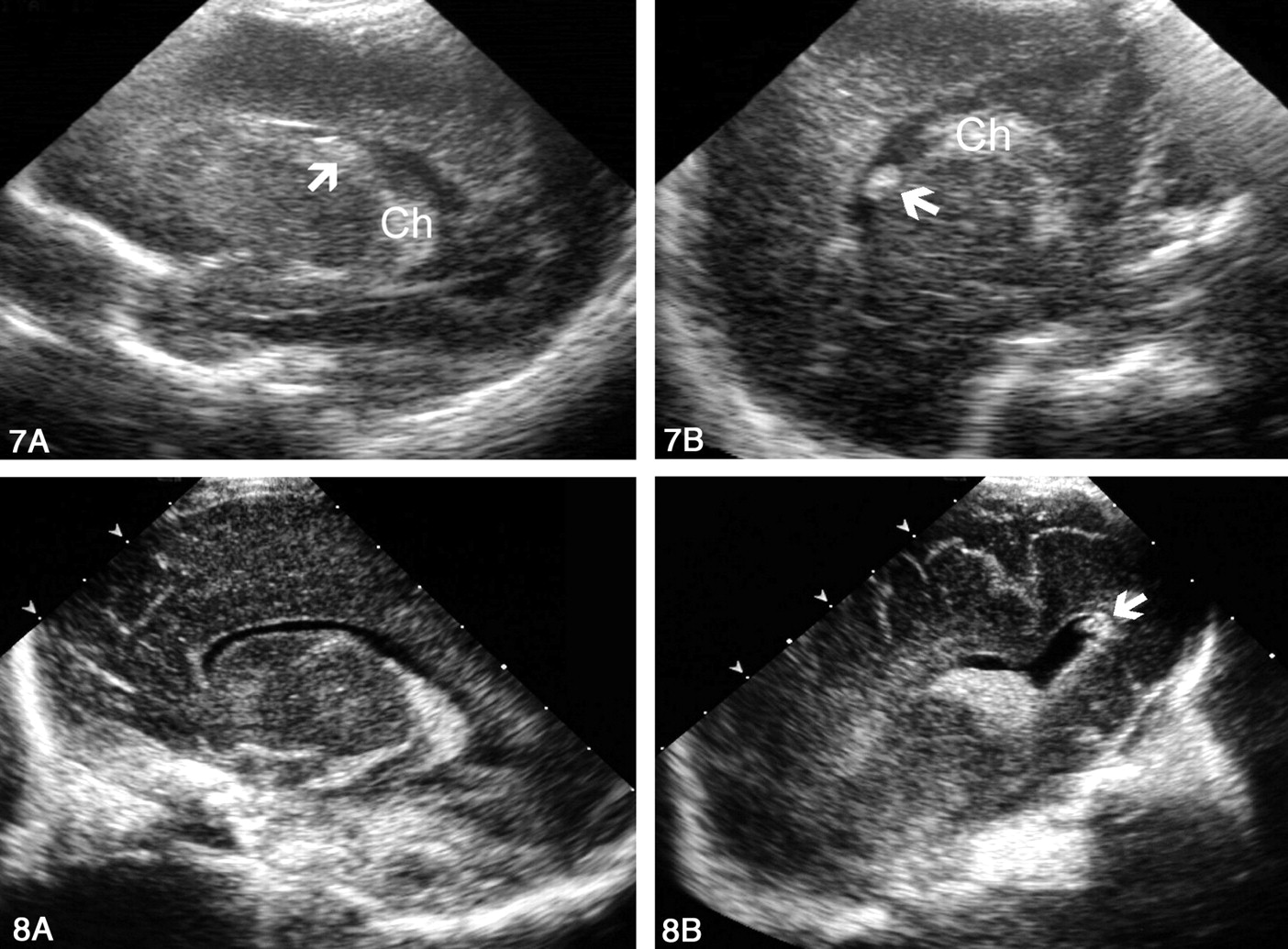
  • Fig 7.
    • Download figure
    • Open in new tab
    • Download powerpoint
    Fig 7.

    Sonograms obtained in a premature neonate with grade I hemorrhage that was better delineated through the PF than through the AF.

    A, AF left parasagittal view shows an equivocally echogenic image (arrow). The choroid plexus (Ch) is not well depicted.

    B, PF left parasagittal view clearly depicts the echogenic lesion (arrow) independent of the choroids (Ch). On follow-up studies, the lesion disappeared.

  • Fig 8.
    • Download figure
    • Open in new tab
    • Download powerpoint
    Fig 8.

    Sonograms obtained in a premature neonate with grade II hemorrhage.

    A, AF left parasagittal view shows a normal-appearing left lateral ventricle.

    B, PF left parasagittal view reveals a small clot in the posteriormost portion of the occipital horn (arrow).
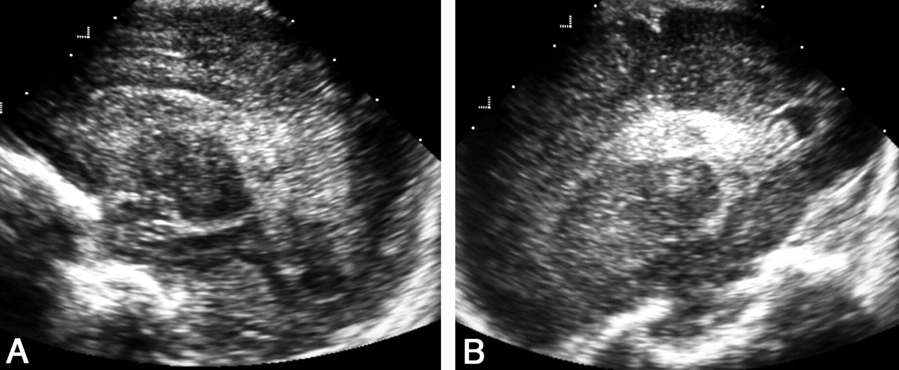
  • Fig 9.
    • Download figure
    • Open in new tab
    • Download powerpoint
    Fig 9.

    Sonograms obtained in a premature neonate with grade II intraventricular hemorrhage.

    A, AF right parasagittal view shows inconclusive appearance of the choroid plexus.

    B, PF right parasagittal view clearly depicts two clots in the occipital horn (arrows).

  • Fig 10.
    • Download figure
    • Open in new tab
    • Download powerpoint
    Fig 10.

    Sonogram of the occipital horn layering. Coronal PF view shows a fluid-fluid level (arrowheads) caused by blood and CSF in a slightly dilated left occipital horn.

  • Fig 11.
    • Download figure
    • Open in new tab
    • Download powerpoint
    Fig 11.

    Sonograms of the calcar avis simulating intraventricular hemorrhage.

    A, AF parasagittal view shows a rounded masslike image mimicking an intraventricular clot within the occipital horn (arrow).

    B, PF parasagittal view demonstrates that the mass is the calcar avis (arrow). Note its continuity with the occipital white matter and the calcarine fissure (CF).

  • Fig 12.
    • Download figure
    • Open in new tab
    • Download powerpoint
    Fig 12.

    Sonograms of bilateral cerebellar hemorrhage obtained in a neonate without intraventricular hemorrhage at 30 weeks’ gestational age.

    A, AF coronal view shows the lateral ventricles to be normal. Echogenic areas in the subtentorial space (arrows) of indistinct cerebellar or extracerebellar origin are seen.

    B, PF coronal view clearly reveals bilateral cerebellar hemorrhages (arrows).

    C, PF coronal view obtained 1 week later shows that the hemorrhages have decreased in size and echogenicity (arrows).

  • Fig 13.
    • Download figure
    • Open in new tab
    • Download powerpoint
    Fig 13.

    Sonograms obtained in a premature neonate with massive intraventricular and unilateral cerebellar hemorrhages. T indicates tentorium.

    A, AF coronal view shows echogenic material occupying the right lateral ventricle (asterisk) that hinders correct visualization of the cerebellum owing to slight shadowing.

    B, PF coronal view clearly depicts left echogenic cerebellar hemorrhage (arrows). OH indicates the occipital horn.

  • Fig 14.
    • Download figure
    • Open in new tab
    • Download powerpoint
    Fig 14.

    Sonograms of abnormal peritrigonal hyperechogenicity in a premature neonate with periventricular leukomalacia.

    A, AF left parasagittal view depicts heterogeneous peritrigonal hyperechogenicity with areas of cavitation.

    B, PF left parasagittal view. Coarse peritrigonal echogenicity and cavities are evident.

  • Fig 15.
    • Download figure
    • Open in new tab
    • Download powerpoint
    Fig 15.

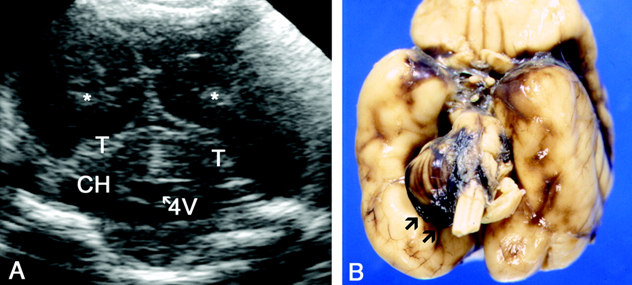
    Sonogram and gross specimen obtained in a patient with discrepant findings at sonography and autopsy.

    A, PF coronal sonogram shows echogenic material in the occipital horns (asterisk) corresponding to intraventricular hemorrhage. The cerebellum and basal subarachnoid space seem normal. CH indicates cerebellar hemispheres; T, tentorium; and 4V, fourth ventricle.

    B, Macroscopic view of the specimen 1 day later shows a hemorrhage surrounding the right cerebellar hemisphere.

  • Fig 16.
    • Download figure
    • Open in new tab
    • Download powerpoint
    Fig 16.

    Sonograms of a hemorrhage originating in the left choroid plexus.

    A, AF left parasagittal view shows an inconclusive choroid appearance.

    B, PF left parasagittal view depicts an enlarged choroid with heterogeneous echogenicity due to internal hemorrhage.

Tables

  • Figures
  • Statistical analysis of the diagnosis of hemorrhagic lesions with AF and PF sonography

    HemorrhageBetter Approach for DiagnosisP Value, McNemar TestKappa Index (95% CI)
    Grade IAF.0010.165 (0.028, 0.301)
    Grade IIPF.0010.517 (0.336, 0.698)
    Grade IIIAF and PF equal>.050.889 (0.764, 1.013)
    Hemorrhagic infarctionAF.0340.647 (0.421, 0.873)
PreviousNext
Back to top

In this issue

American Journal of Neuroradiology: 25 (7)
American Journal of Neuroradiology
Vol. 25, Issue 7
1 Aug 2004
  • Table of Contents
  • Index by author
Advertisement
Print
Download PDF
Email Article

Thank you for your interest in spreading the word on American Journal of Neuroradiology.

NOTE: We only request your email address so that the person you are recommending the page to knows that you wanted them to see it, and that it is not junk mail. We do not capture any email address.

Enter multiple addresses on separate lines or separate them with commas.
Posterior Fontanelle Sonography: An Acoustic Window into the Neonatal Brain
(Your Name) has sent you a message from American Journal of Neuroradiology
(Your Name) thought you would like to see the American Journal of Neuroradiology web site.
CAPTCHA
This question is for testing whether or not you are a human visitor and to prevent automated spam submissions.
Cite this article
Flavia Correa, Goya Enríquez, José Rosselló, Javier Lucaya, Joaquim Piqueras, Celestino Aso, Elida Vázquez, Arantxa Ortega, Alfredo Gallart
Posterior Fontanelle Sonography: An Acoustic Window into the Neonatal Brain
American Journal of Neuroradiology Aug 2004, 25 (7) 1274-1282;

Citation Manager Formats

  • BibTeX
  • Bookends
  • EasyBib
  • EndNote (tagged)
  • EndNote 8 (xml)
  • Medlars
  • Mendeley
  • Papers
  • RefWorks Tagged
  • Ref Manager
  • RIS
  • Zotero
0 Responses
Respond to this article
Share
Bookmark this article
Posterior Fontanelle Sonography: An Acoustic Window into the Neonatal Brain
Flavia Correa, Goya Enríquez, José Rosselló, Javier Lucaya, Joaquim Piqueras, Celestino Aso, Elida Vázquez, Arantxa Ortega, Alfredo Gallart
American Journal of Neuroradiology Aug 2004, 25 (7) 1274-1282;
del.icio.us logo Twitter logo Facebook logo Mendeley logo
  • Tweet Widget
  • Facebook Like
  • Google Plus One

Jump to section

  • Article
    • Abstract
    • Methods
    • Results
    • Discussion
    • Conclusion
    • Acknowledgments
    • Footnotes
    • References
  • Figures & Data
  • Info & Metrics
  • Responses
  • References
  • PDF

Related Articles

  • No related articles found.
  • PubMed
  • Google Scholar

Cited By...

  • AIUM Practice Guideline for the Performance of a Transcranial Doppler Ultrasound Examination for Adults and Children
  • Cranial ultrasound and MRI at term age in extremely preterm infants
  • Cranial ultrasound findings in well newborn Ugandan infants
  • Crossref
  • Google Scholar

This article has not yet been cited by articles in journals that are participating in Crossref Cited-by Linking.

More in this TOC Section

  • Early Ultrasonic Monitoring of Brain Growth and Later Neurodevelopmental Outcome in Very Preterm Infants
  • Diagnostic Value of Sylvian Fissure Hyperechogenicity in Fetal SAH
  • ACTA2-Related Dysgyria: An Under-Recognized Malformation of Cortical Development
Show more Pediatrics

Similar Articles

Advertisement

Indexed Content

  • Current Issue
  • Accepted Manuscripts
  • Article Preview
  • Past Issues
  • Editorials
  • Editor's Choice
  • Fellows' Journal Club
  • Letters to the Editor
  • Video Articles

Cases

  • Case Collection
  • Archive - Case of the Week
  • Archive - Case of the Month
  • Archive - Classic Case

More from AJNR

  • Trainee Corner
  • Imaging Protocols
  • MRI Safety Corner
  • Book Reviews

Multimedia

  • AJNR Podcasts
  • AJNR Scantastics

Resources

  • Turnaround Time
  • Submit a Manuscript
  • Submit a Video Article
  • Submit an eLetter to the Editor/Response
  • Manuscript Submission Guidelines
  • Statistical Tips
  • Fast Publishing of Accepted Manuscripts
  • Graphical Abstract Preparation
  • Imaging Protocol Submission
  • Evidence-Based Medicine Level Guide
  • Publishing Checklists
  • Author Policies
  • Become a Reviewer/Academy of Reviewers
  • News and Updates

About Us

  • About AJNR
  • Editorial Board
  • Editorial Board Alumni
  • Alerts
  • Permissions
  • Not an AJNR Subscriber? Join Now
  • Advertise with Us
  • Librarian Resources
  • Feedback
  • Terms and Conditions
  • AJNR Editorial Board Alumni

American Society of Neuroradiology

  • Not an ASNR Member? Join Now

© 2025 by the American Society of Neuroradiology All rights, including for text and data mining, AI training, and similar technologies, are reserved.
Print ISSN: 0195-6108 Online ISSN: 1936-959X

Powered by HighWire