Advertisement

Main menu

  • Home
  • Content
    • Current Issue
    • Accepted Manuscripts
    • Article Preview
    • Past Issue Archive
    • Video Articles
    • AJNR Case Collection
    • Case of the Week Archive
    • Case of the Month Archive
    • Classic Case Archive
  • Special Collections
    • AJNR Awards
    • Low-Field MRI
    • Alzheimer Disease
    • ASNR Foundation Special Collection
    • Photon-Counting CT
    • View All
  • Multimedia
    • AJNR Podcasts
    • AJNR SCANtastic
    • Trainee Corner
    • MRI Safety Corner
    • Imaging Protocols
  • For Authors
    • Submit a Manuscript
    • Submit a Video Article
    • Submit an eLetter to the Editor/Response
    • Manuscript Submission Guidelines
    • Statistical Tips
    • Fast Publishing of Accepted Manuscripts
    • Graphical Abstract Preparation
    • Imaging Protocol Submission
    • Author Policies
  • About Us
    • About AJNR
    • Editorial Board
    • Editorial Board Alumni
  • More
    • Become a Reviewer/Academy of Reviewers
    • Subscribers
    • Permissions
    • Alerts
    • Feedback
    • Advertisers
    • ASNR Home

User menu

  • Alerts
  • Log in

Search

  • Advanced search
American Journal of Neuroradiology

American Journal of Neuroradiology

ASHNR American Society of Functional Neuroradiology ASHNR American Society of Pediatric Neuroradiology ASSR
  • Alerts
  • Log in

Advanced Search

  • Home
  • Content
    • Current Issue
    • Accepted Manuscripts
    • Article Preview
    • Past Issue Archive
    • Video Articles
    • AJNR Case Collection
    • Case of the Week Archive
    • Case of the Month Archive
    • Classic Case Archive
  • Special Collections
    • AJNR Awards
    • Low-Field MRI
    • Alzheimer Disease
    • ASNR Foundation Special Collection
    • Photon-Counting CT
    • View All
  • Multimedia
    • AJNR Podcasts
    • AJNR SCANtastic
    • Trainee Corner
    • MRI Safety Corner
    • Imaging Protocols
  • For Authors
    • Submit a Manuscript
    • Submit a Video Article
    • Submit an eLetter to the Editor/Response
    • Manuscript Submission Guidelines
    • Statistical Tips
    • Fast Publishing of Accepted Manuscripts
    • Graphical Abstract Preparation
    • Imaging Protocol Submission
    • Author Policies
  • About Us
    • About AJNR
    • Editorial Board
    • Editorial Board Alumni
  • More
    • Become a Reviewer/Academy of Reviewers
    • Subscribers
    • Permissions
    • Alerts
    • Feedback
    • Advertisers
    • ASNR Home
  • Follow AJNR on Twitter
  • Visit AJNR on Facebook
  • Follow AJNR on Instagram
  • Join AJNR on LinkedIn
  • RSS Feeds

AJNR Awards, New Junior Editors, and more. Read the latest AJNR updates

Research ArticleHEAD AND NECK

Pediatric Head and Neck Lesions: Assessment of Vascularity by MR Digital Subtraction Angiography

Weng Kong Chooi, Neil Woodhouse, Stuart C. Coley and Paul D. Griffiths
American Journal of Neuroradiology August 2004, 25 (7) 1251-1255;
Weng Kong Chooi
  • Find this author on Google Scholar
  • Find this author on PubMed
  • Search for this author on this site
Neil Woodhouse
  • Find this author on Google Scholar
  • Find this author on PubMed
  • Search for this author on this site
Stuart C. Coley
  • Find this author on Google Scholar
  • Find this author on PubMed
  • Search for this author on this site
Paul D. Griffiths
  • Find this author on Google Scholar
  • Find this author on PubMed
  • Search for this author on this site
  • Article
  • Figures & Data
  • Info & Metrics
  • Responses
  • References
  • PDF
Loading

Article Figures & Data

Figures

  • Tables
  • Fig 1.
    • Download figure
    • Open in new tab
    • Download powerpoint
    Fig 1.

    Images from the case of a 3-year-old female patient with a suspected vascular abnormality of the scalp.

    A, Unenhanced sagittal T1-weighted MR image shows an extra-calvarial soft tissue mass.

    B, Marked uniform enhancement can be seen after the administration of contrast material. Serpiginous flow void represents an enlarged feeding vessel.

    C, Selected MR digital subtraction angiograms show prominent filling of contrast material in the arterial phase and prominent enhancement of the lesion. This was considered to represent a high-flow lesion.

  • Fig 2.
    • Download figure
    • Open in new tab
    • Download powerpoint
    Fig 2.

    Images from the case of a 14-month-old female patient with left-sided neck swelling.

    A, Unenhanced axial T1-weighted MR image shows an encapsulated mass in the left parotid gland.

    B, After the administration of contrast material, marked contrast enhancement is seen. Note the vascular flow voids within the lesion.

    C, Selected MR digital subtraction angiograms show early arterial filling of the hypervascular lesion and arteriovenous shunting into an early draining vein. This is consistent with a parotid hemangioma in the proliferative phase.
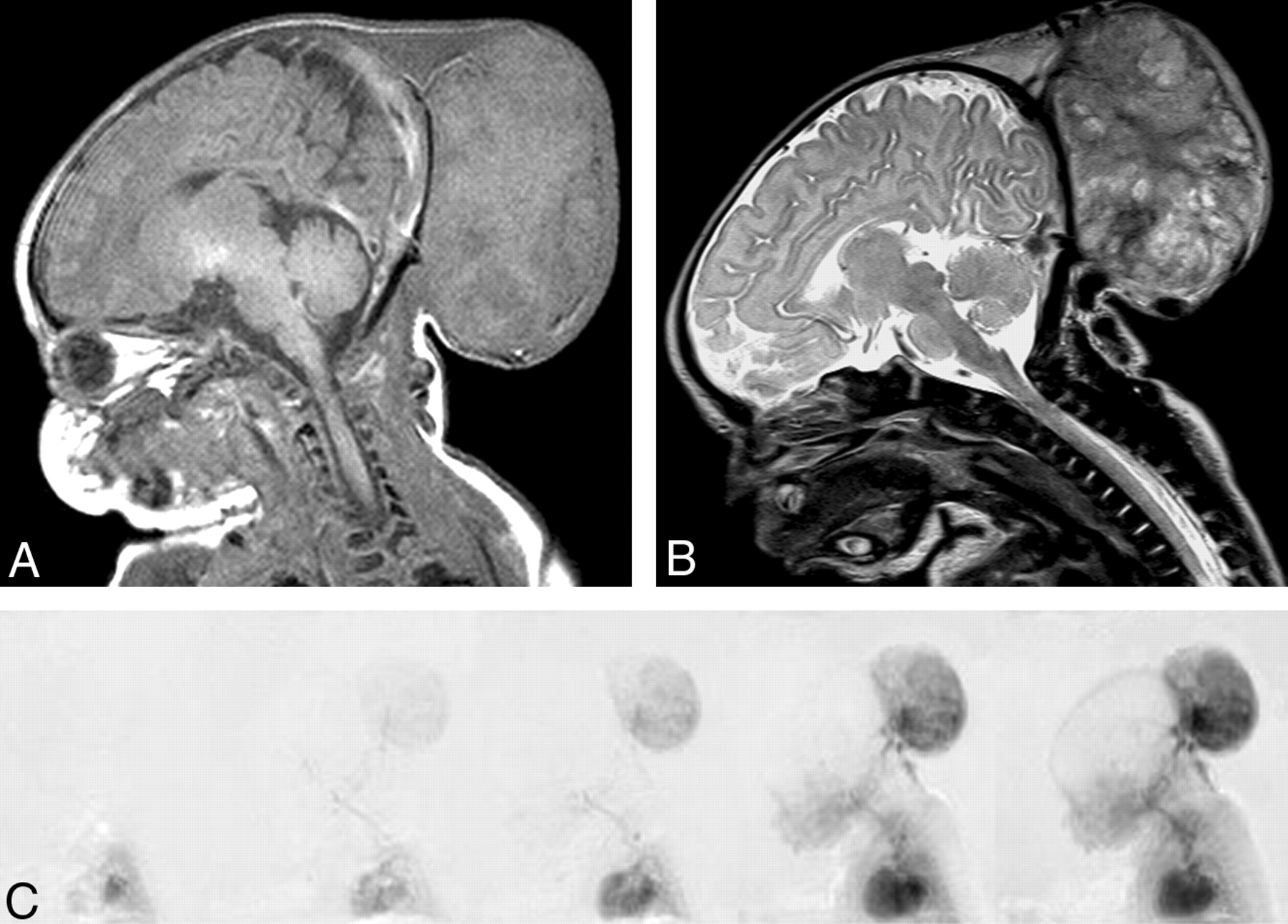
  • Fig 3.
    • Download figure
    • Open in new tab
    • Download powerpoint
    Fig 3.

    Images from the case of a 5-day-old female patient with a large occipital mass thought to be an encephalocele.

    A, Sagittal T1-weighted MR image shows a large occipital mass with intermediate signal intensity.

    B, More heterogeneous appearance can be seen on the T2-weighted image. No intracranial communication is shown.

    C, Selected MR digital subtraction angiograms show rapid opacification of the lesion during the early arterial phase. Flow in this lesion was so high that the intracranial vessels are poorly shown. Pathologic examination revealed an undifferentiated highly vascular tumor.
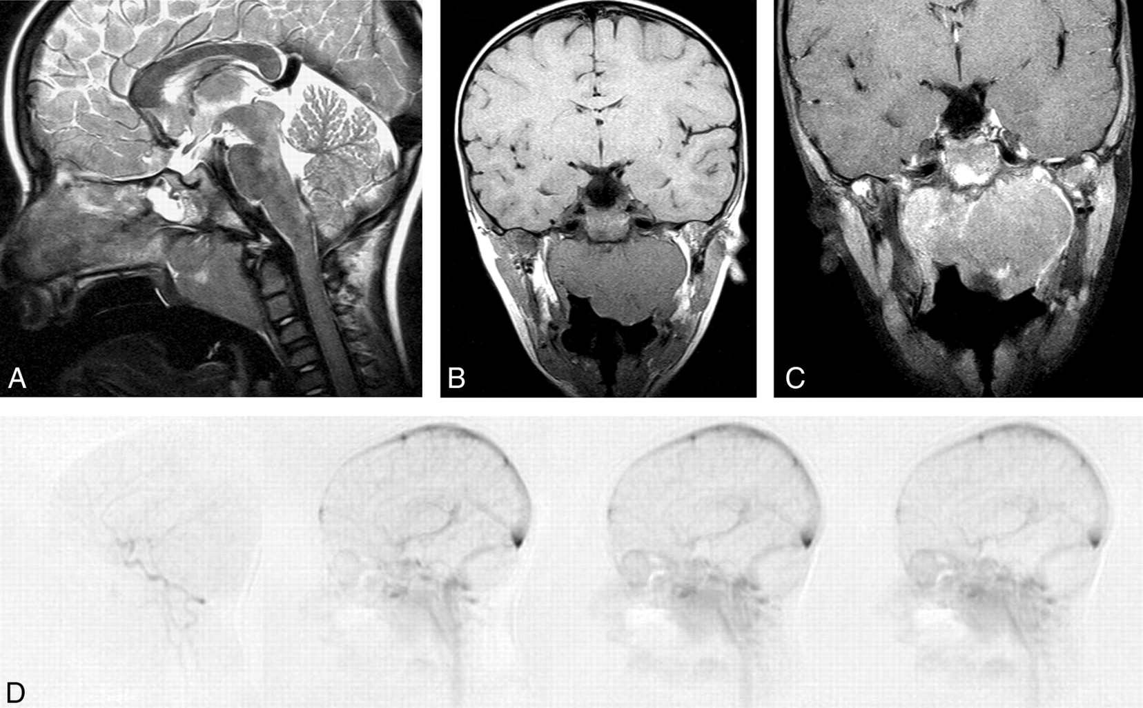
  • Fig 4.
    • Download figure
    • Open in new tab
    • Download powerpoint
    Fig 4.

    Images from the case of a 5-year-old male patient with a nasopharyngeal mass.

    A, Lobulated nasopharyngeal mass is seen on the sagittal T2-weighted MR image.

    B, T1-weighted coronal MR image also shows the lobulated nasopharyngeal mass.

    C, Coronal contrast-enhanced fat-saturated T1-weighted MR image shows minor eccentric contrast enhancement.

    D, No significant contrast material flow was seen within the lesion on MR digital subtraction angiograms. This lesion was diagnosed as Burkitt lymphoma on the basis of biopsy findings.

Tables

  • Figures
  • Clinical details and MR imaging findings

    Case No.Sex, AgeClinical DetailsConventional MR Imaging FindingsMR-DSA VascularityFinal Diagnosis
    1 (Fig. 1)F, 3 yr*Scalp mass, indeterminate vascularityEnhancing soft tissue mass with flow voidHighExtracranial AVM
    2 (Fig. 2)F, 14 moNeck swelling, indeterminate vascularityEnhancing soft tissue mass with flow voidsHighHemangioma (proliferative phase)
    3 (Fig. 3)F, 1 wkLarge occipital mass, presumed occipital encephaloceleEnhancing heterogeneous extra-calvarial massHighUndifferentiated highly vascular tumor (+)
    4F, 2 yr*Eyelid mass, ?dermoid, ?encephaloceleEnhancing mass with flow voidsHighAVM
    5M, 15 moEyelid hemangiomaEnhancing rounded eyelid mass, no flow voidsHighAVM
    6 (Fig. 4)M, 5 yr*Nasopharyngeal massHeterogeneous mass with minor enhancementLowBurkitt lymphoma (+)
    7F, 2 d*Large palatal massLarge enhancing tumorHighCystic teratoma (+)
    8F, 16 yrSturge-Weber syndrome, ?ocular angiomaChoroidal thickening with enhancementLowDiffuse choroidal angioma plus detachment
    9M, 2 yr*Eyelid hemangiomaEnhancing soft tissue mass, no flow voidsLowHemangioma
    10F, 3 yr*Scalp lesionEnhancing soft tissue mass, no flow voidsLowVenous malformation
    11M, 10 yr*?VI naevus flammeus, ?Sturge-Weber syndromeChoroidal thickening with enhancementLowDiffuse choroidal angioma
    12M, 12 yr*Known cervical osteochondromaDisplacement but no invasion of the vertebral artery by bony tumorLowOsteochondroma with no vascular invasion (+)
    • Note.—DSA indicates digital subtraction angiography; F, female; M, male; AVM, arteriovenous malformation; (+), histologic confirmation; ?, possible.

    • * MR imaging examinations performed with the patient under general anesthesia.

PreviousNext
Back to top

In this issue

American Journal of Neuroradiology: 25 (7)
American Journal of Neuroradiology
Vol. 25, Issue 7
1 Aug 2004
  • Table of Contents
  • Index by author
Advertisement
Print
Download PDF
Email Article

Thank you for your interest in spreading the word on American Journal of Neuroradiology.

NOTE: We only request your email address so that the person you are recommending the page to knows that you wanted them to see it, and that it is not junk mail. We do not capture any email address.

Enter multiple addresses on separate lines or separate them with commas.
Pediatric Head and Neck Lesions: Assessment of Vascularity by MR Digital Subtraction Angiography
(Your Name) has sent you a message from American Journal of Neuroradiology
(Your Name) thought you would like to see the American Journal of Neuroradiology web site.
CAPTCHA
This question is for testing whether or not you are a human visitor and to prevent automated spam submissions.
Cite this article
Weng Kong Chooi, Neil Woodhouse, Stuart C. Coley, Paul D. Griffiths
Pediatric Head and Neck Lesions: Assessment of Vascularity by MR Digital Subtraction Angiography
American Journal of Neuroradiology Aug 2004, 25 (7) 1251-1255;

Citation Manager Formats

  • BibTeX
  • Bookends
  • EasyBib
  • EndNote (tagged)
  • EndNote 8 (xml)
  • Medlars
  • Mendeley
  • Papers
  • RefWorks Tagged
  • Ref Manager
  • RIS
  • Zotero
0 Responses
Respond to this article
Share
Bookmark this article
Pediatric Head and Neck Lesions: Assessment of Vascularity by MR Digital Subtraction Angiography
Weng Kong Chooi, Neil Woodhouse, Stuart C. Coley, Paul D. Griffiths
American Journal of Neuroradiology Aug 2004, 25 (7) 1251-1255;
del.icio.us logo Twitter logo Facebook logo Mendeley logo
  • Tweet Widget
  • Facebook Like
  • Google Plus One

Jump to section

  • Article
    • Abstract
    • Methods
    • Results
    • Discussion
    • Conclusion
    • Footnotes
    • References
  • Figures & Data
  • Info & Metrics
  • Responses
  • References
  • PDF

Related Articles

  • No related articles found.
  • PubMed
  • Google Scholar

Cited By...

  • No citing articles found.
  • Crossref
  • Google Scholar

This article has not yet been cited by articles in journals that are participating in Crossref Cited-by Linking.

More in this TOC Section

  • Correlation of Apparent Diffusion Coefficient at 3T with Prognostic Parameters of Retinoblastoma
  • Parathyroid Lesions: Characterization with Dual-Phase Arterial and Venous Enhanced CT of the Neck
  • MR Diagnosis of Facial Neuritis: Diagnostic Performance of Contrast-Enhanced 3D-FLAIR Technique Compared with Contrast-Enhanced 3D-T1-Fast-Field Echo with Fat Suppression
Show more HEAD AND NECK

Similar Articles

Advertisement

Indexed Content

  • Current Issue
  • Accepted Manuscripts
  • Article Preview
  • Past Issues
  • Editorials
  • Editor's Choice
  • Fellows' Journal Club
  • Letters to the Editor
  • Video Articles

Cases

  • Case Collection
  • Archive - Case of the Week
  • Archive - Case of the Month
  • Archive - Classic Case

More from AJNR

  • Trainee Corner
  • Imaging Protocols
  • MRI Safety Corner
  • Book Reviews

Multimedia

  • AJNR Podcasts
  • AJNR Scantastics

Resources

  • Turnaround Time
  • Submit a Manuscript
  • Submit a Video Article
  • Submit an eLetter to the Editor/Response
  • Manuscript Submission Guidelines
  • Statistical Tips
  • Fast Publishing of Accepted Manuscripts
  • Graphical Abstract Preparation
  • Imaging Protocol Submission
  • Evidence-Based Medicine Level Guide
  • Publishing Checklists
  • Author Policies
  • Become a Reviewer/Academy of Reviewers
  • News and Updates

About Us

  • About AJNR
  • Editorial Board
  • Editorial Board Alumni
  • Alerts
  • Permissions
  • Not an AJNR Subscriber? Join Now
  • Advertise with Us
  • Librarian Resources
  • Feedback
  • Terms and Conditions
  • AJNR Editorial Board Alumni

American Society of Neuroradiology

  • Not an ASNR Member? Join Now

© 2025 by the American Society of Neuroradiology All rights, including for text and data mining, AI training, and similar technologies, are reserved.
Print ISSN: 0195-6108 Online ISSN: 1936-959X

Powered by HighWire