
Article Figures & Data
Figures
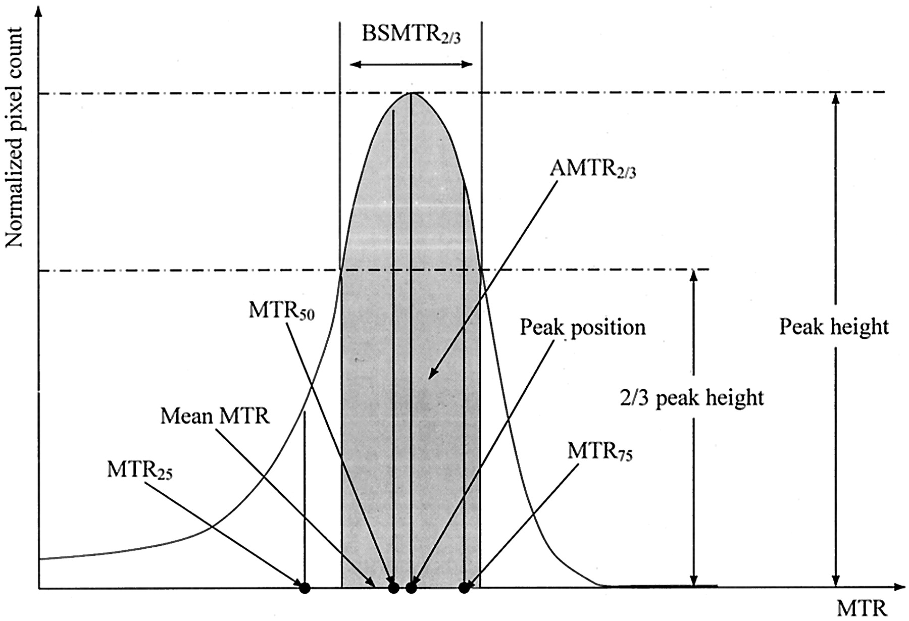
- Fig 1.
Graphical representation of the MTR histogram–derived parameters used in this study.
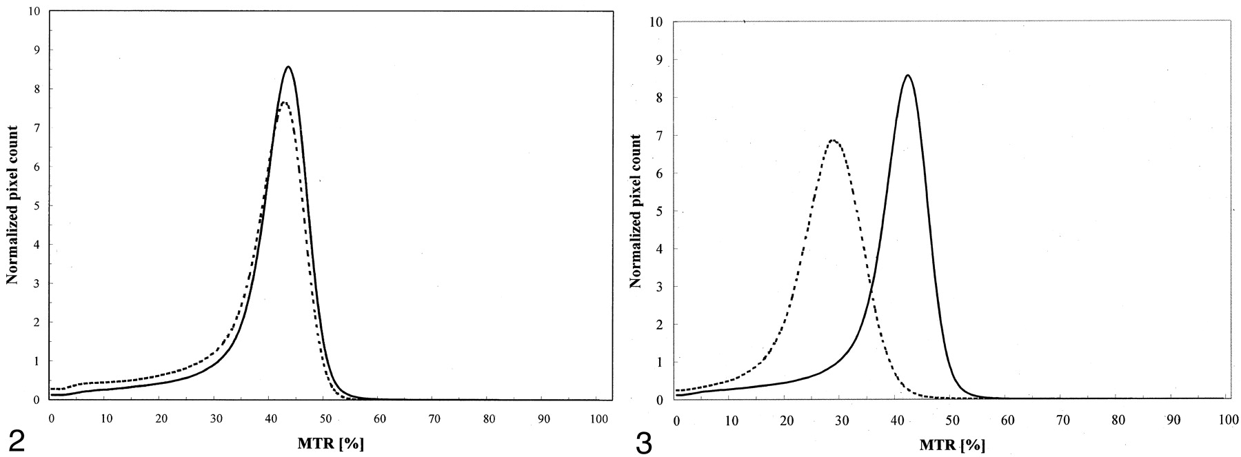
- Fig 2.
Average MTR histograms from 10 patients with MS (dotted line) and 10 healthy control subjects (solid line) obtained by using the same MR imaging unit.
- Fig 3.
Average MTR histograms from groups B (solid line) and C (dotted line), the two control groups, who were imaged with different MR imaging units.
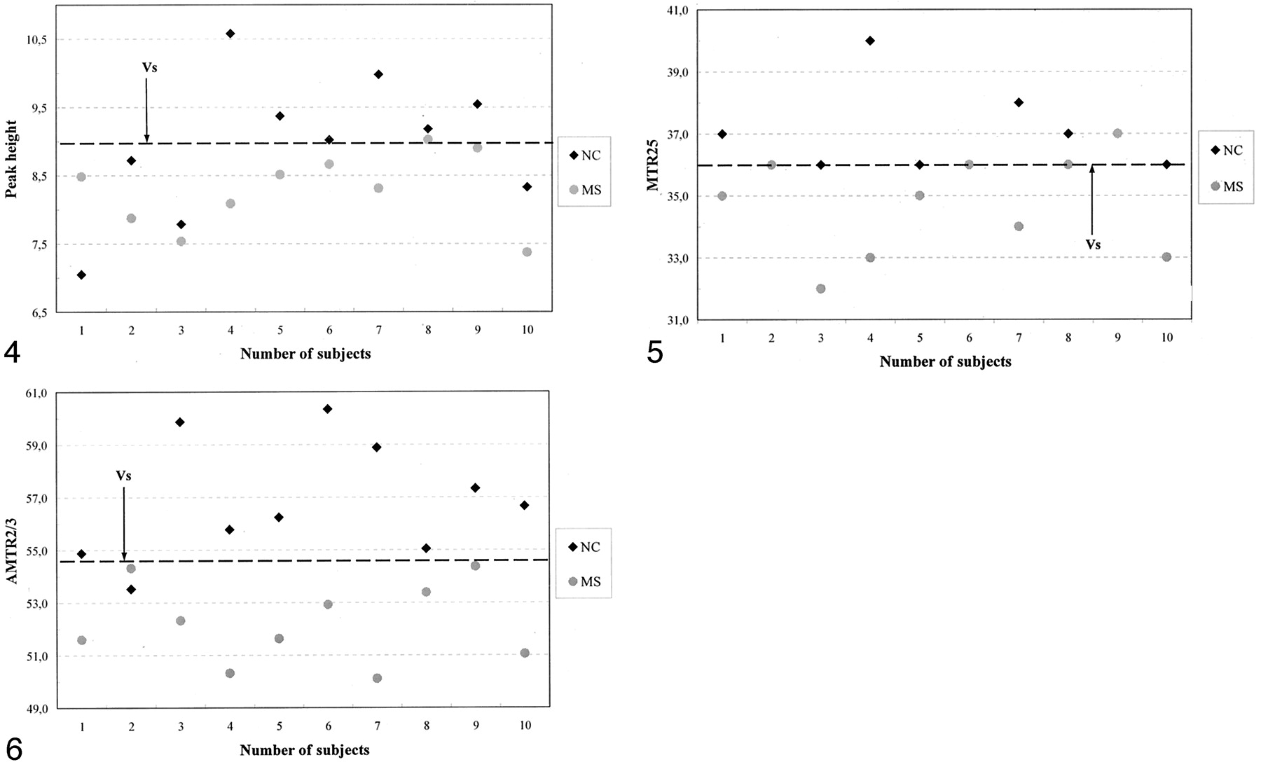
- Fig 4.
Scatterplot of MTR histogram peak heights indicates the value that best distinguished patients with MS and the control group B.
- Fig 5.
Scatterplot of MTR25 values indicates the value that best distinguished patients from the control group B.
- Fig 6.
Scatterplot of AMTR2/3 values indicates that AMTR2/3 was the best parameter for differentiating patients from control subjects.


Tables
Parameter MS Patient Group A (n = 10) Healthy Control Group B (n = 10) Relative Difference P Value Mean ± SD SD/Mean Mean ± SD SD/Mean Global MTR 37.19 ± 1.44 3.87% 38.97 ± 1.07 2.75% −4.69% .006 Mean MTR 37.37 ± 1.43 3.83% 39.11 ± 1.04 2.65% −4.55% .006 MTR histogram peak height 8.28 ± 0.55 6.64% 8.99 ± 1.03 11.45% −8.14% .07 MTR histogram peak position 41.30 ± 1.34 3.24% 42.10 ± 0.88 2.09% −1.92% .10 MTR25 34.70 ± 1.64 4.73% 36.90 ± 1.29 3.49% −6.15% .004 MTR50 39.60 ± 1.35 3.41% 40.70 ± 1.06 2.60% −2.74% .06 MTR75 42.60 ± 1.35 3.17% 43.60 ± 0.97 2.22% −2.32% .07 AMTR2/3 52.23 ± 1.46 2.80% 57.03 ± 2.47 4.33% −8.79% <.001 BSMTR2/3 6.94 ± 0.53 7.64% 6.77 ± 0.86 12.70% +2.40% .60 - TABLE 2:
MT MR imaging–derived parameters in control subjects imaged with different imaging systems
Parameter Control Group B (n = 10) Control Group C (n = 4) Relative Difference P Value Mean ± SD SD/Mean Mean ± SD SD/Mean Global MTR 38.97 ± 1.07 2.75% 27.56 ± 0.97 3.53% 34.31% <.001 Mean MTR 39.11 ± 1.04 2.65% 27.69 ± 0.96 3.45% 34.19% <.001 MTR histogram peak height 8.99 ± 1.03 11.45% 7.23 ± 0.52 7.17% 21.73% .007 MTR histogram peak position 42.10 ± 0.88 2.09% 28.75 ± 0.96 3.34% 37.69% <.001 MTR25 36.90 ± 1.29 3.49% 24.00 ± 1.41 5.89% 42.36% <.001 MTR50 40.70 ± 1.06 2.60% 28.00 ± 0.82 2.92% 36.97% <.001 MTR75 43.60 ± 0.97 2.22% 31.75 ± 0.96 3.02% 31.45% <.001 AMTR2/3 57.03 ± 2.47 4.33% 59.50 ± 1.55 2.62% 4.25% .091 BSMTR2/3 6.77 ± 0.86 12.70% 9.25 ± 0.57 6.16% 30.92% <.001


