Advertisement

Main menu

  • Home
  • Content
    • Current Issue
    • Accepted Manuscripts
    • Article Preview
    • Past Issue Archive
    • Video Articles
    • AJNR Case Collection
    • Case of the Week Archive
    • Case of the Month Archive
    • Classic Case Archive
  • Special Collections
    • AJNR Awards
    • Low-Field MRI
    • Alzheimer Disease
    • ASNR Foundation Special Collection
    • Photon-Counting CT
    • View All
  • Multimedia
    • AJNR Podcasts
    • AJNR SCANtastic
    • Trainee Corner
    • MRI Safety Corner
    • Imaging Protocols
  • For Authors
    • Submit a Manuscript
    • Submit a Video Article
    • Submit an eLetter to the Editor/Response
    • Manuscript Submission Guidelines
    • Statistical Tips
    • Fast Publishing of Accepted Manuscripts
    • Graphical Abstract Preparation
    • Imaging Protocol Submission
    • Author Policies
  • About Us
    • About AJNR
    • Editorial Board
    • Editorial Board Alumni
  • More
    • Become a Reviewer/Academy of Reviewers
    • Subscribers
    • Permissions
    • Alerts
    • Feedback
    • Advertisers
    • ASNR Home

User menu

  • Alerts
  • Log in

Search

  • Advanced search
American Journal of Neuroradiology

American Journal of Neuroradiology

ASHNR American Society of Functional Neuroradiology ASHNR American Society of Pediatric Neuroradiology ASSR
  • Alerts
  • Log in

Advanced Search

  • Home
  • Content
    • Current Issue
    • Accepted Manuscripts
    • Article Preview
    • Past Issue Archive
    • Video Articles
    • AJNR Case Collection
    • Case of the Week Archive
    • Case of the Month Archive
    • Classic Case Archive
  • Special Collections
    • AJNR Awards
    • Low-Field MRI
    • Alzheimer Disease
    • ASNR Foundation Special Collection
    • Photon-Counting CT
    • View All
  • Multimedia
    • AJNR Podcasts
    • AJNR SCANtastic
    • Trainee Corner
    • MRI Safety Corner
    • Imaging Protocols
  • For Authors
    • Submit a Manuscript
    • Submit a Video Article
    • Submit an eLetter to the Editor/Response
    • Manuscript Submission Guidelines
    • Statistical Tips
    • Fast Publishing of Accepted Manuscripts
    • Graphical Abstract Preparation
    • Imaging Protocol Submission
    • Author Policies
  • About Us
    • About AJNR
    • Editorial Board
    • Editorial Board Alumni
  • More
    • Become a Reviewer/Academy of Reviewers
    • Subscribers
    • Permissions
    • Alerts
    • Feedback
    • Advertisers
    • ASNR Home
  • Follow AJNR on Twitter
  • Visit AJNR on Facebook
  • Follow AJNR on Instagram
  • Join AJNR on LinkedIn
  • RSS Feeds

AJNR Awards, New Junior Editors, and more. Read the latest AJNR updates

Getting new auth cookie, if you see this message a lot, tell someone!
OtherPEDIATRICS

Vacuolating Megalencephalic Leukoencephalopathy with Mild Clinical Course Validated by Diffusion Tensor Imaging and MR Spectroscopy

Yi-Fang Tu, Cheng-Yu Chen, Chao-Ching Huang and Chang-Shin Lee
American Journal of Neuroradiology June 2004, 25 (6) 1041-1045;
Yi-Fang Tu
  • Find this author on Google Scholar
  • Find this author on PubMed
  • Search for this author on this site
Cheng-Yu Chen
  • Find this author on Google Scholar
  • Find this author on PubMed
  • Search for this author on this site
Chao-Ching Huang
  • Find this author on Google Scholar
  • Find this author on PubMed
  • Search for this author on this site
Chang-Shin Lee
  • Find this author on Google Scholar
  • Find this author on PubMed
  • Search for this author on this site
  • Article
  • Figures & Data
  • Info & Metrics
  • Responses
  • References
  • PDF
Loading

Article Figures & Data

Figures

  • Tables
  • Fig 1.
    • Download figure
    • Open in new tab
    • Download powerpoint
    Fig 1.

    MR images obtained when the patient was 2 years 10 months old.

    A, Axial fast spin-echo T2-weighted image (TR/TE, 3500/93; two signals acquired) at the level of the basal ganglia, showing diffuse demyelination involving mainly the frontal lobe sparing the occipital subcortical regions and internal capsule.

    B, Coronal T1-weighted contrast-enhanced image (700/14), showing periventricular linear enhancement next to bilateral frontal horns.

    C, Axial T1-weighted contrast-enhanced image (700/14), demonstrating enhanced lesions in bilateral globus pallidi (arrows).

  • Fig 2.
    • Download figure
    • Open in new tab
    • Download powerpoint
    Fig 2.

    Follow-up axial T2-weighted MR image (4000/97.6), obtained when the patient was 5 years old, at the level of the basal ganglia, showing consistent demyelination at the same regions on Fig 1A with thinning of the genu of the corpus callosum and dilatation of the cavum septi pellucidi.
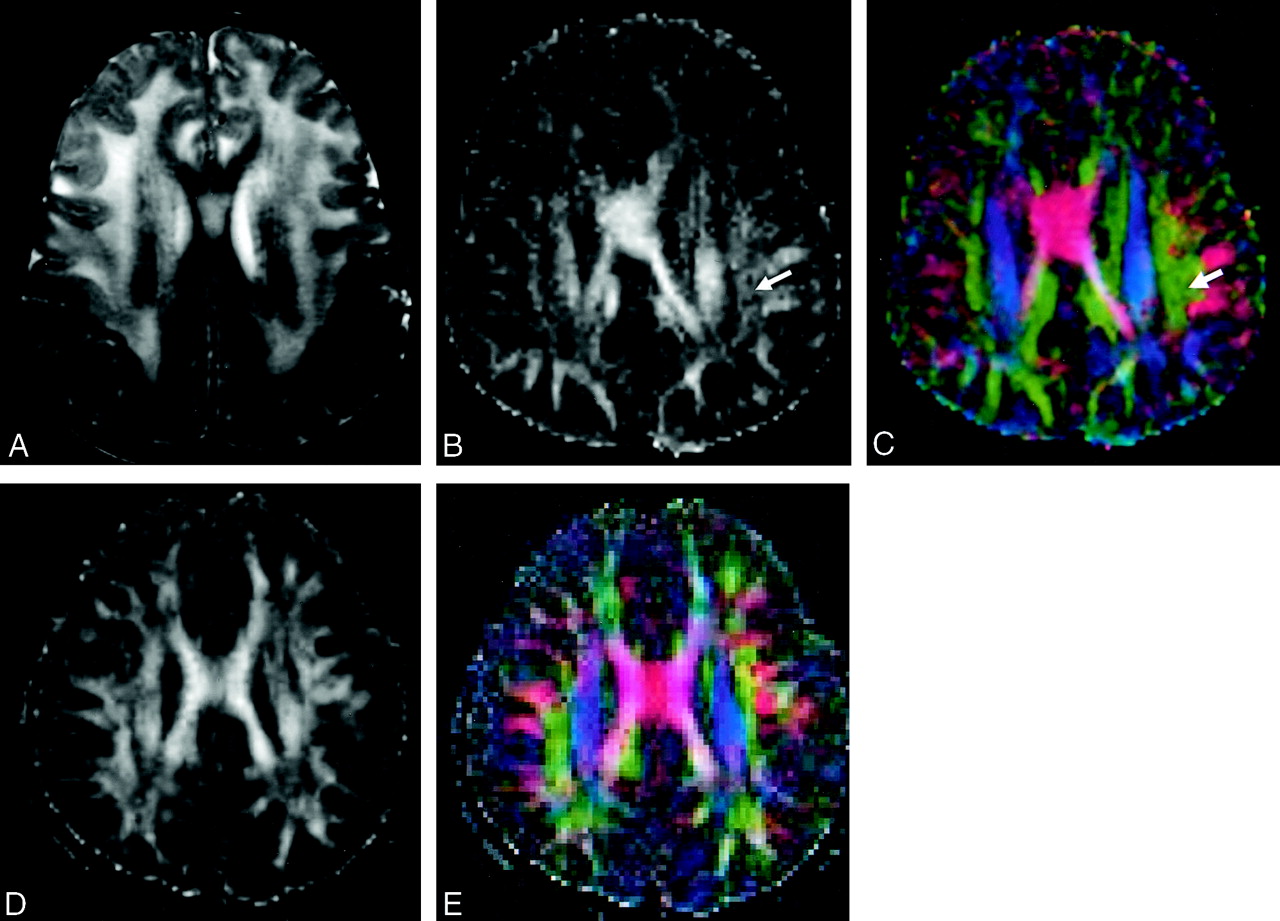
  • Fig 3.
    • Download figure
    • Open in new tab
    • Download powerpoint
    Fig 3.

    MR and DT images obtained when the patient was 5 years 8 months old.

    Axial T2-weighted MR image (A) at the level just above the body of the corpus callosum, showing diffuse high-signal-intensity white matter abnormalities. On FA (B and D) and vector maps (96 × 128 matrix size) (C and E), the white matter anisotropy and directionality were only mildly impaired (arrows) in our patient (B and C) as compared with a healthy control subject (D and E).

  • Fig 4.
    • Download figure
    • Open in new tab
    • Download powerpoint
    Fig 4.

    Depiction of regions of interest used in data analysis obtained when the patient was 5 years old. Each region of interest was placed symmetrically and bilaterally except for region of interest 4.

  • Fig 5.
    • Download figure
    • Open in new tab
    • Download powerpoint
    Fig 5.

    Multivoxel 1H MR spectra, obtained when the patient was 5 years old showing a significant decrease of NAA/Cr + phosphocreatine. Left spectrum, which is a magnified voxel indicated with thick frame in multivoxel 1H MR spectroscopy, shows the presence of lactate peaks in the temporoparietal white matter regions.

Tables

  • Figures
  • Mean FA of a patient with vacuolating megalencephalic leukoencephalopathy and of control subjects

    Region of InterestMean FAFA Decrease (%)
    VML PatientControl Subjects (n = 2)
    10.13 ± 0.040.14 ± 0.017.1
    20.16 ± 0.010.21 ± 0.0723.8
    30.26 ± 0.050.34 ± 0.0423.5
    40.340.40 ± 0.0815
    • Note.—FA indicates fractional anisotropy; VML, vacuolating megalencephalic leukoencephalopathy; locations of regions of interest are depicted in Figure 4.

PreviousNext
Back to top

In this issue

American Journal of Neuroradiology: 25 (6)
American Journal of Neuroradiology
Vol. 25, Issue 6
1 Jun 2004
  • Table of Contents
  • Index by author
Advertisement
Print
Download PDF
Email Article

Thank you for your interest in spreading the word on American Journal of Neuroradiology.

NOTE: We only request your email address so that the person you are recommending the page to knows that you wanted them to see it, and that it is not junk mail. We do not capture any email address.

Enter multiple addresses on separate lines or separate them with commas.
Vacuolating Megalencephalic Leukoencephalopathy with Mild Clinical Course Validated by Diffusion Tensor Imaging and MR Spectroscopy
(Your Name) has sent you a message from American Journal of Neuroradiology
(Your Name) thought you would like to see the American Journal of Neuroradiology web site.
CAPTCHA
This question is for testing whether or not you are a human visitor and to prevent automated spam submissions.
Cite this article
Yi-Fang Tu, Cheng-Yu Chen, Chao-Ching Huang, Chang-Shin Lee
Vacuolating Megalencephalic Leukoencephalopathy with Mild Clinical Course Validated by Diffusion Tensor Imaging and MR Spectroscopy
American Journal of Neuroradiology Jun 2004, 25 (6) 1041-1045;

Citation Manager Formats

  • BibTeX
  • Bookends
  • EasyBib
  • EndNote (tagged)
  • EndNote 8 (xml)
  • Medlars
  • Mendeley
  • Papers
  • RefWorks Tagged
  • Ref Manager
  • RIS
  • Zotero
0 Responses
Respond to this article
Share
Bookmark this article
Vacuolating Megalencephalic Leukoencephalopathy with Mild Clinical Course Validated by Diffusion Tensor Imaging and MR Spectroscopy
Yi-Fang Tu, Cheng-Yu Chen, Chao-Ching Huang, Chang-Shin Lee
American Journal of Neuroradiology Jun 2004, 25 (6) 1041-1045;
del.icio.us logo Twitter logo Facebook logo Mendeley logo
  • Tweet Widget
  • Facebook Like
  • Google Plus One

Jump to section

  • Article
    • Abstract
    • Case Report
    • Discussion
    • References
  • Figures & Data
  • Info & Metrics
  • Responses
  • References
  • PDF

Related Articles

  • No related articles found.
  • PubMed
  • Google Scholar

Cited By...

  • No citing articles found.
  • Crossref
  • Google Scholar

This article has not yet been cited by articles in journals that are participating in Crossref Cited-by Linking.

More in this TOC Section

  • Early Ultrasonic Monitoring of Brain Growth and Later Neurodevelopmental Outcome in Very Preterm Infants
  • Diagnostic Value of Sylvian Fissure Hyperechogenicity in Fetal SAH
  • Feasibility and Added Value of Fetal DTI Tractography in the Evaluation of an Isolated Short Corpus Callosum: Preliminary Results
Show more Pediatrics

Similar Articles

Advertisement

Indexed Content

  • Current Issue
  • Accepted Manuscripts
  • Article Preview
  • Past Issues
  • Editorials
  • Editor's Choice
  • Fellows' Journal Club
  • Letters to the Editor
  • Video Articles

Cases

  • Case Collection
  • Archive - Case of the Week
  • Archive - Case of the Month
  • Archive - Classic Case

More from AJNR

  • Trainee Corner
  • Imaging Protocols
  • MRI Safety Corner
  • Book Reviews

Multimedia

  • AJNR Podcasts
  • AJNR Scantastics

Resources

  • Turnaround Time
  • Submit a Manuscript
  • Submit a Video Article
  • Submit an eLetter to the Editor/Response
  • Manuscript Submission Guidelines
  • Statistical Tips
  • Fast Publishing of Accepted Manuscripts
  • Graphical Abstract Preparation
  • Imaging Protocol Submission
  • Evidence-Based Medicine Level Guide
  • Publishing Checklists
  • Author Policies
  • Become a Reviewer/Academy of Reviewers
  • News and Updates

About Us

  • About AJNR
  • Editorial Board
  • Editorial Board Alumni
  • Alerts
  • Permissions
  • Not an AJNR Subscriber? Join Now
  • Advertise with Us
  • Librarian Resources
  • Feedback
  • Terms and Conditions
  • AJNR Editorial Board Alumni

American Society of Neuroradiology

  • Not an ASNR Member? Join Now

© 2025 by the American Society of Neuroradiology All rights, including for text and data mining, AI training, and similar technologies, are reserved.
Print ISSN: 0195-6108 Online ISSN: 1936-959X

Powered by HighWire