Advertisement

Main menu

  • Home
  • Content
    • Current Issue
    • Accepted Manuscripts
    • Article Preview
    • Past Issue Archive
    • Video Articles
    • AJNR Case Collection
    • Case of the Week Archive
    • Case of the Month Archive
    • Classic Case Archive
  • Special Collections
    • AJNR Awards
    • Low-Field MRI
    • Alzheimer Disease
    • ASNR Foundation Special Collection
    • Photon-Counting CT
    • View All
  • Multimedia
    • AJNR Podcasts
    • AJNR SCANtastic
    • Trainee Corner
    • MRI Safety Corner
    • Imaging Protocols
  • For Authors
    • Submit a Manuscript
    • Submit a Video Article
    • Submit an eLetter to the Editor/Response
    • Manuscript Submission Guidelines
    • Statistical Tips
    • Fast Publishing of Accepted Manuscripts
    • Graphical Abstract Preparation
    • Imaging Protocol Submission
    • Author Policies
  • About Us
    • About AJNR
    • Editorial Board
    • Editorial Board Alumni
  • More
    • Become a Reviewer/Academy of Reviewers
    • Subscribers
    • Permissions
    • Alerts
    • Feedback
    • Advertisers
    • ASNR Home

User menu

  • Alerts
  • Log in

Search

  • Advanced search
American Journal of Neuroradiology

American Journal of Neuroradiology

ASHNR American Society of Functional Neuroradiology ASHNR American Society of Pediatric Neuroradiology ASSR
  • Alerts
  • Log in

Advanced Search

  • Home
  • Content
    • Current Issue
    • Accepted Manuscripts
    • Article Preview
    • Past Issue Archive
    • Video Articles
    • AJNR Case Collection
    • Case of the Week Archive
    • Case of the Month Archive
    • Classic Case Archive
  • Special Collections
    • AJNR Awards
    • Low-Field MRI
    • Alzheimer Disease
    • ASNR Foundation Special Collection
    • Photon-Counting CT
    • View All
  • Multimedia
    • AJNR Podcasts
    • AJNR SCANtastic
    • Trainee Corner
    • MRI Safety Corner
    • Imaging Protocols
  • For Authors
    • Submit a Manuscript
    • Submit a Video Article
    • Submit an eLetter to the Editor/Response
    • Manuscript Submission Guidelines
    • Statistical Tips
    • Fast Publishing of Accepted Manuscripts
    • Graphical Abstract Preparation
    • Imaging Protocol Submission
    • Author Policies
  • About Us
    • About AJNR
    • Editorial Board
    • Editorial Board Alumni
  • More
    • Become a Reviewer/Academy of Reviewers
    • Subscribers
    • Permissions
    • Alerts
    • Feedback
    • Advertisers
    • ASNR Home
  • Follow AJNR on Twitter
  • Visit AJNR on Facebook
  • Follow AJNR on Instagram
  • Join AJNR on LinkedIn
  • RSS Feeds

AJNR Awards, New Junior Editors, and more. Read the latest AJNR updates

OtherPediatric Neuroimaging

Appearance of an Interhemispheric Cyst Associated with Agenesis of the Corpus Callosum

Annemarie Stroustrup Smith and Deborah Levine
American Journal of Neuroradiology June 2004, 25 (6) 1037-1040;
Annemarie Stroustrup Smith
  • Find this author on Google Scholar
  • Find this author on PubMed
  • Search for this author on this site
Deborah Levine
  • Find this author on Google Scholar
  • Find this author on PubMed
  • Search for this author on this site
  • Article
  • Figures & Data
  • Info & Metrics
  • Responses
  • References
  • PDF
Loading

Article Figures & Data

Figures

  • Tables
  • Fig 1.
    • Download figure
    • Open in new tab
    • Download powerpoint
    Fig 1.

    Transabdominal sonogram obtained in a fetus with ACC and DWM at 27 weeks’ gestation. Coronal view shows elevation of the third ventricle, vertical orientation of the frontal horns (arrows), and absence of the corpus callosum.
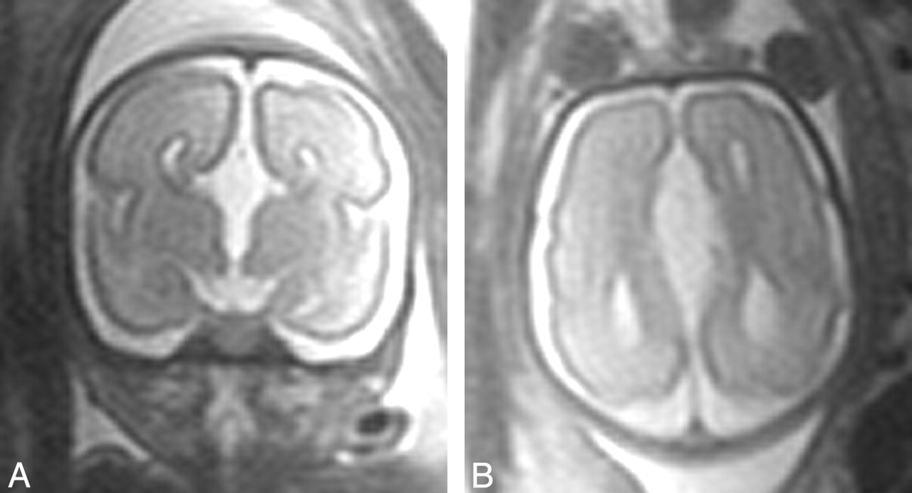
  • Fig 2.
    • Download figure
    • Open in new tab
    • Download powerpoint
    Fig 2.

    Single-shot half-Fourier turbo spin-echo MR images of the fetal brain at 27 weeks’ gestation. Coronal (A) and axial (B) images demonstrate agenesis of the corpus callosum and absence of any frontal cyst.

  • Fig 3.
    • Download figure
    • Open in new tab
    • Download powerpoint
    Fig 3.

    Neonatal brain MR image taken when the patient was 1 day old. Axial T1-weighted image (similar plane to that of Figure 2B) shows the increased size of the frontal and occipital horns (compare with prenatal scan) in addition to the new finding of a frontal para-midline cyst (C).

  • Fig 4.
    • Download figure
    • Open in new tab
    • Download powerpoint
    Fig 4.

    Neonatal cranial sonogram obtained when the patient was 5 days old. Coronal view in plane similar to that of Figures 2B and 3 demonstrates agenesis of the corpus callosum. In addition, a large frontal paramidline cyst is present (C).

Tables

  • Figures
  • Classification of agenesis of the corpus callosum with interhemispheric cyst after Barkovich et al (3)

    Type 1*
    SubtypeCyst CharacteristicsCommunicationAssociated Abnormalities
    Type 1a: Presumed communicating hydrocephalusIsointense to CSF (MR), unilocularCommunication with lateral ventricles onlyMacrocephaly, hydrocephalus, Dandy-Walker malformation
    Type 1b: Hydrocephalus secondary to diencephalic anomalyIsointense to CSF (MR), unilocularCommunication with and obstruction of third ventricleMacrocephaly, diencephalic malformation (eg, thalamic fusion without subcortical heterotopia)
    Type 1c: Small head size and cerebral hypoplasiaIsointense to CSF (MR), unilocularCommunication with lateral and third ventriclesMicrocephaly, cerebral dysplasia or hypoplasia
    Type 2†
    Type 2a: No abnormality apart from ACCIsointense to CSF (MR), multilocularNo communication with lateral or third ventriclesMacrocephaly, hydrocephalus
    Type 2b: Aicardi syndromeHyperattenuation (CT), hyperintense (T1W MR), multilocularNo communication with lateral or third ventriclesFemale predominance, subependymal heterotopia, polymicrogyria, seizures, hypoplastic falx cerebri, uni- or bilateral ventriculomegaly, developmental delay
    Type 2c: Subcortical heterotopiaIsointense to CSF (MR), multilocularNo communication with lateral or third ventriclesSubcortical heterotopia, developmental delay
    • * Extension or diverticulation of third or lateral ventricles.

    • † Loculated, lack of communication with ventricular system.

PreviousNext
Back to top

In this issue

American Journal of Neuroradiology: 25 (6)
American Journal of Neuroradiology
Vol. 25, Issue 6
1 Jun 2004
  • Table of Contents
  • Index by author
Advertisement
Print
Download PDF
Email Article

Thank you for your interest in spreading the word on American Journal of Neuroradiology.

NOTE: We only request your email address so that the person you are recommending the page to knows that you wanted them to see it, and that it is not junk mail. We do not capture any email address.

Enter multiple addresses on separate lines or separate them with commas.
Appearance of an Interhemispheric Cyst Associated with Agenesis of the Corpus Callosum
(Your Name) has sent you a message from American Journal of Neuroradiology
(Your Name) thought you would like to see the American Journal of Neuroradiology web site.
CAPTCHA
This question is for testing whether or not you are a human visitor and to prevent automated spam submissions.
Cite this article
Annemarie Stroustrup Smith, Deborah Levine
Appearance of an Interhemispheric Cyst Associated with Agenesis of the Corpus Callosum
American Journal of Neuroradiology Jun 2004, 25 (6) 1037-1040;

Citation Manager Formats

  • BibTeX
  • Bookends
  • EasyBib
  • EndNote (tagged)
  • EndNote 8 (xml)
  • Medlars
  • Mendeley
  • Papers
  • RefWorks Tagged
  • Ref Manager
  • RIS
  • Zotero
0 Responses
Respond to this article
Share
Bookmark this article
Appearance of an Interhemispheric Cyst Associated with Agenesis of the Corpus Callosum
Annemarie Stroustrup Smith, Deborah Levine
American Journal of Neuroradiology Jun 2004, 25 (6) 1037-1040;
del.icio.us logo Twitter logo Facebook logo Mendeley logo
  • Tweet Widget
  • Facebook Like
  • Google Plus One

Jump to section

  • Article
    • Abstract
    • Case Report
    • Discussion
    • Footnotes
    • References
  • Figures & Data
  • Info & Metrics
  • Responses
  • References
  • PDF

Related Articles

  • No related articles found.
  • PubMed
  • Google Scholar

Cited By...

  • Dandy-Walker Phenotype with Brainstem Involvement: 2 Distinct Subgroups with Different Prognosis
  • Agenesis of the corpus callosum with interhemispheric cyst: clinical implications and outcome
  • Asymmetric Ventriculomegaly, Interhemispheric Cyst, and Dysgenesis of the Corpus Callosum (AVID): An Imaging Triad
  • Crossref
  • Google Scholar

This article has not yet been cited by articles in journals that are participating in Crossref Cited-by Linking.

More in this TOC Section

  • Frontal Paraventricular Cysts
  • Sodium MRI in Pediatric Brain Tumors
  • FRACTURE MR in Congenital Vertebral Anomalies
Show more Pediatric Neuroimaging

Similar Articles

Advertisement

Indexed Content

  • Current Issue
  • Accepted Manuscripts
  • Article Preview
  • Past Issues
  • Editorials
  • Editor's Choice
  • Fellows' Journal Club
  • Letters to the Editor
  • Video Articles

Cases

  • Case Collection
  • Archive - Case of the Week
  • Archive - Case of the Month
  • Archive - Classic Case

More from AJNR

  • Trainee Corner
  • Imaging Protocols
  • MRI Safety Corner
  • Book Reviews

Multimedia

  • AJNR Podcasts
  • AJNR Scantastics

Resources

  • Turnaround Time
  • Submit a Manuscript
  • Submit a Video Article
  • Submit an eLetter to the Editor/Response
  • Manuscript Submission Guidelines
  • Statistical Tips
  • Fast Publishing of Accepted Manuscripts
  • Graphical Abstract Preparation
  • Imaging Protocol Submission
  • Evidence-Based Medicine Level Guide
  • Publishing Checklists
  • Author Policies
  • Become a Reviewer/Academy of Reviewers
  • News and Updates

About Us

  • About AJNR
  • Editorial Board
  • Editorial Board Alumni
  • Alerts
  • Permissions
  • Not an AJNR Subscriber? Join Now
  • Advertise with Us
  • Librarian Resources
  • Feedback
  • Terms and Conditions
  • AJNR Editorial Board Alumni

American Society of Neuroradiology

  • Not an ASNR Member? Join Now

© 2025 by the American Society of Neuroradiology All rights, including for text and data mining, AI training, and similar technologies, are reserved.
Print ISSN: 0195-6108 Online ISSN: 1936-959X

Powered by HighWire