Advertisement

Main menu

  • Home
  • Content
    • Current Issue
    • Accepted Manuscripts
    • Article Preview
    • Past Issue Archive
    • Video Articles
    • AJNR Case Collection
    • Case of the Week Archive
    • Case of the Month Archive
    • Classic Case Archive
  • Special Collections
    • AJNR Awards
    • Low-Field MRI
    • Alzheimer Disease
    • ASNR Foundation Special Collection
    • Photon-Counting CT
    • View All
  • Multimedia
    • AJNR Podcasts
    • AJNR SCANtastic
    • Trainee Corner
    • MRI Safety Corner
    • Imaging Protocols
  • For Authors
    • Submit a Manuscript
    • Submit a Video Article
    • Submit an eLetter to the Editor/Response
    • Manuscript Submission Guidelines
    • Statistical Tips
    • Fast Publishing of Accepted Manuscripts
    • Graphical Abstract Preparation
    • Imaging Protocol Submission
    • Author Policies
  • About Us
    • About AJNR
    • Editorial Board
    • Editorial Board Alumni
  • More
    • Become a Reviewer/Academy of Reviewers
    • Subscribers
    • Permissions
    • Alerts
    • Feedback
    • Advertisers
    • ASNR Home

User menu

  • Alerts
  • Log in

Search

  • Advanced search
American Journal of Neuroradiology

American Journal of Neuroradiology

ASHNR American Society of Functional Neuroradiology ASHNR American Society of Pediatric Neuroradiology ASSR
  • Alerts
  • Log in

Advanced Search

  • Home
  • Content
    • Current Issue
    • Accepted Manuscripts
    • Article Preview
    • Past Issue Archive
    • Video Articles
    • AJNR Case Collection
    • Case of the Week Archive
    • Case of the Month Archive
    • Classic Case Archive
  • Special Collections
    • AJNR Awards
    • Low-Field MRI
    • Alzheimer Disease
    • ASNR Foundation Special Collection
    • Photon-Counting CT
    • View All
  • Multimedia
    • AJNR Podcasts
    • AJNR SCANtastic
    • Trainee Corner
    • MRI Safety Corner
    • Imaging Protocols
  • For Authors
    • Submit a Manuscript
    • Submit a Video Article
    • Submit an eLetter to the Editor/Response
    • Manuscript Submission Guidelines
    • Statistical Tips
    • Fast Publishing of Accepted Manuscripts
    • Graphical Abstract Preparation
    • Imaging Protocol Submission
    • Author Policies
  • About Us
    • About AJNR
    • Editorial Board
    • Editorial Board Alumni
  • More
    • Become a Reviewer/Academy of Reviewers
    • Subscribers
    • Permissions
    • Alerts
    • Feedback
    • Advertisers
    • ASNR Home
  • Follow AJNR on Twitter
  • Visit AJNR on Facebook
  • Follow AJNR on Instagram
  • Join AJNR on LinkedIn
  • RSS Feeds

AJNR Awards, New Junior Editors, and more. Read the latest AJNR updates

Research ArticlePEDIATRICS

Diffusion-Weighted MR Imaging of Subdural Empyemas in Children

Alex M. Wong, Robert A. Zimmerman, Erin M. Simon, Avrum N. Pollock and Larissa T. Bilaniuk
American Journal of Neuroradiology June 2004, 25 (6) 1016-1021;
Alex M. Wong
  • Find this author on Google Scholar
  • Find this author on PubMed
  • Search for this author on this site
Robert A. Zimmerman
  • Find this author on Google Scholar
  • Find this author on PubMed
  • Search for this author on this site
Erin M. Simon
  • Find this author on Google Scholar
  • Find this author on PubMed
  • Search for this author on this site
Avrum N. Pollock
  • Find this author on Google Scholar
  • Find this author on PubMed
  • Search for this author on this site
Larissa T. Bilaniuk
  • Find this author on Google Scholar
  • Find this author on PubMed
  • Search for this author on this site
  • Article
  • Figures & Data
  • Info & Metrics
  • Responses
  • References
  • PDF
Loading

Article Figures & Data

Figures

  • Tables
  • Fig 1.
    • Download figure
    • Open in new tab
    • Download powerpoint
    Fig 1.

    Patient 8. Bifrontal and right parafalcine SDE.

    A. Axial T1WI (650/14) shows slight hypointensity in the SDE (arrows).

    B, Axial T2WI (6000/99) shows intermediate signal intensity in the bifrontal collections (arrows) and hyperintensity in the right parafalcine collection (arrowheads).

    C, Coronal contrast-enhanced T1WI (800/20) with MT shows irregular capsular enhancement of the collections (arrows).

    D and E, Axial DWI (4000/110, b = 1000 s/mm2) and ADC map show reduced water diffusion in the SDE.
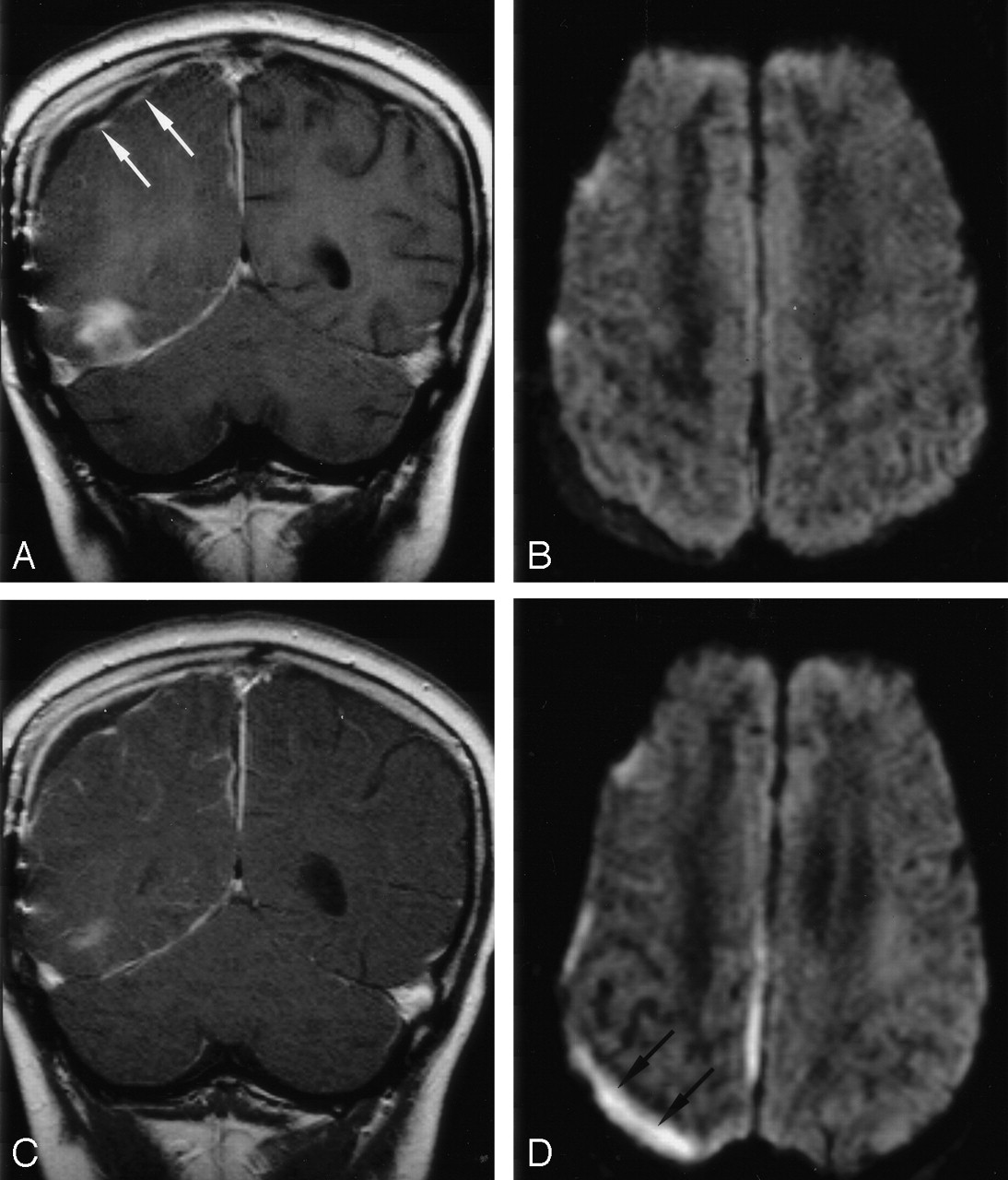
  • Fig 2.
    • Download figure
    • Open in new tab
    • Download powerpoint
    Fig 2.

    Patient 1. Right SDE and left RSE.

    A, Axial T1WI (650/14) shows intermediate signal intensity in the right SDE and hypointensity in the left RSE.

    B, Axial T2WI (6000/99) shows high signal intensity in both subdural collections. Strands of intermediate signal intensity (arrows) are noted in the right SDE.

    C, Axial contrast-enhanced T1WI (800/20) with MT shows peripheral enhancement of both subdural collections. The enhancement is thicker in the right SDE (arrows) than in the left RSE (arrowheads).

    D, Axial DWI (4000/110, b = 1000 s/mm2) shows mixed high (arrows) and low signal intensities in the right SDE.

    E, Corresponding ADC map shows mixed low (arrows) and high signal intensities in the right SDE, indicating heterogeneous water motion. Increased water motion is noted in the left RSE.

  • Fig 3.
    • Download figure
    • Open in new tab
    • Download powerpoint
    Fig 3.

    Patient 7. Follow-up DWI show superimposed infection of a right RSE in this 10-year-old boy with right temporal and parafalcine SDEs.

    A, Coronal contrast-enhanced T1WI (800/20) with MT shows capsular enhancement (arrows) of the right parietal subdural collection and the anterior aspect of the right temporal base SDE.

    B, Axial DWI (4000/110, b = 1000 s/mm2) shows increased diffusion in the collections.

    C, Follow-up coronal contrast-enhanced T1WI (800/20) with MT shows no substantial change in the capsular contrast enhancement of the collection.

    D, Follow-up axial DWI (4000/110, b = 1000 s/mm2) shows increased signal intensity in the collection (arrows) and the interhemispheric fissure, suggesting superimposed infection.

Tables

  • Figures
    • View popup
    TABLE 1:

    Clinical features of 10 patients with SDE

    Patient (No)/ Sex/AgeSource of InfectionClinical Findings at Admission*/Duration, dWBC Count, ×103/dLPathogenTreatment
    1/M/4 moMeningitisFever; irritability; stiff neck/154.4Escherichia coli (CSF)Antibiotics, burr hole drainage
    1/M/14 ySinusitis S, E, MFever; swollen, painful L eye/412.6Moraxella species (sinus)Antibiotics, FESS, orbital drainage
    3/F/13 ySinusitis F, S, E, MFever; headache; slurred speech, R extremity weakness/1417.4Group A Streptococcus (subdural collection)Antibiotics, Craniotomy
    4/M/5 wkMeningitisFever; vomiting/424.9Pasteurella multocida (CSF)Antibiotics
    5/M/16 ySinusitis F, S, E, MFever; swollen, painful L eye, photophobia, impaired gaze; seizure/412.6Streptococcus milleri species (subdural collection)Antibiotics, FESS, craniotomy, preseptal drainage
    6/M/13 yMeningitisFever; hallucinations/430.6Peptostreptococcus anaerobius, Prevotella melaninogenica (CSF)Antibiotics, FESS
    7/M/10 yRight mastoiditisFever; headache; seizure/106.1Streptococcus equiinus, Micrococcus species (subdural collection)Antibiotics, Craniotomy
    8/F/17 ySinusitis FFever; headache; L extremity weakness/313.4S milleri species (subdural collection)Antibiotics, FESS, craniotomy
    9/F/8 ySinusitis S, E, MFever; seizure, R hemiparesis, R facial palsy/519.8Streptococcus mitis (blood)Antibiotics, FESS, craniotomy
    10/M/13 ySinusitis F, S, E, MFever; headache; painful neck; photophobia/722.1Veillonella species, Propionibacterium granulosum (sinus)Antibiotics, FESS, anticoagulants
    • Note.—Patient 6 died; all others recovered. E indicates ethmoid; F, frontal; FESS, functional endoscopic sinus surgery; M, maxillary; and S, sphenoid.

    • * Systemic and neurologic signs.

    • View popup
    TABLE 2:

    Initial and follow-up imaging findings in 10 patients with SDE

    Patient (No.)Location of SDEInitial MR Study*ADC, ×10−3 mm2/sAssociated AbnormalitiesDuration between Initial and Follow-up MR Studies, dClinical Features at Follow-up MR ImagingFollow-up MR Study*
    T1WIT2WIEnhanced†DWIEnhanced†DWI
    1R FPHypoMild hyperThick, linearHyper, hypo0.892L RSE (thin, linear)†21FeverThick, linearHyper, hypo
    2L FHypoHyperThick, linearHyperNAL orbital cellulites16FeverThick, linearHyper
    3L FPIsoHyperThick, irregularHyperNANANANANANA
    4B FPOIsoMild hyperThick, linearHyper0.644NA12Fever, seizureThick, linearHyper
    5L FIsoHyperThick, linearHyper0.306L orbital cellulitesNANANANA
    6Mid F baseHypoHyperThick, linearHyperNANANANANA
    7R pF, R PO, R TMild hypoIsoThick, linearHyper0.642R RSE (thin, linear)† infected later5FeverThick, linearHyper
    8B F, R pFIsoMild hyperThick, linearHyper0.698NA23Fever, chillsThick, linearHyper
    9L FP, L pFIsoMild hyperThin, linearHyper0.438NA6Fever, R hemiparesisThin, linearHyper
    10L CPAHypoHyperThick, linearHyper0.608Cavernous sinus infection10Afebrile, leukocytosis (blood)Thick, linearHyper
    • Note.—B indicates bilateral; CPA, cerebellopontine angle; F, frontal; FP, frontoparietal; FPO, fronto-occipitoparietal; Hyper, hyperintense; hypo, hypointense; Iso, isointense; NA, not available or not applicable; pF, parafalcine; PO, parieto-occipital; T, temporal; T1WI, T1-weighted image; and T2WI, T2-weighted image.

    • * Signal intensity relative to that of gray matter.

    • † Morphology of the enhancing membrane.

PreviousNext
Back to top

In this issue

American Journal of Neuroradiology: 25 (6)
American Journal of Neuroradiology
Vol. 25, Issue 6
1 Jun 2004
  • Table of Contents
  • Index by author
Advertisement
Print
Download PDF
Email Article

Thank you for your interest in spreading the word on American Journal of Neuroradiology.

NOTE: We only request your email address so that the person you are recommending the page to knows that you wanted them to see it, and that it is not junk mail. We do not capture any email address.

Enter multiple addresses on separate lines or separate them with commas.
Diffusion-Weighted MR Imaging of Subdural Empyemas in Children
(Your Name) has sent you a message from American Journal of Neuroradiology
(Your Name) thought you would like to see the American Journal of Neuroradiology web site.
CAPTCHA
This question is for testing whether or not you are a human visitor and to prevent automated spam submissions.
Cite this article
Alex M. Wong, Robert A. Zimmerman, Erin M. Simon, Avrum N. Pollock, Larissa T. Bilaniuk
Diffusion-Weighted MR Imaging of Subdural Empyemas in Children
American Journal of Neuroradiology Jun 2004, 25 (6) 1016-1021;

Citation Manager Formats

  • BibTeX
  • Bookends
  • EasyBib
  • EndNote (tagged)
  • EndNote 8 (xml)
  • Medlars
  • Mendeley
  • Papers
  • RefWorks Tagged
  • Ref Manager
  • RIS
  • Zotero
0 Responses
Respond to this article
Share
Bookmark this article
Diffusion-Weighted MR Imaging of Subdural Empyemas in Children
Alex M. Wong, Robert A. Zimmerman, Erin M. Simon, Avrum N. Pollock, Larissa T. Bilaniuk
American Journal of Neuroradiology Jun 2004, 25 (6) 1016-1021;
del.icio.us logo Twitter logo Facebook logo Mendeley logo
  • Tweet Widget
  • Facebook Like
  • Google Plus One

Jump to section

  • Article
    • Abstract
    • Methods
    • Results
    • Discussion
    • Acknowledgments
    • References
  • Figures & Data
  • Info & Metrics
  • Responses
  • References
  • PDF

Related Articles

  • No related articles found.
  • PubMed
  • Google Scholar

Cited By...

  • Subdural and subarachnoid empyema as a complication of an acute meningococcal meningitis
  • Subdural empyema in bacterial meningitis
  • Role of imaging in the diagnosis of acute bacterial meningitis and its complications
  • The clinical challenge of recognizing infratentorial empyema
  • Unusual findings in cerebral abscess: report of two cases.
  • Crossref
  • Google Scholar

This article has not yet been cited by articles in journals that are participating in Crossref Cited-by Linking.

More in this TOC Section

  • Early Ultrasonic Monitoring of Brain Growth and Later Neurodevelopmental Outcome in Very Preterm Infants
  • Diagnostic Value of Sylvian Fissure Hyperechogenicity in Fetal SAH
  • Feasibility and Added Value of Fetal DTI Tractography in the Evaluation of an Isolated Short Corpus Callosum: Preliminary Results
Show more Pediatrics

Similar Articles

Advertisement

Indexed Content

  • Current Issue
  • Accepted Manuscripts
  • Article Preview
  • Past Issues
  • Editorials
  • Editor's Choice
  • Fellows' Journal Club
  • Letters to the Editor
  • Video Articles

Cases

  • Case Collection
  • Archive - Case of the Week
  • Archive - Case of the Month
  • Archive - Classic Case

More from AJNR

  • Trainee Corner
  • Imaging Protocols
  • MRI Safety Corner
  • Book Reviews

Multimedia

  • AJNR Podcasts
  • AJNR Scantastics

Resources

  • Turnaround Time
  • Submit a Manuscript
  • Submit a Video Article
  • Submit an eLetter to the Editor/Response
  • Manuscript Submission Guidelines
  • Statistical Tips
  • Fast Publishing of Accepted Manuscripts
  • Graphical Abstract Preparation
  • Imaging Protocol Submission
  • Evidence-Based Medicine Level Guide
  • Publishing Checklists
  • Author Policies
  • Become a Reviewer/Academy of Reviewers
  • News and Updates

About Us

  • About AJNR
  • Editorial Board
  • Editorial Board Alumni
  • Alerts
  • Permissions
  • Not an AJNR Subscriber? Join Now
  • Advertise with Us
  • Librarian Resources
  • Feedback
  • Terms and Conditions
  • AJNR Editorial Board Alumni

American Society of Neuroradiology

  • Not an ASNR Member? Join Now

© 2025 by the American Society of Neuroradiology All rights, including for text and data mining, AI training, and similar technologies, are reserved.
Print ISSN: 0195-6108 Online ISSN: 1936-959X

Powered by HighWire