
Article Figures & Data
Figures
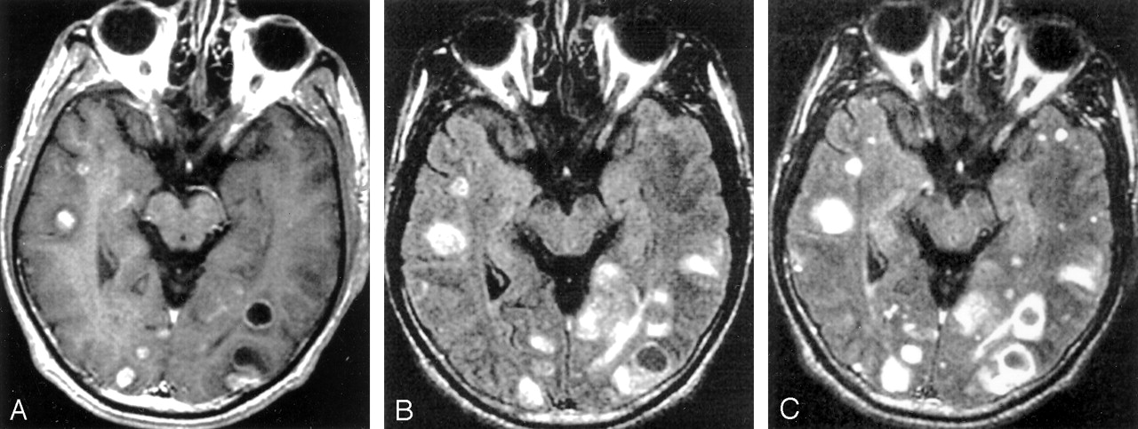
- Fig 1.
Images obtained in a 57-year-old-man with small-cell lung cancer reveal multiple, enhancing, solid and cystic parenchymal metastases. The number, conspicuity, and contrast enhancement are superior on the postcontrast FLAIR image compared with the postcontrast T1W image. In this patient, FLAIR was performed as the second postcontrast sequence.
A, Contrast-enhanced T1W image.
B, Nonenhanced FLAIR image.
C, Contrast-enhanced FLAIR image.
- Fig 2.
Images obtained in a 45-year-old-woman with breast cancer. The conspicuity, extension, and contrast enhancement of the lesions are superior on the postcontrast FLAIR image (C) than on the postcontrast T1W image (A). The leptomeningeal enhancement of frontal lobes (white arrow) is not conspicuous in A as compared with C. For this patient, T1W imaging was performed as the second postcontrast sequence.
A, Contrast-enhanced T1W image shows abnormal enhancement in the basal cisterns (arrow) and leptomeninges consistent with metastases.
B, Nonenhanced FLAIR image.
C, Contrast-enhanced FLAIR image also shows the abnormal enhancement of the leptomeninges (white arrow) and basal cisterns (black arrow).
- Fig 3.
Images obtained in a 54-year-old-man with small-cell lung cancer. In this patient, FLAIR imaging was the second postcontrast sequence.
A and B, Nonenhanced (A) and contrast-enhanced (B) FLAIR images show involvement of the prechiasmal portion of the right optic nerve (white arrow) and extension to the optic chiasm (black arrow) and right optic tract (arrowhead). These findings appear as abnormal hyperintensity in A and as abnormal contrast enhancement in B.
C, Contrast-enhanced T1W image. Although involvement of optic chiasm is shown as abnormal contrast enhancement (arrow), involvement of the right optic nerve and right optic tract could not be detected.


Tables
Localization No. of Patients Isolated parenchymal metastases 24 Parenchymal and cranial-nerve metastases 4 Parenchymal and leptomeningeal-cisternal metastases 5 Isolated leptomeningeal-cisternal metastases 6 Feature No. of Patients FLAIR Superior to T1W* FLAIR Equal to T1W FLAIR Inferior to T1W Number of lesions 5 20† 8‡ Lesion conspicuity 5 1* 27§ Degree of contrast enhancement 5 5* 23‖ * In all patients, FLAIR was the second postcontrast sequence.
† In 19 patients, FLAIR was the second postcontrast sequence, and in one, the T1W sequence was second.
‡ In four patients, FLAIR was the second postcontrast sequence, and in the other four patients, the T1W sequence was second.
§ In 18 patients, FLAIR was the second postcontrast sequence, and in nine, the T1W sequence was second.
‖ In 10 patients, FLAIR was the second postcontrast sequence, and in 13, the T1W sequence was second.
Feature No. of Patients FLAIR Superior to T1W* FLAIR Equal to T1W FLAIR Inferior to T1W Lesion extension 8* 3† 0 Lesion conspicuity 8* 0 3† Degree of contrast enhancement 8* 0 3† * In five patients, FLAIR was the second postcontrast sequence, and in three, the T1W sequence was second.
† In two patients, FLAIR was the second postcontrast sequence, and in one, the T1W sequence was second.
Patient and Involvement FLAIR T1W Patient 1* Right optic nerve Positive Negative Optic chiasm Positive Positive Right optic tract Positive Negative Patient 2† Right optic tract Positive Negative Patient 3* Optic chiasm Positive Negative Left optic tract Positive Negative Patient 4† Bilateral CNs VII, VIII Positive Positive * FLAIR was the second postcontrast sequence.
† T1W imaging was the second postcontrast sequence.
In this issue


Jump to section
Related Articles
Cited By...
- Compressed Sensitivity Encoding Artificial Intelligence Accelerates Brain Metastasis Imaging by Optimizing Image Quality and Reducing Scan Time
- Utility of Contrast-Enhanced T2 FLAIR for Imaging Brain Metastases Using a Half-dose High-Relaxivity Contrast Agent
- Leptomeningeal contrast enhancement and blood-CSF barrier dysfunction in aseptic meningitis