
Article Figures & Data
Figures
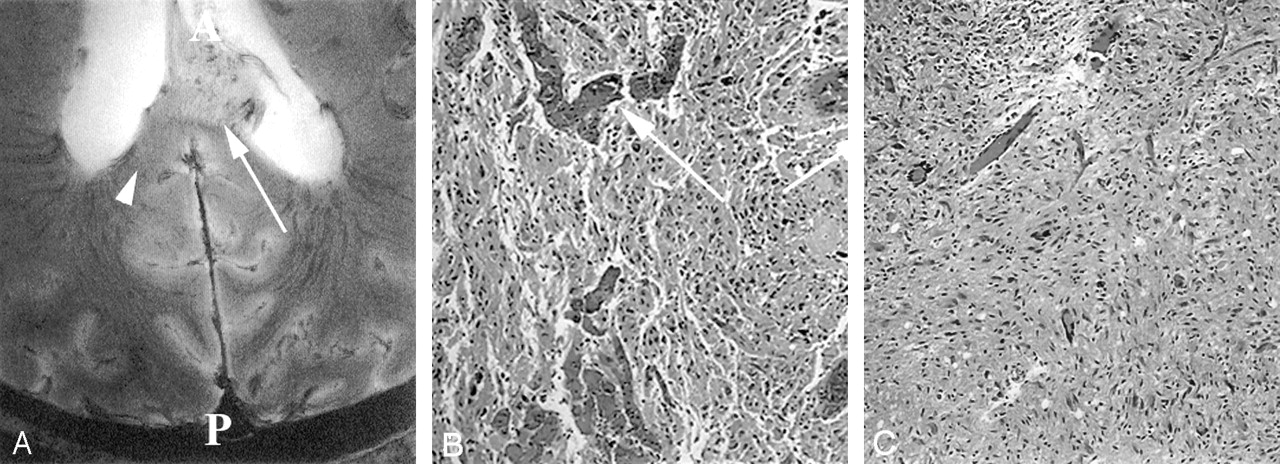
- Fig 1.
Images from the intact cadaver of a patient with known GBM.
A, Axial 8-T GE MR image (matrix = 1024 × 1024;TR/TE = 600/12; flip angle = 20°) in the patient with GBM at the level of the trigone of the lateral ventricles (T) and the level of the splenium (S) demonstrates an exophytic mass extending into the left lateral ventricle. A = anterior, P = posterior.
B–D, Reticulin-stained sections (original magnification ×100) correspond to an area of normal gray matter (B), the focus depicted by the arrow in A (C), and the focus depicted by the arrowhead in A (D). The arrow in B depicts the sulcus. Image in C shows an area of high vascular density and large vessels. Image in D corresponds to an area of vascularity and vessel size similar to that of the gray matter in the tumor bed. Vessels are depicted by arrows in C and D.
- Fig 2.
Additional images from the intact cadaver of a patient with known GBM.
A, Axial 8-T GE MR image (matrix = 1024 × 1024;TR/TE = 600/12; flip angle = 20°) in the patient with known GBM involving the splenium of the corpus callosum. This is 2 mm superior to the image in Figure 1A. Numerous small vessels are visible in the tumor bed. A = anterior, P = posterior.
B and C, Hematoxylin-eosin-stained sections (original magnification ×200) to the foci depicted by the arrow in A (B) and the arrowhead in A (C). B, Histologic specimen shows larger vessels (arrow) in dense concentration. Vessels in this region of exophytic tumor are also severely deformed in A, with loss of normal organization. Vessels can be seen crossing from the corpus callosum into the tumor. C, Image shows tumor infiltration with vascular density similar to white matter.


Tables
This table compares findings on 8T gradient echo MRI to histopathologic findings from corresponding foci within the patient’s brain studied here. Comparisons include whether tumor was identified in each of the modalities (8T and histopathology), vessel size relative to normal brain tissues and vessel density relative to normal tissues
Identification of tumor Vessel size Vessel density 8T Pathology 8T vessel size Histopathologic vessel size 8T vessel density Histopathologic vessel density No Yes small small low low Yes Yes large large high high Yes Yes small small low medium Yes Yes medium medium medium medium Yes Yes medium medium low low Yes Yes large large high high No No large large low low No No large large low low Yes Yes large large low medium Yes Yes necrosis necrosis necrosis necrosis


