Advertisement

Main menu

  • Home
  • Content
    • Current Issue
    • Accepted Manuscripts
    • Article Preview
    • Past Issue Archive
    • Video Articles
    • AJNR Case Collection
    • Case of the Week Archive
    • Case of the Month Archive
    • Classic Case Archive
  • Special Collections
    • AJNR Awards
    • Low-Field MRI
    • Alzheimer Disease
    • ASNR Foundation Special Collection
    • Photon-Counting CT
    • View All
  • Multimedia
    • AJNR Podcasts
    • AJNR SCANtastic
    • Trainee Corner
    • MRI Safety Corner
    • Imaging Protocols
  • For Authors
    • Submit a Manuscript
    • Submit a Video Article
    • Submit an eLetter to the Editor/Response
    • Manuscript Submission Guidelines
    • Statistical Tips
    • Fast Publishing of Accepted Manuscripts
    • Graphical Abstract Preparation
    • Imaging Protocol Submission
    • Author Policies
  • About Us
    • About AJNR
    • Editorial Board
    • Editorial Board Alumni
  • More
    • Become a Reviewer/Academy of Reviewers
    • Subscribers
    • Permissions
    • Alerts
    • Feedback
    • Advertisers
    • ASNR Home

User menu

  • Alerts
  • Log in

Search

  • Advanced search
American Journal of Neuroradiology

American Journal of Neuroradiology

ASHNR American Society of Functional Neuroradiology ASHNR American Society of Pediatric Neuroradiology ASSR
  • Alerts
  • Log in

Advanced Search

  • Home
  • Content
    • Current Issue
    • Accepted Manuscripts
    • Article Preview
    • Past Issue Archive
    • Video Articles
    • AJNR Case Collection
    • Case of the Week Archive
    • Case of the Month Archive
    • Classic Case Archive
  • Special Collections
    • AJNR Awards
    • Low-Field MRI
    • Alzheimer Disease
    • ASNR Foundation Special Collection
    • Photon-Counting CT
    • View All
  • Multimedia
    • AJNR Podcasts
    • AJNR SCANtastic
    • Trainee Corner
    • MRI Safety Corner
    • Imaging Protocols
  • For Authors
    • Submit a Manuscript
    • Submit a Video Article
    • Submit an eLetter to the Editor/Response
    • Manuscript Submission Guidelines
    • Statistical Tips
    • Fast Publishing of Accepted Manuscripts
    • Graphical Abstract Preparation
    • Imaging Protocol Submission
    • Author Policies
  • About Us
    • About AJNR
    • Editorial Board
    • Editorial Board Alumni
  • More
    • Become a Reviewer/Academy of Reviewers
    • Subscribers
    • Permissions
    • Alerts
    • Feedback
    • Advertisers
    • ASNR Home
  • Follow AJNR on Twitter
  • Visit AJNR on Facebook
  • Follow AJNR on Instagram
  • Join AJNR on LinkedIn
  • RSS Feeds

AJNR Awards, New Junior Editors, and more. Read the latest AJNR updates

Research ArticleBrain

Volumetric Proton Spectroscopic Imaging of Mild Traumatic Brain Injury

Varanavasi Govindaraju, Grant E. Gauger, Geoffrey T. Manley, Andreas Ebel, Michele Meeker and Andrew A. Maudsley
American Journal of Neuroradiology May 2004, 25 (5) 730-737;
Varanavasi Govindaraju
  • Find this author on Google Scholar
  • Find this author on PubMed
  • Search for this author on this site
Grant E. Gauger
  • Find this author on Google Scholar
  • Find this author on PubMed
  • Search for this author on this site
Geoffrey T. Manley
  • Find this author on Google Scholar
  • Find this author on PubMed
  • Search for this author on this site
Andreas Ebel
  • Find this author on Google Scholar
  • Find this author on PubMed
  • Search for this author on this site
Michele Meeker
  • Find this author on Google Scholar
  • Find this author on PubMed
  • Search for this author on this site
Andrew A. Maudsley
  • Find this author on Google Scholar
  • Find this author on PubMed
  • Search for this author on this site
  • Article
  • Figures & Data
  • Info & Metrics
  • Responses
  • References
  • PDF
Loading

Article Figures & Data

Figures

  • Tables
  • Fig 1.
    • Download figure
    • Open in new tab
    • Download powerpoint
    Fig 1.

    Sections for data analysis and corresponding MRSI sections. Central five sections (numbered 3–7) selected for data analysis in the 140-mm-thick slab over which the MRSI acquisition was carried out.

  • Fig 2.
    • Download figure
    • Open in new tab
    • Download powerpoint
    Fig 2.

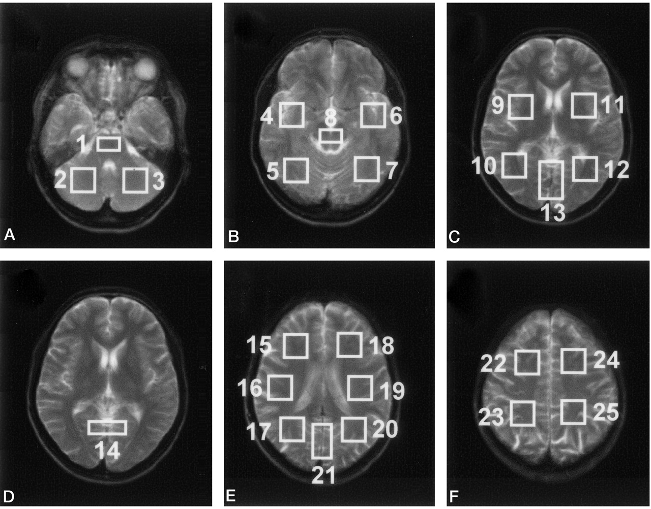
    A–F, MR images corresponding to the full thickness of each MRSI section (C and D are the same), with overlays indicating the 25 regions used for data analysis.

  • Fig 3.
    • Download figure
    • Open in new tab
    • Download powerpoint
    Fig 3.

    Mean regional NAA/Cho ratios of control (black) and TBI (gray) subjects. Numbers above each histogram pair refer to regions identified in Figure 2; multiple numbers indicate that data from the regions were grouped. Average SD for control and TBI subjects were 16.7% and 16.3%, respectively.

  • Fig 4.
    • Download figure
    • Open in new tab
    • Download powerpoint
    Fig 4.

    Mean global metabolite ratios in subjects with TBI (open symbols) plotted against GCS score at admission. Horizontal lines and solid symbols indicate ratios for controls, and error bars show 2 SD. Triangles, circles, and diamonds indicate NAA/Cho, NAA/Cr, and Cho/Cr ratios, respectively.

  • Fig 5.
    • Download figure
    • Open in new tab
    • Download powerpoint
    Fig 5.

    Mean global metabolite ratios in TBI subjects obtained shortly after injury plotted against GOS scores at discharge (open symbols) and at 6 months after injury (solid symbols). Data are separated according to moderate (M) and good (G) outcomes. NAA/Cr (circles), Cho/Cr (diamonds), and NAA/Cho (triangles) ratios are indicated. Horizontal lines indicate ratios for controls, and error bars show 2 SD.

Tables

  • Figures
    • View popup
    TABLE 1:

    Summary of demographic data and imaging findings in subjects with TBI

    Subject/Age (yr)/SexAdmission GCS ScoreTime to MR Imaging (days)Cause of Injury*CT and MR Findings†
    1/21/M142Skateboard accidentL frontal SDH, L posterior SAH, L parietal parenchymal contusion
    2/25/M158Skateboard accidentR parietal occipital skull fx, R tentorial SDH, pneumocephalus
    3/53/F1524MVANormal
    4/41/M153Bike vs auto accidentR temporal and R frontal mild cerebral contusion
    5/36/F1411Ped vs auto accidentDiffuse SAH, L ear laceration
    6/37/M157Ped vs auto accidentMidline SDH
    7/31/M146MVANormal
    8/36/M1414Bike vs auto accidentNormal
    9/18/F158MVANormal
    10/22/M1424MVASmall L SDH
    11/22/M1323MVAR SAH, L SDH, L noncompressed temporal skull fx
    12/49/F1512FallSAH at interhemispheric astula, frontal brain contusion
    13/25/M1414Ped vs auto accidentL frontal SAH, R SDH
    14/28/M1430AssaultL frontotemporal SDH, R SDH, bilateral temporal and L frontal contusion
    • * MVA indicates motor vehicle accident; Ped, pedestrian.

    • † fx indicates fracture; SAH, subarachnoid hematoma; SDH, subdural hematoma.

    • View popup
    TABLE 2:

    Metabolite ratios

    RegionNAA/CrCho/CrNAA/Cho
    ControlTBIControlTBIControlTBI
    51.41 (0.23)1.41 (0.27)0.66 (0.07)0.73 (0.08)*2.17 (0.37)1.94 (0.41)
    101.75 (0.19)1.67 (0.14)0.72 (0.10)0.77 (0.10)2.47 (0.41)2.21 (0.24)*
    121.72 (0.19)1.60 (0.11)0.70 (0.11)0.78 (0.15)2.48 (0.38)2.11 (0.38)*
    131.57 (0.23)1.53 (0.23)0.55 (0.06)0.63 (0.09)*2.89 (0.60)2.50 (0.51)
    161.85 (0.16)1.72 (0.12)*0.86 (0.12)0.80 (0.11)2.19 (0.30)2.18 (0.37)
    191.86 (0.17)1.70 (0.12)*0.90 (0.11)0.86 (0.10)2.10 (0.27)2.00 (0.21)
    201.84 (0.17)1.73 (0.14)0.70 (0.09)0.74 (0.12)2.67 (0.35)2.37 (0.33)*
    211.71 (0.18)1.62 (0.13)0.56 (0.09)0.60 (0.07)3.11 (0.47)2.74 (0.43)*
    • Note.—Data are the mean (SD) and were obtained from eight regions across three contiguous MRSI sections of 15-mm thickness.

    • * P < .05

PreviousNext
Back to top

In this issue

American Journal of Neuroradiology: 25 (5)
American Journal of Neuroradiology
Vol. 25, Issue 5
1 May 2004
  • Table of Contents
  • Index by author
Advertisement
Print
Download PDF
Email Article

Thank you for your interest in spreading the word on American Journal of Neuroradiology.

NOTE: We only request your email address so that the person you are recommending the page to knows that you wanted them to see it, and that it is not junk mail. We do not capture any email address.

Enter multiple addresses on separate lines or separate them with commas.
Volumetric Proton Spectroscopic Imaging of Mild Traumatic Brain Injury
(Your Name) has sent you a message from American Journal of Neuroradiology
(Your Name) thought you would like to see the American Journal of Neuroradiology web site.
CAPTCHA
This question is for testing whether or not you are a human visitor and to prevent automated spam submissions.
Cite this article
Varanavasi Govindaraju, Grant E. Gauger, Geoffrey T. Manley, Andreas Ebel, Michele Meeker, Andrew A. Maudsley
Volumetric Proton Spectroscopic Imaging of Mild Traumatic Brain Injury
American Journal of Neuroradiology May 2004, 25 (5) 730-737;

Citation Manager Formats

  • BibTeX
  • Bookends
  • EasyBib
  • EndNote (tagged)
  • EndNote 8 (xml)
  • Medlars
  • Mendeley
  • Papers
  • RefWorks Tagged
  • Ref Manager
  • RIS
  • Zotero
0 Responses
Respond to this article
Share
Bookmark this article
Volumetric Proton Spectroscopic Imaging of Mild Traumatic Brain Injury
Varanavasi Govindaraju, Grant E. Gauger, Geoffrey T. Manley, Andreas Ebel, Michele Meeker, Andrew A. Maudsley
American Journal of Neuroradiology May 2004, 25 (5) 730-737;
del.icio.us logo Twitter logo Facebook logo Mendeley logo
  • Tweet Widget
  • Facebook Like
  • Google Plus One

Jump to section

  • Article
    • Abstract
    • Methods
    • Results
    • Discussion
    • Conclusions
    • Acknowledgments
    • Footnotes
    • References
  • Figures & Data
  • Info & Metrics
  • Responses
  • References
  • PDF

Related Articles

  • No related articles found.
  • PubMed
  • Google Scholar

Cited By...

  • Imaging Evidence and Recommendations for Traumatic Brain Injury: Advanced Neuro- and Neurovascular Imaging Techniques
  • Evaluation of Delayed Neuronal and Axonal Damage Secondary to Moderate and Severe Traumatic Brain Injury Using Quantitative MR Imaging Techniques
  • Acute metabolic brain changes following traumatic brain injury and their relevance to clinical severity and outcome
  • Crossref
  • Google Scholar

This article has not yet been cited by articles in journals that are participating in Crossref Cited-by Linking.

More in this TOC Section

  • SWI or T2*: Which MRI Sequence to Use in the Detection of Cerebral Microbleeds? The Karolinska Imaging Dementia Study
  • Progression of Microstructural Damage in Spinocerebellar Ataxia Type 2: A Longitudinal DTI Study
  • Statin Therapy Does Not Affect the Radiographic and Clinical Profile of Patients with TIA and Minor Stroke
Show more Brain

Similar Articles

Advertisement

Indexed Content

  • Current Issue
  • Accepted Manuscripts
  • Article Preview
  • Past Issues
  • Editorials
  • Editor's Choice
  • Fellows' Journal Club
  • Letters to the Editor
  • Video Articles

Cases

  • Case Collection
  • Archive - Case of the Week
  • Archive - Case of the Month
  • Archive - Classic Case

More from AJNR

  • Trainee Corner
  • Imaging Protocols
  • MRI Safety Corner
  • Book Reviews

Multimedia

  • AJNR Podcasts
  • AJNR Scantastics

Resources

  • Turnaround Time
  • Submit a Manuscript
  • Submit a Video Article
  • Submit an eLetter to the Editor/Response
  • Manuscript Submission Guidelines
  • Statistical Tips
  • Fast Publishing of Accepted Manuscripts
  • Graphical Abstract Preparation
  • Imaging Protocol Submission
  • Evidence-Based Medicine Level Guide
  • Publishing Checklists
  • Author Policies
  • Become a Reviewer/Academy of Reviewers
  • News and Updates

About Us

  • About AJNR
  • Editorial Board
  • Editorial Board Alumni
  • Alerts
  • Permissions
  • Not an AJNR Subscriber? Join Now
  • Advertise with Us
  • Librarian Resources
  • Feedback
  • Terms and Conditions
  • AJNR Editorial Board Alumni

American Society of Neuroradiology

  • Not an ASNR Member? Join Now

© 2025 by the American Society of Neuroradiology All rights, including for text and data mining, AI training, and similar technologies, are reserved.
Print ISSN: 0195-6108 Online ISSN: 1936-959X

Powered by HighWire