Advertisement

Main menu

  • Home
  • Content
    • Current Issue
    • Accepted Manuscripts
    • Article Preview
    • Past Issue Archive
    • Video Articles
    • AJNR Case Collection
    • Case of the Week Archive
    • Case of the Month Archive
    • Classic Case Archive
  • Special Collections
    • AJNR Awards
    • Low-Field MRI
    • Alzheimer Disease
    • ASNR Foundation Special Collection
    • Photon-Counting CT
    • View All
  • Multimedia
    • AJNR Podcasts
    • AJNR SCANtastic
    • Trainee Corner
    • MRI Safety Corner
    • Imaging Protocols
  • For Authors
    • Submit a Manuscript
    • Submit a Video Article
    • Submit an eLetter to the Editor/Response
    • Manuscript Submission Guidelines
    • Statistical Tips
    • Fast Publishing of Accepted Manuscripts
    • Graphical Abstract Preparation
    • Imaging Protocol Submission
    • Author Policies
  • About Us
    • About AJNR
    • Editorial Board
    • Editorial Board Alumni
  • More
    • Become a Reviewer/Academy of Reviewers
    • Subscribers
    • Permissions
    • Alerts
    • Feedback
    • Advertisers
    • ASNR Home

User menu

  • Alerts
  • Log in

Search

  • Advanced search
American Journal of Neuroradiology

American Journal of Neuroradiology

ASHNR American Society of Functional Neuroradiology ASHNR American Society of Pediatric Neuroradiology ASSR
  • Alerts
  • Log in

Advanced Search

  • Home
  • Content
    • Current Issue
    • Accepted Manuscripts
    • Article Preview
    • Past Issue Archive
    • Video Articles
    • AJNR Case Collection
    • Case of the Week Archive
    • Case of the Month Archive
    • Classic Case Archive
  • Special Collections
    • AJNR Awards
    • Low-Field MRI
    • Alzheimer Disease
    • ASNR Foundation Special Collection
    • Photon-Counting CT
    • View All
  • Multimedia
    • AJNR Podcasts
    • AJNR SCANtastic
    • Trainee Corner
    • MRI Safety Corner
    • Imaging Protocols
  • For Authors
    • Submit a Manuscript
    • Submit a Video Article
    • Submit an eLetter to the Editor/Response
    • Manuscript Submission Guidelines
    • Statistical Tips
    • Fast Publishing of Accepted Manuscripts
    • Graphical Abstract Preparation
    • Imaging Protocol Submission
    • Author Policies
  • About Us
    • About AJNR
    • Editorial Board
    • Editorial Board Alumni
  • More
    • Become a Reviewer/Academy of Reviewers
    • Subscribers
    • Permissions
    • Alerts
    • Feedback
    • Advertisers
    • ASNR Home
  • Follow AJNR on Twitter
  • Visit AJNR on Facebook
  • Follow AJNR on Instagram
  • Join AJNR on LinkedIn
  • RSS Feeds

AJNR Awards, New Junior Editors, and more. Read the latest AJNR updates

Research ArticleHead and Neck Imaging

3-T Imaging of the Cochlear Nerve and Labyrinth in Cochlear-Implant Candidates: 3D Fast Recovery Fast Spin-Echo versus 3D Constructive Interference in the Steady State Techniques

John I. Lane, Heidi Ward, Robert J. Witte, Matt A. Bernstein and Colin L. W. Driscoll
American Journal of Neuroradiology April 2004, 25 (4) 618-622;
John I. Lane
  • Find this author on Google Scholar
  • Find this author on PubMed
  • Search for this author on this site
Heidi Ward
  • Find this author on Google Scholar
  • Find this author on PubMed
  • Search for this author on this site
Robert J. Witte
  • Find this author on Google Scholar
  • Find this author on PubMed
  • Search for this author on this site
Matt A. Bernstein
  • Find this author on Google Scholar
  • Find this author on PubMed
  • Search for this author on this site
Colin L. W. Driscoll
  • Find this author on Google Scholar
  • Find this author on PubMed
  • Search for this author on this site
  • Article
  • Figures & Data
  • Info & Metrics
  • Responses
  • References
  • PDF
Loading

Article Figures & Data

Figures

  • Tables
  • Fig 1.
    • Download figure
    • Open in new tab
    • Download powerpoint
    Fig 1.

    Comparison of the IAC on oblique sagittal images.

    A, Two-phase–cycled, 3D CISS reconstructed image oriented perpendicular to the long axis of the cochlear nerve demonstrates excellent signal intensity in the IAC good definition of all four nerves.

    B, 3D FRFSE image demonstrates less intense CSF signal with greater background noise. Nerve definition is satisfactory.

  • Fig 2.
    • Download figure
    • Open in new tab
    • Download powerpoint
    Fig 2.

    Comparison of a small IAC on oblique sagittal images.

    A, 3D CISS image demonstrates satisfactory nerve definition within the IAC despite the small caliber of the bony canal. Note CSF surrounding the cochlear nerve (arrow).

    B, 3D FRFSE image shows relatively low CSF signal intensity and high background noise. Note the loss of CSF signal intensity around the cochlear nerve (arrow).
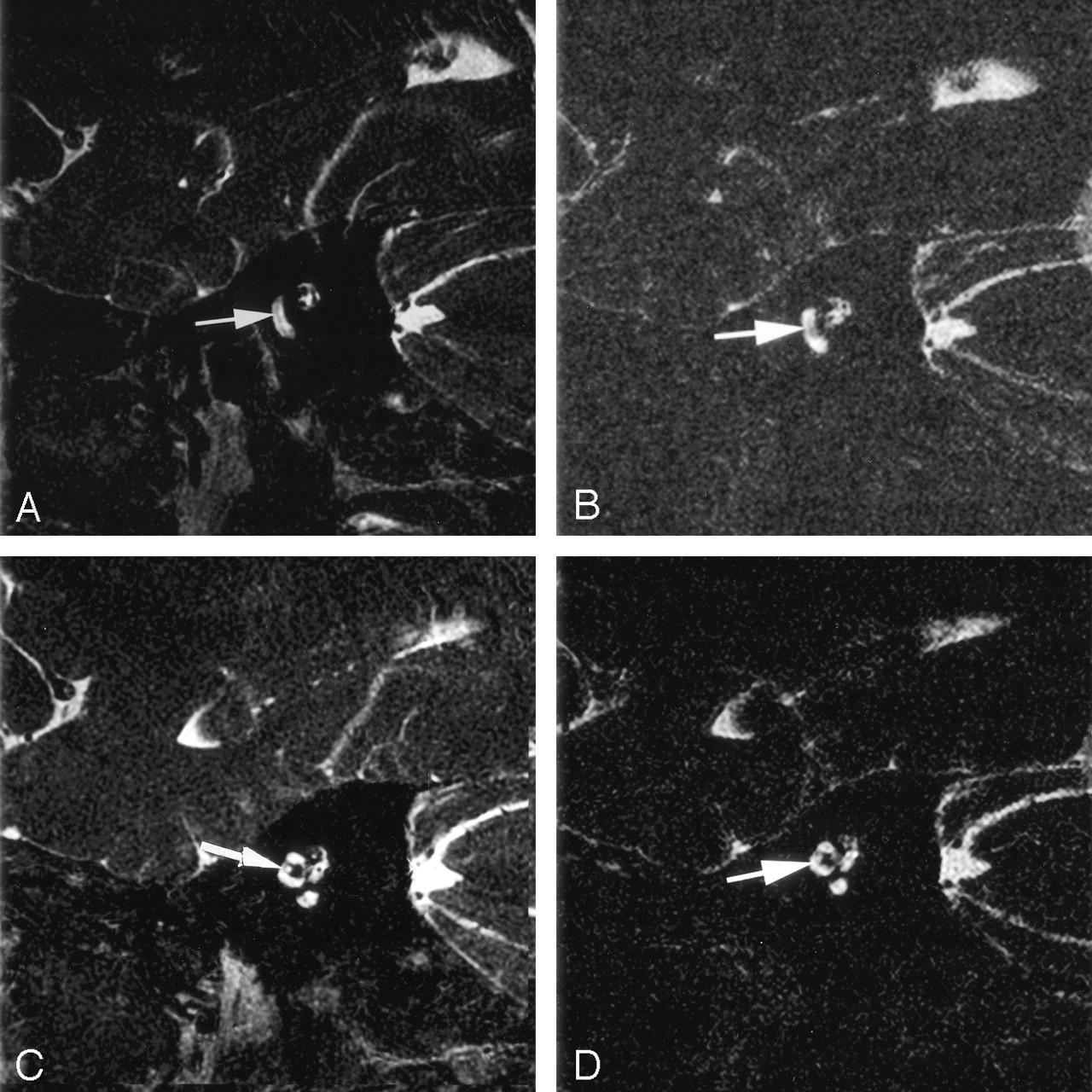
  • Fig 3.
    • Download figure
    • Open in new tab
    • Download powerpoint
    Fig 3.

    Comparison of the spiral lamina and the modiolus of the cochlea on oblique sagittal images..

    A and B, 3D CISS (A) and 3D FRFSE (B) images demonstrate a linear focus of decreased signal intensity that represents the spiral lamina (arrow).

    C and D, 3D CISS (C) and 3D FRFSE (D) images demonstrate the modiolus (arrow).

  • Fig 4.
    • Download figure
    • Open in new tab
    • Download powerpoint
    Fig 4.

    Comparison of the semicircular canal and its apex (arrow) on oblique sagittal images.

    A, 3D CISS reformatted in the plane of the superior semicircular canal (SSC) demonstrates excellent definition of fluid in the apex.

    B, Volume-rendered maximum intensity projection (MIP) of A shows intact fluid rings in all semicircular canals.

    C, 3D FRFSE image also reformatted in the plane of the SCC demonstrates signal intensity loss at the apex.

    D, Volume-rendered MIP of D also depicts this finding.

  • Fig 5.
    • Download figure
    • Open in new tab
    • Download powerpoint
    Fig 5.

    Comparison of the FNC (arrow) on oblique sagittal images.

    A, 3D CISS image adequately displays the normal course of the FNC.

    B, Corresponding 3D FRFSE image shows considerably less signal intensity.

  • Fig 6.
    • Download figure
    • Open in new tab
    • Download powerpoint
    Fig 6.

    Comparison of the endolymphatic duct on oblique sagittal images.

    A, 3D CISS demonstrates modest signal intensity in the normal small endolymphatic duct in the vestibular aqueduct, which is seen just medial to the common crus of the superior and posterior SSCs (arrow).

    B, The duct (arrow) is barely identifiable on the corresponding 3D-FRFSE image. The window and level may need to be adjusted to visualize this normally small duct, which is often not visualized with either technique.

  • Fig 7.
    • Download figure
    • Open in new tab
    • Download powerpoint
    Fig 7.

    3D CISS banding artifact.

    A, Oblique sagittal source image from two-phase–cycled 3D CISS demonstrates marked banding artifact through the SSC and vestibule (arrow).

    B, Postprocessed MIP image at the same level demonstrates subtle irregularity of the contour of the SCC, which represents incomplete averaging of the banding artifact (arrow).

  • Fig 8.
    • Download figure
    • Open in new tab
    • Download powerpoint
    Fig 8.

    Motion artifact between phase cycles on 3D CISS images. Postprocessed MIP image fails to remove banding artifact secondary to patient motion during data acquisition (arrow).

Tables

  • Figures
    • View popup
    TABLE 1:

    Contrast-to-Noise Ratios

    TechniqueFacial NerveIAC CSFBackgroundCNR
    3D CISS96 (51.3–130.7)475.0 (313.1–843.8)47.2 (37.1–60.0)7.9 (0.70–11.9)
    3D FRFSE117.8 (65.6–315.7)269.0 (126.9–603.0)44.6 (31.3–73.3)3.3 (2.0–5.4)
    • Note.—Data are the mean (range). The ratio of CNRs for 3D CISS to 3D FRFSE was 2.5 (2.2–3.5).

    • View popup
    TABLE 2:

    Subjective evaluation of the structures in the IAC and membranous labyrinth

    Anatomic StructureNo. of Patients
    3D CISS3D FRFSE
    IAC
     Facial nerve8/88/8
     Cochlear nerve8/88/8
     Superior vestibular nerve8/88/8
     Inferior vestibular nerve8/88/8
    Cochlea
     2.5 turns8/88/8
     Modiolus8/88/8
     Spiral lamina8/88/8
    Semicircular canals
     Superior8/84/8
     Posterior8/87/8
     Horizontal8/87/8
    Endolymphatic duct3/82/8
    Facial nerve canal6/84/8
    • Note.—Structures were defined as adequately visualized or absent. Note loss of fluid signal intensity in the SCC on one-half of the FRFSE images. Visualized endolymphatic ducts were all normal in caliber.

PreviousNext
Back to top

In this issue

American Journal of Neuroradiology: 25 (4)
American Journal of Neuroradiology
Vol. 25, Issue 4
1 Apr 2004
  • Table of Contents
  • Index by author
Advertisement
Print
Download PDF
Email Article

Thank you for your interest in spreading the word on American Journal of Neuroradiology.

NOTE: We only request your email address so that the person you are recommending the page to knows that you wanted them to see it, and that it is not junk mail. We do not capture any email address.

Enter multiple addresses on separate lines or separate them with commas.
3-T Imaging of the Cochlear Nerve and Labyrinth in Cochlear-Implant Candidates: 3D Fast Recovery Fast Spin-Echo versus 3D Constructive Interference in the Steady State Techniques
(Your Name) has sent you a message from American Journal of Neuroradiology
(Your Name) thought you would like to see the American Journal of Neuroradiology web site.
CAPTCHA
This question is for testing whether or not you are a human visitor and to prevent automated spam submissions.
Cite this article
John I. Lane, Heidi Ward, Robert J. Witte, Matt A. Bernstein, Colin L. W. Driscoll
3-T Imaging of the Cochlear Nerve and Labyrinth in Cochlear-Implant Candidates: 3D Fast Recovery Fast Spin-Echo versus 3D Constructive Interference in the Steady State Techniques
American Journal of Neuroradiology Apr 2004, 25 (4) 618-622;

Citation Manager Formats

  • BibTeX
  • Bookends
  • EasyBib
  • EndNote (tagged)
  • EndNote 8 (xml)
  • Medlars
  • Mendeley
  • Papers
  • RefWorks Tagged
  • Ref Manager
  • RIS
  • Zotero
0 Responses
Respond to this article
Share
Bookmark this article
3-T Imaging of the Cochlear Nerve and Labyrinth in Cochlear-Implant Candidates: 3D Fast Recovery Fast Spin-Echo versus 3D Constructive Interference in the Steady State Techniques
John I. Lane, Heidi Ward, Robert J. Witte, Matt A. Bernstein, Colin L. W. Driscoll
American Journal of Neuroradiology Apr 2004, 25 (4) 618-622;
del.icio.us logo Twitter logo Facebook logo Mendeley logo
  • Tweet Widget
  • Facebook Like
  • Google Plus One

Jump to section

  • Article
    • Abstract
    • Methods
    • Results
    • Discussion
    • Conclusion
    • References
  • Figures & Data
  • Info & Metrics
  • Responses
  • References
  • PDF

Related Articles

  • No related articles found.
  • PubMed
  • Google Scholar

Cited By...

  • Correlation between Histopathology and Signal Loss on Spin-Echo T2-Weighted MR Images of the Inner Ear: Distinguishing Artifacts from Anatomy
  • Diagnostic Accuracy of Screening MR Imaging Using Unenhanced Axial CISS and Coronal T2WI for Detection of Small Internal Auditory Canal Lesions
  • High-Resolution MRI of the Intraparotid Facial Nerve Based on a Microsurface Coil and a 3D Reversed Fast Imaging with Steady-State Precession DWI Sequence at 3T
  • 3D Double-Echo Steady-State with Water Excitation MR Imaging of the Intraparotid Facial Nerve at 1.5T: A Pilot Study
  • Is All "Communicating" Hydrocephalus Really Communicating? Prospective Study on the Value of 3D-Constructive Interference in Steady State Sequence at 3T
  • Detailed MR Imaging Anatomy of the Cisternal Segments of the Glossopharyngeal, Vagus, and Spinal Accessory Nerves in the Posterior Fossa: The Use of 3D Balanced Fast-Field Echo MR Imaging
  • Crossref
  • Google Scholar

This article has not yet been cited by articles in journals that are participating in Crossref Cited-by Linking.

More in this TOC Section

  • MRI of the Parasellar Ligaments
  • ASL Sensitivity for Head and Neck Paraganglioma
  • Post SRS Peritumoral Hyperintense Signal of VSs
Show more Head and Neck Imaging

Similar Articles

Advertisement

Indexed Content

  • Current Issue
  • Accepted Manuscripts
  • Article Preview
  • Past Issues
  • Editorials
  • Editor's Choice
  • Fellows' Journal Club
  • Letters to the Editor
  • Video Articles

Cases

  • Case Collection
  • Archive - Case of the Week
  • Archive - Case of the Month
  • Archive - Classic Case

More from AJNR

  • Trainee Corner
  • Imaging Protocols
  • MRI Safety Corner
  • Book Reviews

Multimedia

  • AJNR Podcasts
  • AJNR Scantastics

Resources

  • Turnaround Time
  • Submit a Manuscript
  • Submit a Video Article
  • Submit an eLetter to the Editor/Response
  • Manuscript Submission Guidelines
  • Statistical Tips
  • Fast Publishing of Accepted Manuscripts
  • Graphical Abstract Preparation
  • Imaging Protocol Submission
  • Evidence-Based Medicine Level Guide
  • Publishing Checklists
  • Author Policies
  • Become a Reviewer/Academy of Reviewers
  • News and Updates

About Us

  • About AJNR
  • Editorial Board
  • Editorial Board Alumni
  • Alerts
  • Permissions
  • Not an AJNR Subscriber? Join Now
  • Advertise with Us
  • Librarian Resources
  • Feedback
  • Terms and Conditions
  • AJNR Editorial Board Alumni

American Society of Neuroradiology

  • Not an ASNR Member? Join Now

© 2025 by the American Society of Neuroradiology All rights, including for text and data mining, AI training, and similar technologies, are reserved.
Print ISSN: 0195-6108 Online ISSN: 1936-959X

Powered by HighWire