
Article Figures & Data
Figures
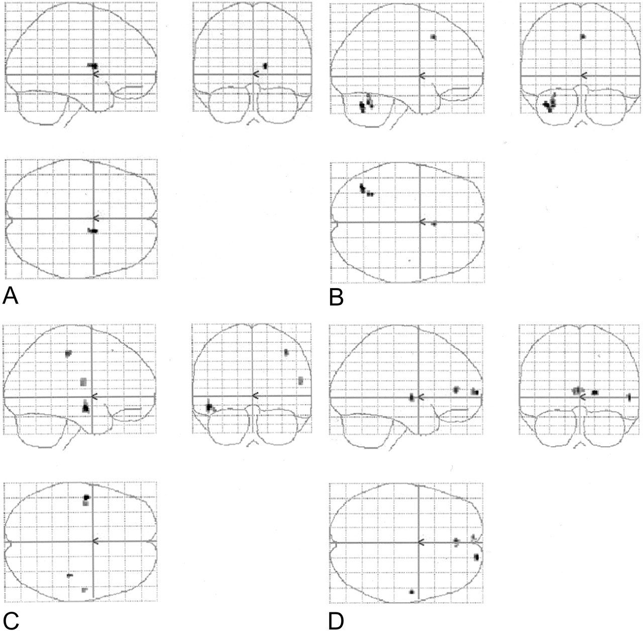
- Fig 1.
Four sets of orthogonal SPM99 glass-brain MIP images depict the normalized group-averaged results of all eight individuals (P = .001, 10-voxel clustering threshold). On the coronal images, the left side represents the left hemisphere. On the axial images, the left side is posterior, and the lower half represents the right hemisphere.
A, Spanish noun-verb task subtracted from the English noun-verb task.
B, Spanish phonological task subtracted from the English phonological task.
C, English noun-verb task subtracted from the Spanish noun-verb task.
D, English phonological task subtracted from the Spanish phonological task.
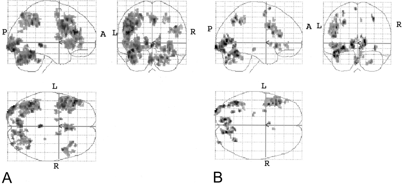
- Fig 2.
Combined (phonological and noun-verb) task subtractions. Orthogonal glass-brain MIP images display group-averaged normalized results (P < .001, 10-voxel clustering threshold).
A, English minus Spanish.
B, Spanish minus English.
- Fig 3.
Group-averaged normalized data for all eight subjects. Activated voxels are overlaid on standard T1-weighted axial anatomic images, which demonstrate the difference in lateralization between the tasks, with greater left lateralization in the English task. The datasets were normalized to the standard MNI space within SPM99. Top row, Spanish noun-verb task. Bottom row, English noun-verb task.
- Fig 4.
Combined tasks. These results are essentially based on a single sample t test based on a consideration of individual normalized data for each of the two tasks (P < .001 with 10-voxel clustering).
A, Combined English tasks versus control.
B, Combined Spanish tasks versus control.
- Fig 5.
Anatomic MPRAGE images show the functional data overlays on the anatomic sections through the cerebellum. The results from Figure 4A (top) and B (bottom) are depicted. The right side of each image is the anatomic left side, and the left side of each image, the anatomic right side.
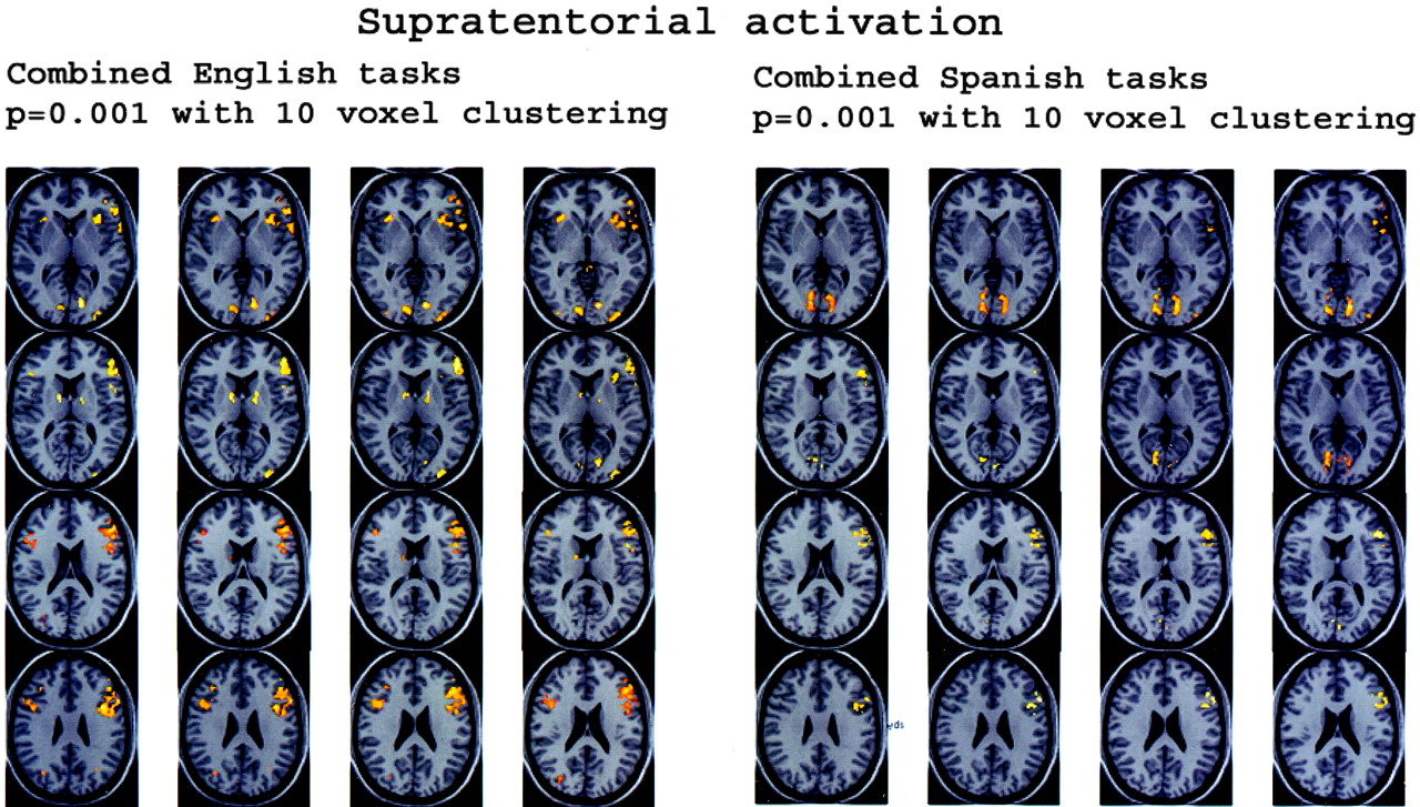
- Fig 6.
Anatomic overlays of the group-averaged normalized combined English task and combined Spanish task functional data on supratentorial anatomic MPRAGE sections. Functional data from Figure 5 are shown (P < .001 with 10 voxel clustering).


Tables
Cerebellar ROI fMRI activation data
Task Voxel Count Left ROI Right ROI Coordinates of Voxel with Maximal T Value Maximal T Value Modified Voxel Count Coordinates of Voxel with Maximal T Value Maximal T Value Modified Voxel Count Left Right x y z x y z Subject A ENG NnVb 368 169 −34 −84 −28 8.61 199 6 −78 −38 7.02 51 ENG Phono 403 100 −40 −62 −26 8.11 256 8 −76 −28 5.31 22 SPN NnVb 155 68 −32 −84 −26 6.75 85 18 −80 −40 4.85 30 SPN Phono 194 0 −40 −60 −26 6.31 157 0 0 Subject B ENG NnVb 1223 1078 −40 −76 −22 17.81 1084 36 −74 −20 11.9 669 ENG Phono 1175 1018 −40 −68 −18 15.55 1067 38 −74 −26 10.31 683 SPN NnVb 387 186 −38 −76 −20 12.59 272 28 −82 −28 5.33 21 SPN Phono 383 210 −28 −82 −20 11.89 357 36 −74 −16 6.59 210 Subject C ENG NnVb 148 55 −28 −70 −22 5.79 50 12 −74 −24 4.31 26 ENG Phono 185 59 −48 −58 −30 6.85 74 18 −54 −12 4.96 6 SPN NnVb 240 87 −18 −58 −12 5.66 26 40 −52 −22 4.53 36 SPN Phono 86 43 −40 −76 −30 4.88 31 34 −70 −26 4.47 14 Subject D ENG NnVb 542 414 −26 −84 −32 9.23 359 26 −84 −26 11.25 276 ENG Phono 309 426 −24 −86 −34 7.23 262 28 −84 −26 11.91 223 SPN NnVb 227 32 −42 −74 −26 6.28 139 44 −60 −20 4.12 5 SPN Phono 46 131 −42 −76 −24 4.25 29 24 −84 −25 6.92 110 Subject E ENG NnVb 402 115 −36 −52 −30 7.94 388 16 −78 −24 4.9 92 ENG Phono 346 163 −36 −78 −22 9.06 172 22 −76 −26 4.85 117 SPN NnVb 167 13 −34 −76 −24 5.99 99 26 −80 −32 4.53 13 SPN Phono 87 18 −46 −66 −24 5.08 22 14 −82 −24 3.9 17 Subject F ENG NnVb 1 0 −42 −48 −24 3.64 1 0 0 ENG Phono 285 248 −36 −64 −30 6.55 124 22 −74 −20 6.69 175 SPN NnVb 120 118 −38 −48 −32 5.45 77 40 −74 −22 6.46 71 SPN Phono 1 0 −42 −48 −24 3.64 1 0 0 Subject G ENG NnVb 985 448 −44 −64 −26 10.56 840 20 −80 −50 8.02 63 ENG Phono 401 391 −46 −58 −26 7.36 327 38 −76 −36 7.27 110 SPN NnVb 71 12 −46 −58 −26 5.33 38 6 −76 −32 4 12 SPN Phono 90 26 −46 −60 −28 4.98 86 40 −58 −24 4.21 23 Subject H ENG NnVb 233 118 −38 −80 −24 5.71 206 16 −74 −40 4.74 76 ENG Phono 338 112 −42 −74 −24 6.72 310 20 −72 −36 5.84 83 SPN NnVb 279 139 −46 −66 −26 5.68 150 28 −74 −30 6.07 73 SPN Phono 17 0 −42 −66 −22 4.61 17 0 0 Note.—ENG = English, SPN = Spanish, NnVb = noun-verb association task, Phono = phonological task.


