
Article Figures & Data
Figures
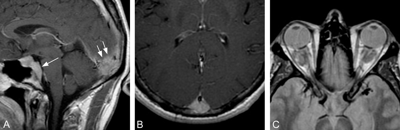
- Fig 1.
Selected MR images through the head and orbits obtained at initial presentation, 3 years before histologic diagnosis.
A, Sagittal T1-weighted postcontrast image shows abnormal homogeneous enhancement extending posteriorly from the sella, along the clivus (single arrow). There is a 2.5-cm enhancing epidural mass seen at the level of the torcula (double arrows).
B, Axial T1-weighted postcontrast image reveals a focal enhancing epidural lesion displacing the posterior portion of the superior sagittal sinus anteriorly.
C, Axial proton attenuation-weighted image through the orbits shows hypointense bilateral intraconal mass lesions.
- Fig 2.
Selected MR images obtained through the brain and orbits 3.0 and 2.5 years after presentation, respectively.
A, Axial T1-weighted postcontrast image through the level of the sella shows a homogeneously enhancing sellar mass lesion extending posteriorly encasing the basilar artery (single arrow). Lateral extension encases the cavernous internal carotid arteries. Patchy enhancement is present within the dorsal aspect of the pons (arrowheads). There has been a significant interval increase in size of the enhancing midline epidural lesion identified at the level of the torcula (double arrows).
B, Sagittal midline postcontrast image shows significant compression of the pons by the prepontine component of the sellar mass. Hazy enhancement within the pons is visualized (white arrow). The posterior falx cerebri is thickened, and the posterior aspect of the superior sagittal sinus is encased by enhancing tissue (black arrow).
C, Axial T2-weighted image obtained through the level of the fourth ventricle reveals confluent hyperintense signal intensity within the central and dorsal pons (arrows). The sellar and torcula mass lesions, as well as the intraconal orbital masses, are markedly hypointense.
D, Axial T1-weighted fat-suppressed postcontrast image through the orbits shows diffuse replacement of the intraconal fat by the large, homogeneously enhancing orbital masses. Patchy enhancement within the ethmoid sinuses reflects sinus disease, not considered a component of the patient’s disease burden.
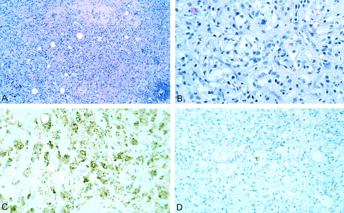
- Fig 3.
Non-Langerhans histocytosis.
A, Histiocytic and chronic inflammatory lesion with extensive fibrosis.
B, Much of the lesion was foamy histiocytes without emperipolesis or giant cells.
C and D, Tumor cells exhibited extensive CD68 (C) but only extremely rare S-100 protein (D) and no CD1a immunoreactivity.


In this issue


Jump to section
Related Articles
Cited By...
- Erdheim-Chester Disease
- Erdheim-Chester Disease
- Erdheim-Chester disease associated with intramedullary spinal cord lesion
- Erdheim-Chester Disease of the Central Nervous System: New Manifestations of a Rare Disease
- Imaging Lesions of the Cavernous Sinus
- Successful treatment of Erdheim-Chester disease, a non-Langerhans-cell histiocytosis, with interferon-{alpha}