Advertisement

Main menu

  • Home
  • Content
    • Current Issue
    • Accepted Manuscripts
    • Article Preview
    • Past Issue Archive
    • Video Articles
    • AJNR Case Collection
    • Case of the Week Archive
    • Case of the Month Archive
    • Classic Case Archive
  • Special Collections
    • AJNR Awards
    • Low-Field MRI
    • Alzheimer Disease
    • ASNR Foundation Special Collection
    • Photon-Counting CT
    • View All
  • Multimedia
    • AJNR Podcasts
    • AJNR SCANtastic
    • Trainee Corner
    • MRI Safety Corner
    • Imaging Protocols
  • For Authors
    • Submit a Manuscript
    • Submit a Video Article
    • Submit an eLetter to the Editor/Response
    • Manuscript Submission Guidelines
    • Statistical Tips
    • Fast Publishing of Accepted Manuscripts
    • Graphical Abstract Preparation
    • Imaging Protocol Submission
    • Author Policies
  • About Us
    • About AJNR
    • Editorial Board
    • Editorial Board Alumni
  • More
    • Become a Reviewer/Academy of Reviewers
    • Subscribers
    • Permissions
    • Alerts
    • Feedback
    • Advertisers
    • ASNR Home

User menu

  • Alerts
  • Log in

Search

  • Advanced search
American Journal of Neuroradiology

American Journal of Neuroradiology

ASHNR American Society of Functional Neuroradiology ASHNR American Society of Pediatric Neuroradiology ASSR
  • Alerts
  • Log in

Advanced Search

  • Home
  • Content
    • Current Issue
    • Accepted Manuscripts
    • Article Preview
    • Past Issue Archive
    • Video Articles
    • AJNR Case Collection
    • Case of the Week Archive
    • Case of the Month Archive
    • Classic Case Archive
  • Special Collections
    • AJNR Awards
    • Low-Field MRI
    • Alzheimer Disease
    • ASNR Foundation Special Collection
    • Photon-Counting CT
    • View All
  • Multimedia
    • AJNR Podcasts
    • AJNR SCANtastic
    • Trainee Corner
    • MRI Safety Corner
    • Imaging Protocols
  • For Authors
    • Submit a Manuscript
    • Submit a Video Article
    • Submit an eLetter to the Editor/Response
    • Manuscript Submission Guidelines
    • Statistical Tips
    • Fast Publishing of Accepted Manuscripts
    • Graphical Abstract Preparation
    • Imaging Protocol Submission
    • Author Policies
  • About Us
    • About AJNR
    • Editorial Board
    • Editorial Board Alumni
  • More
    • Become a Reviewer/Academy of Reviewers
    • Subscribers
    • Permissions
    • Alerts
    • Feedback
    • Advertisers
    • ASNR Home
  • Follow AJNR on Twitter
  • Visit AJNR on Facebook
  • Follow AJNR on Instagram
  • Join AJNR on LinkedIn
  • RSS Feeds

AJNR Awards, New Junior Editors, and more. Read the latest AJNR updates

OtherHead and Neck Imaging

Imaging Characteristics of Diffuse Primary Cutaneous B-Cell Lymphoma of the Cranial Vault with Orbital and Brain Invasion

Mecit Kantarci, Teoman Erdem, Fatih Alper, Cemal Gundogdu, Adnan Okur and Akin Aktas
American Journal of Neuroradiology August 2003, 24 (7) 1324-1326;
Mecit Kantarci
aDepartment of Radiology, Atatürk University, School of Medicine, Erzurum, Turkey
  • Find this author on Google Scholar
  • Find this author on PubMed
  • Search for this author on this site
Teoman Erdem
bDepartment of Dermatology, Atatürk University, School of Medicine, Erzurum, Turkey
  • Find this author on Google Scholar
  • Find this author on PubMed
  • Search for this author on this site
Fatih Alper
aDepartment of Radiology, Atatürk University, School of Medicine, Erzurum, Turkey
  • Find this author on Google Scholar
  • Find this author on PubMed
  • Search for this author on this site
Cemal Gundogdu
cDepartment of Pathology, Atatürk University, School of Medicine, Erzurum, Turkey
  • Find this author on Google Scholar
  • Find this author on PubMed
  • Search for this author on this site
Adnan Okur
aDepartment of Radiology, Atatürk University, School of Medicine, Erzurum, Turkey
  • Find this author on Google Scholar
  • Find this author on PubMed
  • Search for this author on this site
Akin Aktas
bDepartment of Dermatology, Atatürk University, School of Medicine, Erzurum, Turkey
  • Find this author on Google Scholar
  • Find this author on PubMed
  • Search for this author on this site
  • Article
  • Figures & Data
  • Info & Metrics
  • Responses
  • References
  • PDF
Loading

Abstract

Summary: We herein present the imaging findings in a case of diffuse primary cutaneous B-cell lymphoma of the cranial vault with orbital and brain invasion. MR imaging revealed first concomitant orbital and parenchymal invasion in a case of primary diffuse non-Hodgkin lymphoma of the cranial vault. Contrast-enhanced MR imaging revealed a diffuse mass in the scalp of the frontoparietal region bilaterally and invasion of the right orbit and left frontoparietal lobe. Histologic and immunopathologic examinations revealed diffuse primary cutaneous B-cell lymphoma.

Non-Hodgkin lymphoma represents only 3% to 4% of all neoplasms in the general population; however, it occurs more frequently in patients with AIDS. Direct involvement of the CNS occurs in only 1% to 2% of patients with lymphoma (1, 2). A literature search revealed that none of the reported cases included unilateral proptosis and diffuse involvement of cranial vault and brain parenchyma, as did the case reported herein.

Case Report

A 65-year-old man was admitted to the dermatology department because of a right frontoparietal nodule that included ulcers. He had had the lesions for 6 months and swelling on the right lid for 1 month. He had no other signs or symptoms. A dermatologic examination revealed a firm, nontender (10 × 8 cm) nodule that included several ulcers in the right frontoparietal region. Diffuse edema and erythema were observed on the right lid. He had several mobile lymph nodes measuring approximately 1 × 2 cm at the cervical region. The results of another dermatologic examination were normal. Corneal infiltration, orbital invasion, and proptosis were revealed by ophthalmologic examination. A laboratory examination showed that serum lactate dehydrogenase activity was 1135 U/L (normal range, 225–450 U/L). The results of other laboratory examinations were normal. The patient had no history of HIV definition and had not received any medication for this disease. CT of the thorax and abdomen showed no abnormal findings. Bone marrow biopsy showed hypocellularity but no findings of lymphoma or leukemia infiltrations.

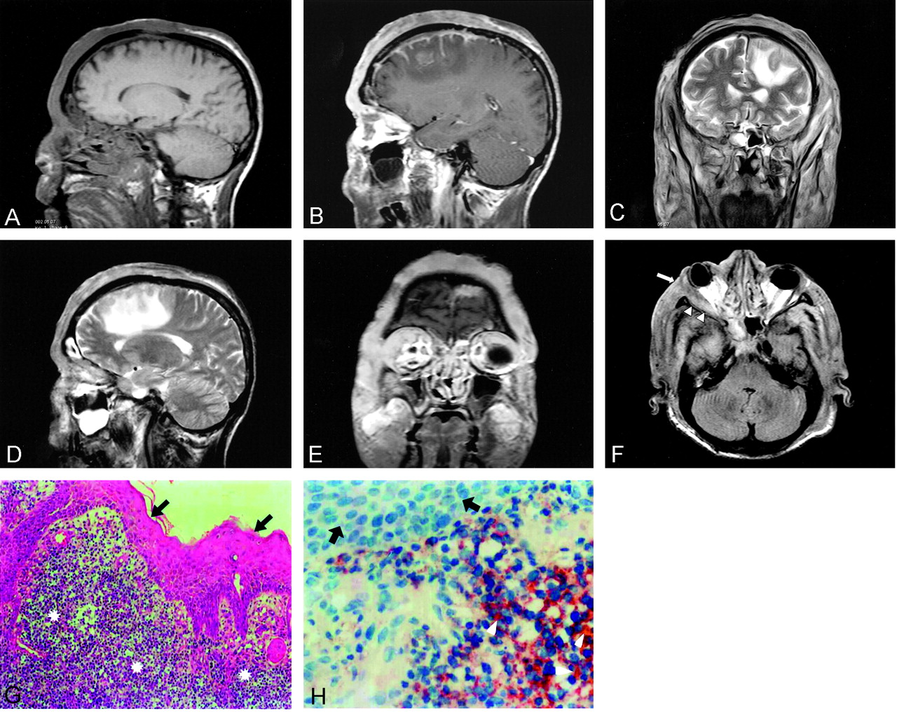
Unenhanced and contrast-enhanced sagittal T1-weighted MR imaging were performed. Marked contrast enhancement of a diffuse mass in the bilateral frontoparietal region of the scalp was shown. It was attached to the dura and vascular and medial frontal gyrus (Fig 1A and B). The coronal and sagittal T2-weighted images showed an isointense lesion and surrounding hyperintense edema on the left medial frontal gyrus (Fig 1C and D). Contrast-enhanced coronal T1-weighted images showed marked enhancement in the right lateral orbital wall and lateral rectus muscle, and axial fluid-attenuated inversion recovery images showed invasion of the orbital wall and the muscle (Fig 1E and F). We performed a wedge biopsy for further analysis under hematoxylin and eosin microscopy. The biopsy revealed a diffuse, large cell type of lymphoma. Histologic sections showed skin with a dermal and focal subcutaneous infiltrate composed of large cells with oval to irregular nuclei and vesicular chromatin. Immunohistochemistry revealed numerous positive cells reacting with CD20, which is a marker for B cells (Fig 1G and H).

Fig 1.
  • Download figure
  • Open in new tab
  • Download powerpoint
Fig 1.

Images of a 65-year-old man who presented with a 6-month history of a right frontoparietal nodule that included ulcers and a 1-month history of swelling on the right lid.

A, Unenhanced sagittal T1-weighted MR image does not show any cranial invasion.

B, Contrast-enhanced sagittal T1-weighted MR image shows strong enhancement of a diffuse mass in the scalp bilateral frontoparietal region and invasion of the dura and left gyrus frontalis medius.

C, Coronal view T2-weighted image shows isointense lesion and peripheral hyperintense edema on the left medial frontal gyrus.

D, Sagittal T2-weighted image shows hyperintense edema on the frontal lobe.

E, Contrast-enhanced coronal T1-weighted image shows marked enhancement at the right lateral orbital wall and muscle rectus lateralis.

F, Axial fluid-attenuated inversion recovery image shows invasion of orbital wall (arrow) and muscle (arrowheads).

G, Photomicrograph of a specimen shows tumoral cells (stars) in stratified squamous epithelium (arrows) (hematoxylin and eosin; original magnification, ×200).

H, Photomicrograph shows CD20-positive tumoral cells (arrowheads) (strep ABC; original magnification, ×100) and stratified squamous epithelium (arrows).

Discussion

The incidence of CNS lymphoma has increased in both immunocompromised and immunocompetent persons (3). Non-Hodgkin lymphoma represents only 3% to 4% of all neoplasms in the general population; however, it is more common in patients with AIDS. Diffuse large B-cell lymphoma is an adult lymphoma accounting for 30% to 40% of all non-Hodgkin lymphomas, and the peak age at onset is during the 6th decade.

According to the Kiel classification, centroblastic lymphoma, immunoblastic B-cell lymphoma, and large cell anaplastic B-cell lymphoma are synonyms of diffuse large B-cell lymphoma. According to Revised European-American Classification of Lymphoid Neoplasms, the large B-cell tumors were formerly described as centroblastic, immunoblastic, and anaplastic together. Clinically, a rapidly growing, often painful mass can be seen to be of single nodal or extranodal size. The disease may primarily involve the skin, particularly in the head and neck region (4).

According to age, sex, and involved region, our case is similar to other cases reported in the literature (5–7). However, the case reported herein included the diverse features of unilateral proptosis and diffuse involvement of cranial vault and brain parenchyma. The initial symptoms and signs of lymphoma in the skull include a painless scalp lump, headache due to bone destruction or tumor infiltration of meninges, seizures, and focal neurologic deficits resulting from the cortex infiltration. The disease involves the pericranium, underlying meninges, and subcutaneous tissue. In most of the cases, no bone destruction occurs. Bone destruction may not be seen in some cases because of the characteristic permeating growth pattern of lymphoma with a large soft-tissue component and very little bone destruction (7,8).

The signal intensity on MR images is nonspecific; most reported cases of skull lymphomas report the signal intensity to be isointense on unenhanced MR images and to show marked enhancement on contrast-enhanced images. Even when cranial vault lymphoma is suspected, MR imaging is not definitively diagnostic because the appearance can mimic those of metastatic carcinoma, osteomyelitis, or meningioma. Cancer metastasis to the skull may show osteolytic bone lesion with extradural or intraparenchymal spread. Patients with osteomyelitis have lytic bone lesions and soft-tissue masses; however, the systemic signs are usually present by the time soft-tissue masses develop. Meningiomas would have associated hyperostosis (9). One case of primary non-Hodgkin lymphoma of cranial vault with associated proptosis is reported in the literature. This case was diagnosed as diffuse high grade large B-cell lymphoma of follicle center cell origin. The present case is the first report of proptosis related to B-cell type diffuse non-Hodgkin lymphoma. A literature search revealed that none of the reported cases included unilateral proptosis and diffuse involvement of cranial vault and brain parenchyma, as did the case reported herein.

Our extensive literature search indicated that this is the first case of orbital and parenchymal invasion associated with primary diffuse non-Hodgkin lymphoma of the cranial vault to be revealed by MR imaging. Although the prognostic factors of this rare tumor are unknown, invasion of the CNS is likely to indicate a poor prognosis (10).

Conclusion

MR imaging is helpful in showing diffuse primary cutaneous lymphoma of the cranial vault with orbital and brain invasion and can thus aid in the decision making regarding different treatment strategies by revealing the invasion of tumor.

References

  1. ↵
    Curty B, Kernan J, Favre J. Malignant non-Hodgkin’s lymphoma of the cranial vault: a case report. Br J Neurosurg 1997;11:433–436
    PubMed
  2. ↵
    Braus DF, Schwechheimer K, Muller-Hermelink HK, Schwarzkopf G, Volk B, Mundinger F. Primary cerebral malignant non-Hodgkin’s lymphomas: a retrospective clinical study. J Neurol 1992;239:117–124
    CrossRefPubMed
  3. ↵
    Baleydier F, Galambrun C, Manel AM, Guibaud L, Nicolino M, Bertrand Y. Primary lymphoma of the pituitary stalk in an immunocompetent 9-year-old child. Med Pediatr Oncol 2001;36:392–395
    CrossRefPubMed
  4. ↵
    Braun-Falco O, Pleving G, Wolff HH, Burgdarf WH. Dermatology. 2nd ed. Berlin: Springer-Verlag;2000 :1611–1640
  5. ↵
    Agbi CB, Bannister CM, Turnbull IW. Primary cranial vault lymphoma mimicking a meningioma. Neurochirurgia (Stuttg) 1983;26:130–132
    PubMed
  6. Maiuri F, Corriero G, Giamundo A. Primary lymphoma of the cranial vault. J Neurosurg Sci 1987;31:183–186
    PubMed
  7. ↵
    Holtas S, Monajati A, Utz R. Computed tomography of malignant lymphoma involving the skull. J Comput Assist Tomogr 1985;9:725–727
    PubMed
  8. ↵
    Parekh HC, Sharma RR, Keogh AJ, Prabhu SS. Primary malignant non-Hodgkin’s lymphoma of cranial vault: a case report. Surg Neurol 1993;39:286–289
    CrossRefPubMed
  9. ↵
    Jamjoom AB, Jamjoom ZA, Naim-Ur-Rahman, Cheema MA. Primary midline cranial vault lymphoma simulating a parasagittal meningioma: the role of angiography in preoperative diagnosis. Neurosurg Rev 1998;21:202–205
    PubMed
  10. ↵
    Wendum D, Sebban C, Gaulard P, et al. Follicular large-cell lymphoma treated with intensive chemotherapy: an analysis of 89 cases included in the LNH87 trial and comparison with the outcome of diffuse large B-cell lymphoma: Groupe d’Etude des Lymphomes de l’Adulte. J Clin Oncol 1997;15:1654–1663
    Abstract
  • Received October 22, 2002.
  • Accepted after revision January 24, 2003.
  • Copyright © American Society of Neuroradiology
View Abstract
PreviousNext
Back to top

In this issue

American Journal of Neuroradiology: 24 (7)
American Journal of Neuroradiology
Vol. 24, Issue 7
1 Aug 2003
  • Table of Contents
  • Index by author
Advertisement
Print
Download PDF
Email Article

Thank you for your interest in spreading the word on American Journal of Neuroradiology.

NOTE: We only request your email address so that the person you are recommending the page to knows that you wanted them to see it, and that it is not junk mail. We do not capture any email address.

Enter multiple addresses on separate lines or separate them with commas.
Imaging Characteristics of Diffuse Primary Cutaneous B-Cell Lymphoma of the Cranial Vault with Orbital and Brain Invasion
(Your Name) has sent you a message from American Journal of Neuroradiology
(Your Name) thought you would like to see the American Journal of Neuroradiology web site.
CAPTCHA
This question is for testing whether or not you are a human visitor and to prevent automated spam submissions.
Cite this article
Mecit Kantarci, Teoman Erdem, Fatih Alper, Cemal Gundogdu, Adnan Okur, Akin Aktas
Imaging Characteristics of Diffuse Primary Cutaneous B-Cell Lymphoma of the Cranial Vault with Orbital and Brain Invasion
American Journal of Neuroradiology Aug 2003, 24 (7) 1324-1326;

Citation Manager Formats

  • BibTeX
  • Bookends
  • EasyBib
  • EndNote (tagged)
  • EndNote 8 (xml)
  • Medlars
  • Mendeley
  • Papers
  • RefWorks Tagged
  • Ref Manager
  • RIS
  • Zotero
0 Responses
Respond to this article
Share
Bookmark this article
Imaging Characteristics of Diffuse Primary Cutaneous B-Cell Lymphoma of the Cranial Vault with Orbital and Brain Invasion
Mecit Kantarci, Teoman Erdem, Fatih Alper, Cemal Gundogdu, Adnan Okur, Akin Aktas
American Journal of Neuroradiology Aug 2003, 24 (7) 1324-1326;
del.icio.us logo Twitter logo Facebook logo Mendeley logo
  • Tweet Widget
  • Facebook Like
  • Google Plus One

Jump to section

  • Article
    • Abstract
    • Case Report
    • Discussion
    • Conclusion
    • References
  • Figures & Data
  • Info & Metrics
  • Responses
  • References
  • PDF

Related Articles

  • No related articles found.
  • PubMed
  • Google Scholar

Cited By...

  • Massive transcalvarial lymphoma
  • Massive transcalvarial lymphoma
  • Crossref
  • Google Scholar

This article has not yet been cited by articles in journals that are participating in Crossref Cited-by Linking.

More in this TOC Section

  • MRI of the Parasellar Ligaments
  • ASL Sensitivity for Head and Neck Paraganglioma
  • Post SRS Peritumoral Hyperintense Signal of VSs
Show more Head and Neck Imaging

Similar Articles

Advertisement

Indexed Content

  • Current Issue
  • Accepted Manuscripts
  • Article Preview
  • Past Issues
  • Editorials
  • Editor's Choice
  • Fellows' Journal Club
  • Letters to the Editor
  • Video Articles

Cases

  • Case Collection
  • Archive - Case of the Week
  • Archive - Case of the Month
  • Archive - Classic Case

More from AJNR

  • Trainee Corner
  • Imaging Protocols
  • MRI Safety Corner
  • Book Reviews

Multimedia

  • AJNR Podcasts
  • AJNR Scantastics

Resources

  • Turnaround Time
  • Submit a Manuscript
  • Submit a Video Article
  • Submit an eLetter to the Editor/Response
  • Manuscript Submission Guidelines
  • Statistical Tips
  • Fast Publishing of Accepted Manuscripts
  • Graphical Abstract Preparation
  • Imaging Protocol Submission
  • Evidence-Based Medicine Level Guide
  • Publishing Checklists
  • Author Policies
  • Become a Reviewer/Academy of Reviewers
  • News and Updates

About Us

  • About AJNR
  • Editorial Board
  • Editorial Board Alumni
  • Alerts
  • Permissions
  • Not an AJNR Subscriber? Join Now
  • Advertise with Us
  • Librarian Resources
  • Feedback
  • Terms and Conditions
  • AJNR Editorial Board Alumni

American Society of Neuroradiology

  • Not an ASNR Member? Join Now

© 2025 by the American Society of Neuroradiology All rights, including for text and data mining, AI training, and similar technologies, are reserved.
Print ISSN: 0195-6108 Online ISSN: 1936-959X

Powered by HighWire