
Article Figures & Data
Figures
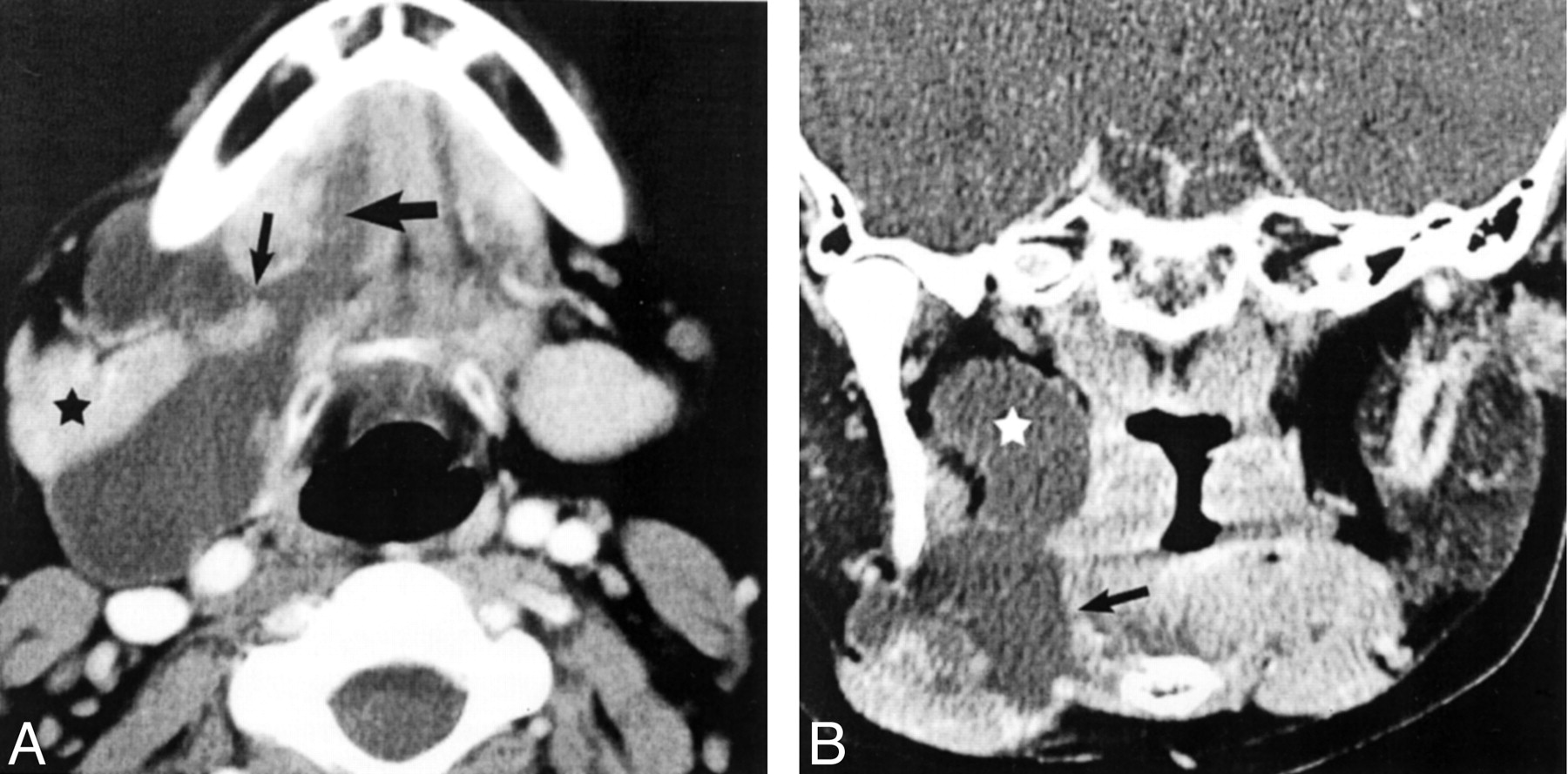
- Fig 1.
Typical imaging findings of giant ranula.
A, Axial view contrast-enhanced CT scan shows a homogenous, fluid attenuation mass centered in the submandibular space with a tail of extension anteriorly into the sublingual space (large arrow). Note the location both anterior and posterior to the submandibular gland (asterisk) due to extension into the submandibular space both posterior to the mylohyoid muscle and laterally through a congenital mylohyoid defect (small arrow).
B, Coronal view image shows cranial extension into the parapharyngeal space (asterisk), without significant mass effect on the surrounding structures. Note the relation of the submandibular component to the mylohyoid muscle (arrow). Reprinted from Harnsberger (17) with permission from the Electronic Medical Education Resource Group.
- Fig 2.
Atypical imaging findings of giant ranulas.
A, Axial view contrast-enhanced CT scan shows peripheral enhancement of this infected giant ranula. Note mild dilation of the submandibular gland duct (arrow) lateral to the sublingual tail in the sublingual space.
B and C, Contiguous (B slightly superior to C) axial view contrast-enhanced CT scans of a patient who had undergone seven failed neck operations. Mild septation is present (white arrows). Subsequent resection of the left sublingual gland resulted in complete resolution. Note the smooth, tapered tail extending into the sublingual space (black arrow). The lesion was pathologically proved to be a giant ranula. Reprinted from Harnsberger (17) with permission from the Electronic Medical Education Resource Group.
- Fig 3.
Axial view contrast-enhanced CT scans of three patients with cystic hygromas show helpful differentiating features.
A, Typical cystic hygroma shows lobulated involvement of the sublingual space (black arrow), mild heterogeneity (white arrow), and extension into the retropharyngeal space (asterisk).
B, Smooth, well-circumscribed cystic hygroma and septation (white arrow) and posterior extension into the posterior cervical space (asterisk) are present.
C, Subtle septation (arrow) is present in this well-circumscribed, homogenous cystic hygroma. Reprinted from Harnsberger (17) with permission from the Electronic Medical Education Resource Group.
- Fig 4.
Giant ranula.
Diagram of axial view shows the anatomic relations of giant ranula (white asterisk), centered in the submandibular space with posterior extension into the parapharyngeal space and an anterior tail extending into the sublingual space. Note the mylohyoid muscle (large arrow) and hyoglossus muscle (small arrow), which define the margins of the sublingual space. Normal anatomic spaces are shown on the right side of the diagram: sublingual space (long thin arrow), submandibular space (black asterisk), and parapharyngeal space (curved arrow). Modified from Coit et al (12) with permission from the Radiological Society of North America.


In this issue


Jump to section
Related Articles
Cited By...
- No citing articles found.