
Article Figures & Data
Figures
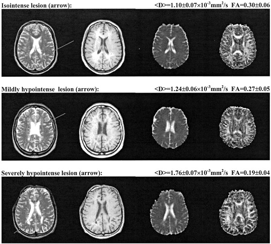
- Fig 1.
Three examples of MS lesions (arrows) with different T1 appearance. Top, isointense lesion (FA=0.30 ± 0.06, coefficient Dav=1.10 ± 0.07 × 10⁻³ mm2/s); middle, mildly hypointense lesion (FA=0.27 ± 0.05, coefficient Dav=1.24 ± 0.06 × 10⁻³ mm2/s); bottom, severely hypointense lesion (FA=0.19 ± 0.04, coefficient Dav = 1.76 ± 0.07 × 10⁻³ mm2/s). For each row, from left to right: axial view fast spin-echo T2-weighted image, spin-echo T1-weighted image, coefficient Dav map, and FA map.
- Fig 2.
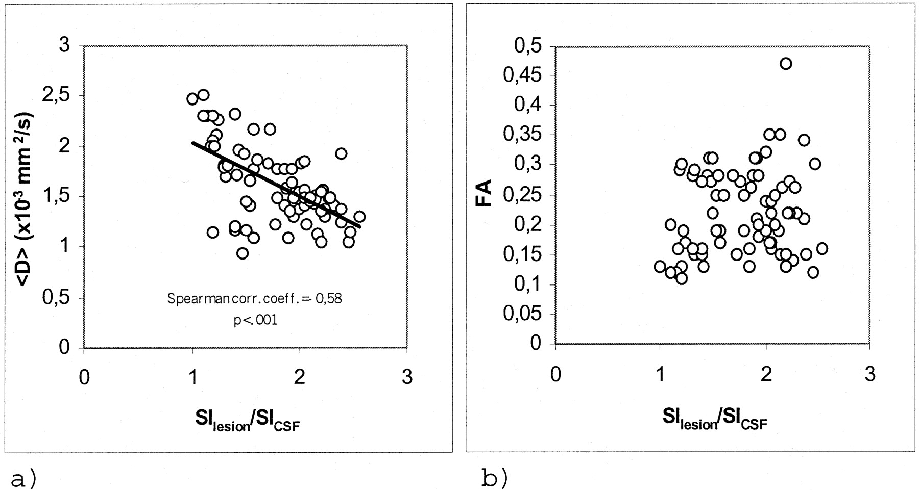
Scatter plots for the 76 lesions studied.
A, Coefficient Dav versus SIlesion/SICSF ratio. Significant inverse correlation was shown.
B, FA versus SIlesion/SICSF ratio. No correlation was shown.


Tables
- TABLE 1:
Mean values (± SD) for fractional anisotropy and coefficient Dav in the various investigated white matter brain regions
FA Dav (×10−3 mm2/s) All plaques (n = 76) 0.22 ± 0.07 1.54 ± 0.33 NAWM regions (n=76) 0.32 ± 0.09 1.00 ± 0.14 NWM regions (n=76) 0.37 ± 0.09 0.89 ± 0.08 Note.—FA indicates fractional anisotropy; Dav, average diffusivity; NAWM, normal appearing white matter; NWM, normal white matter. Note that FA is expressed as a unitless number that may range from 0 to 1 and that Dav is equal to one-third of the trace of the diffusion tensor.
The P value was <.001 for all paired t tests between groups (plaques and NAWM regions, plaques and NWM regions, NAWM and NWM regions).
- TABLE 2:
Mean values (± SD) for absolute and relative fractional anisotropy and coefficient Dav in the three groups of lesions of increasing hypointensity on the T1-weighted images
FA Dav (×10−3 mm2/s) ΔFA T1 isointense (n = 25) 0.26 ± 0.07 1.29 ± 0.17 19 ± 4 T1 mildly hypointense (n=26) 0.21 ± 0.06 1.54 ± 0.31 23 ± 6 T1 severely hypointense (n=25) 0.19 ± 0.06 1.80 ± 0.32 36 ± 4 Note.—FA indicates fractional anisotropy; Dav, averaged diffusivity; ΔFA, percentage FA variation in the lesion, obtained in relation to a symmetric normal appearing white matter area in the contralateral hemisphere.
In this issue


Jump to section
Related Articles
Cited By...
- Short-term surrogate biomarkers of chronic lesion expansion
- Choroid plexus volume is enlarged in clinically isolated syndrome patients with optic neuritis
- Mechanisms of central brain atrophy in multiple sclerosis
- Choroid plexus volume predicts expansion of chronic lesions and brain atrophy
- Loss of corticospinal tract integrity in early MS disease stages
- Increased diffusivity in acute multiple sclerosis lesions predicts risk of black hole
- Diffusion MRI in multiple sclerosis
- A review of structural magnetic resonance neuroimaging