Advertisement

Main menu

  • Home
  • Content
    • Current Issue
    • Accepted Manuscripts
    • Article Preview
    • Past Issue Archive
    • Video Articles
    • AJNR Case Collection
    • Case of the Week Archive
    • Case of the Month Archive
    • Classic Case Archive
  • Special Collections
    • AJNR Awards
    • Low-Field MRI
    • Alzheimer Disease
    • ASNR Foundation Special Collection
    • Photon-Counting CT
    • View All
  • Multimedia
    • AJNR Podcasts
    • AJNR SCANtastic
    • Trainee Corner
    • MRI Safety Corner
    • Imaging Protocols
  • For Authors
    • Submit a Manuscript
    • Submit a Video Article
    • Submit an eLetter to the Editor/Response
    • Manuscript Submission Guidelines
    • Statistical Tips
    • Fast Publishing of Accepted Manuscripts
    • Graphical Abstract Preparation
    • Imaging Protocol Submission
    • Author Policies
  • About Us
    • About AJNR
    • Editorial Board
    • Editorial Board Alumni
  • More
    • Become a Reviewer/Academy of Reviewers
    • Subscribers
    • Permissions
    • Alerts
    • Feedback
    • Advertisers
    • ASNR Home

User menu

  • Alerts
  • Log in

Search

  • Advanced search
American Journal of Neuroradiology

American Journal of Neuroradiology

ASHNR American Society of Functional Neuroradiology ASHNR American Society of Pediatric Neuroradiology ASSR
  • Alerts
  • Log in

Advanced Search

  • Home
  • Content
    • Current Issue
    • Accepted Manuscripts
    • Article Preview
    • Past Issue Archive
    • Video Articles
    • AJNR Case Collection
    • Case of the Week Archive
    • Case of the Month Archive
    • Classic Case Archive
  • Special Collections
    • AJNR Awards
    • Low-Field MRI
    • Alzheimer Disease
    • ASNR Foundation Special Collection
    • Photon-Counting CT
    • View All
  • Multimedia
    • AJNR Podcasts
    • AJNR SCANtastic
    • Trainee Corner
    • MRI Safety Corner
    • Imaging Protocols
  • For Authors
    • Submit a Manuscript
    • Submit a Video Article
    • Submit an eLetter to the Editor/Response
    • Manuscript Submission Guidelines
    • Statistical Tips
    • Fast Publishing of Accepted Manuscripts
    • Graphical Abstract Preparation
    • Imaging Protocol Submission
    • Author Policies
  • About Us
    • About AJNR
    • Editorial Board
    • Editorial Board Alumni
  • More
    • Become a Reviewer/Academy of Reviewers
    • Subscribers
    • Permissions
    • Alerts
    • Feedback
    • Advertisers
    • ASNR Home
  • Follow AJNR on Twitter
  • Visit AJNR on Facebook
  • Follow AJNR on Instagram
  • Join AJNR on LinkedIn
  • RSS Feeds

AJNR Awards, New Junior Editors, and more. Read the latest AJNR updates

Research ArticleBrain

Cortical Blood Flow during Cerebral Vasospasm after Aneurysmal Subarachnoid Hemorrhage: Three-Dimensional N-Isopropyl-p-[123I]Iodoamphetamine Single Photon Emission CT Findings

Hiroki Ohkuma, Shigeharu Suzuki, Kanae Kudo, Shafiqul Islam and Tomonari Kikkawa
American Journal of Neuroradiology March 2003, 24 (3) 444-450;
Hiroki Ohkuma
  • Find this author on Google Scholar
  • Find this author on PubMed
  • Search for this author on this site
Shigeharu Suzuki
  • Find this author on Google Scholar
  • Find this author on PubMed
  • Search for this author on this site
Kanae Kudo
  • Find this author on Google Scholar
  • Find this author on PubMed
  • Search for this author on this site
Shafiqul Islam
  • Find this author on Google Scholar
  • Find this author on PubMed
  • Search for this author on this site
Tomonari Kikkawa
  • Find this author on Google Scholar
  • Find this author on PubMed
  • Search for this author on this site
  • Article
  • Figures & Data
  • Info & Metrics
  • Responses
  • References
  • PDF
Loading

Article Figures & Data

Figures

  • Tables
  • Fig 1.
    • Download figure
    • Open in new tab
    • Download powerpoint
    Fig 1.

    Methods of rCBF assessment.

    Top, 3D images were made by reconstructing the transaxial SPECT data.

    Middle, The surface threshold value was set at 30 mL/100 g/min, and the image defects, which indicate the region with rCBF less than 30 mL/100 g/min, were outlined and calculated by using the image-analysis system (arrows).

    Bottom, Transaxial images were obtained with the IMP-ARG method. ROIs of 30 × 15 mm were set in the lesion with decreased CBF along the cerebral cortex (arrows), and its value was measured.

  • Fig 2.
    • Download figure
    • Open in new tab
    • Download powerpoint
    Fig 2.

    Characteristic findings in cases with DIND.

    Top, Image shows widely decreased cortical rCBF at the left MCA region (arrows).

    Bottom, Image shows widely decreased cortical rCBF at the left ACA region (arrows).
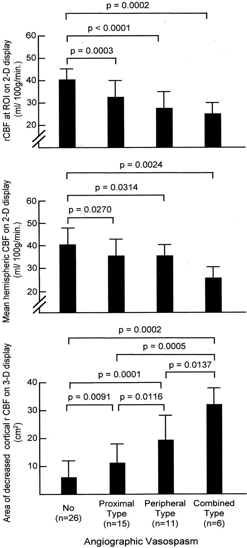
  • Fig 3.
    • Download figure
    • Open in new tab
    • Download powerpoint
    Fig 3.

    CBF data in all cases compared according to the type of angiographic vasospasm.

    Top, rCBF in the ROI on 2D images.

    Middle, Mean hemispheric CBF calculated by using 2D images.

    Bottom, The area of decreased cortical rCBF measured on 3D images.

  • Fig 4.
    • Download figure
    • Open in new tab
    • Download powerpoint
    Fig 4.

    Serial changes in cortical rCBF in a case with DIND.

    Top left, Six days after the onset of SAH, 3D images show multiple, patchy areas of decreased cortical rCBF.

    Top right, cerebral angiography performed on the same day reveals no angiographic vasospasm.

    Bottom left, Ten days after the onset of SAH, 3D images show the widely decreased cortical rCBF.

    Bottom right, Diffuse angiographic vasospasm is seen on an cerebral angiogram obtained on the same day.

Tables

  • Figures
    • View popup
    TABLE 1:

    Clinical parameters in cases with DIND and cases without DIND

    ParameterCases without DIND (n = 43)Cases with DIND (n = 15)
    Mean age ± SD, y56.8 ± 10.554.2 ± 11.6
    Sex ratio (male/female)14/294/11
    Site of aneurysms*
     ICA14 (33)6 (40)
     MCA13 (30)5 (33)
     ACA and ACoA16 (37)4 (27)
    Hunt and Hess grade*
     1 and 222 (51)7 (47)
     318 (35)5 (33)
     4 and 53 (7)3 (20)
    Fisher classification of SAH on CT scans*
     Group 211 (26)2 (13)
     Group 332 (74)13 (87)
    • * Data in parentheses are percentages.

    • View popup
    TABLE 2:

    CBF data

    MeasurerCBF in ROI on 2D Displays, mL/100g/minMean Hemispheric CBF on 2D Displays, mL/100g/minArea of Decreased Cortical CBF on 3D Displays, cm2
    Cases*
     With DIND (n = 15)25.0 ± 5.1†35.5 ± 5.522.6 ± 10.0†
     Without DIND (n = 43)37.0 ± 4.838.0 ± 5.37.4 ± 6.6
    Size of infarction‡
     Large (n = 5)21.8 ± 4.332.6 ± 3.832.3 ± 6.6§
     Small (n = 10)26.6 ± 4.636.9 ± 5.817.8 ± 7.5
    Prognosis at 3 mo‡
     Poor (n = 6)22.2 ± 4.233.7 ± 4.829.8 ± 7.9‖
     Fair (n = 9)26.9 ± 4.536.7 ± 5.817.8 ± 8.4
    • * The CBF data obtained at the onset of DIND in the 15 DIND cases were compared with the data on routine studies in the 43 cases without DIND.

    • † P < .001 compared with cases without DIND.

    • ‡ The CBF data obtained at the onset of DIND in the 15 DIND cases were compared.

    • § P = .006 compared with small size of infarction.

    • ‖ P = .043 compared with fair prognosis.

PreviousNext
Back to top

In this issue

American Journal of Neuroradiology: 24 (3)
American Journal of Neuroradiology
Vol. 24, Issue 3
1 Mar 2003
  • Table of Contents
  • Index by author
Advertisement
Print
Download PDF
Email Article

Thank you for your interest in spreading the word on American Journal of Neuroradiology.

NOTE: We only request your email address so that the person you are recommending the page to knows that you wanted them to see it, and that it is not junk mail. We do not capture any email address.

Enter multiple addresses on separate lines or separate them with commas.
Cortical Blood Flow during Cerebral Vasospasm after Aneurysmal Subarachnoid Hemorrhage: Three-Dimensional N-Isopropyl-p-[123I]Iodoamphetamine Single Photon Emission CT Findings
(Your Name) has sent you a message from American Journal of Neuroradiology
(Your Name) thought you would like to see the American Journal of Neuroradiology web site.
CAPTCHA
This question is for testing whether or not you are a human visitor and to prevent automated spam submissions.
Cite this article
Hiroki Ohkuma, Shigeharu Suzuki, Kanae Kudo, Shafiqul Islam, Tomonari Kikkawa
Cortical Blood Flow during Cerebral Vasospasm after Aneurysmal Subarachnoid Hemorrhage: Three-Dimensional N-Isopropyl-p-[123I]Iodoamphetamine Single Photon Emission CT Findings
American Journal of Neuroradiology Mar 2003, 24 (3) 444-450;

Citation Manager Formats

  • BibTeX
  • Bookends
  • EasyBib
  • EndNote (tagged)
  • EndNote 8 (xml)
  • Medlars
  • Mendeley
  • Papers
  • RefWorks Tagged
  • Ref Manager
  • RIS
  • Zotero
0 Responses
Respond to this article
Share
Bookmark this article
Cortical Blood Flow during Cerebral Vasospasm after Aneurysmal Subarachnoid Hemorrhage: Three-Dimensional N-Isopropyl-p-[123I]Iodoamphetamine Single Photon Emission CT Findings
Hiroki Ohkuma, Shigeharu Suzuki, Kanae Kudo, Shafiqul Islam, Tomonari Kikkawa
American Journal of Neuroradiology Mar 2003, 24 (3) 444-450;
del.icio.us logo Twitter logo Facebook logo Mendeley logo
  • Tweet Widget
  • Facebook Like
  • Google Plus One

Jump to section

  • Article
    • Abstract
    • Methods
    • Results
    • Discussion
    • Conclusion
    • References
  • Figures & Data
  • Info & Metrics
  • Responses
  • References
  • PDF

Related Articles

  • No related articles found.
  • PubMed
  • Google Scholar

Cited By...

  • Does Prevention of Vasospasm in Subarachnoid Hemorrhage Improve Clinical Outcome? No
  • Diagnostic Threshold Values of Cerebral Perfusion Measured With Computed Tomography for Delayed Cerebral Ischemia After Aneurysmal Subarachnoid Hemorrhage
  • Crossref
  • Google Scholar

This article has not yet been cited by articles in journals that are participating in Crossref Cited-by Linking.

More in this TOC Section

  • White Matter Alterations in the Brains of Patients with Active, Remitted, and Cured Cushing Syndrome: A DTI Study
  • Qualitative and Quantitative Analysis of MR Imaging Findings in Patients with Middle Cerebral Artery Stroke Implanted with Mesenchymal Stem Cells
  • Fast Contrast-Enhanced 4D MRA and 4D Flow MRI Using Constrained Reconstruction (HYPRFlow): Potential Applications for Brain Arteriovenous Malformations
Show more Brain

Similar Articles

Advertisement

Indexed Content

  • Current Issue
  • Accepted Manuscripts
  • Article Preview
  • Past Issues
  • Editorials
  • Editor's Choice
  • Fellows' Journal Club
  • Letters to the Editor
  • Video Articles

Cases

  • Case Collection
  • Archive - Case of the Week
  • Archive - Case of the Month
  • Archive - Classic Case

More from AJNR

  • Trainee Corner
  • Imaging Protocols
  • MRI Safety Corner
  • Book Reviews

Multimedia

  • AJNR Podcasts
  • AJNR Scantastics

Resources

  • Turnaround Time
  • Submit a Manuscript
  • Submit a Video Article
  • Submit an eLetter to the Editor/Response
  • Manuscript Submission Guidelines
  • Statistical Tips
  • Fast Publishing of Accepted Manuscripts
  • Graphical Abstract Preparation
  • Imaging Protocol Submission
  • Evidence-Based Medicine Level Guide
  • Publishing Checklists
  • Author Policies
  • Become a Reviewer/Academy of Reviewers
  • News and Updates

About Us

  • About AJNR
  • Editorial Board
  • Editorial Board Alumni
  • Alerts
  • Permissions
  • Not an AJNR Subscriber? Join Now
  • Advertise with Us
  • Librarian Resources
  • Feedback
  • Terms and Conditions
  • AJNR Editorial Board Alumni

American Society of Neuroradiology

  • Not an ASNR Member? Join Now

© 2025 by the American Society of Neuroradiology All rights, including for text and data mining, AI training, and similar technologies, are reserved.
Print ISSN: 0195-6108 Online ISSN: 1936-959X

Powered by HighWire