Advertisement

Main menu

  • Home
  • Content
    • Current Issue
    • Accepted Manuscripts
    • Article Preview
    • Past Issue Archive
    • Video Articles
    • AJNR Case Collection
    • Case of the Week Archive
    • Case of the Month Archive
    • Classic Case Archive
  • Special Collections
    • AJNR Awards
    • Low-Field MRI
    • Alzheimer Disease
    • ASNR Foundation Special Collection
    • Photon-Counting CT
    • View All
  • Multimedia
    • AJNR Podcasts
    • AJNR SCANtastic
    • Trainee Corner
    • MRI Safety Corner
    • Imaging Protocols
  • For Authors
    • Submit a Manuscript
    • Submit a Video Article
    • Submit an eLetter to the Editor/Response
    • Manuscript Submission Guidelines
    • Statistical Tips
    • Fast Publishing of Accepted Manuscripts
    • Graphical Abstract Preparation
    • Imaging Protocol Submission
    • Author Policies
  • About Us
    • About AJNR
    • Editorial Board
    • Editorial Board Alumni
  • More
    • Become a Reviewer/Academy of Reviewers
    • Subscribers
    • Permissions
    • Alerts
    • Feedback
    • Advertisers
    • ASNR Home

User menu

  • Alerts
  • Log in

Search

  • Advanced search
American Journal of Neuroradiology

American Journal of Neuroradiology

ASHNR American Society of Functional Neuroradiology ASHNR American Society of Pediatric Neuroradiology ASSR
  • Alerts
  • Log in

Advanced Search

  • Home
  • Content
    • Current Issue
    • Accepted Manuscripts
    • Article Preview
    • Past Issue Archive
    • Video Articles
    • AJNR Case Collection
    • Case of the Week Archive
    • Case of the Month Archive
    • Classic Case Archive
  • Special Collections
    • AJNR Awards
    • Low-Field MRI
    • Alzheimer Disease
    • ASNR Foundation Special Collection
    • Photon-Counting CT
    • View All
  • Multimedia
    • AJNR Podcasts
    • AJNR SCANtastic
    • Trainee Corner
    • MRI Safety Corner
    • Imaging Protocols
  • For Authors
    • Submit a Manuscript
    • Submit a Video Article
    • Submit an eLetter to the Editor/Response
    • Manuscript Submission Guidelines
    • Statistical Tips
    • Fast Publishing of Accepted Manuscripts
    • Graphical Abstract Preparation
    • Imaging Protocol Submission
    • Author Policies
  • About Us
    • About AJNR
    • Editorial Board
    • Editorial Board Alumni
  • More
    • Become a Reviewer/Academy of Reviewers
    • Subscribers
    • Permissions
    • Alerts
    • Feedback
    • Advertisers
    • ASNR Home
  • Follow AJNR on Twitter
  • Visit AJNR on Facebook
  • Follow AJNR on Instagram
  • Join AJNR on LinkedIn
  • RSS Feeds

AJNR Awards, New Junior Editors, and more. Read the latest AJNR updates

Research ArticleSpine Imaging and Spine Image-Guided Interventions

Percutaneous Translumbar Spinal Cord Compression Injury in a Dog Model That Uses Angioplasty Balloons: MR Imaging and Histopathologic Findings

Phillip D. Purdy, Robert T. Duong, Charles L. White, Donna L. Baer, R. Ross Reichard, G. Lee Pride, Christina Adams, Susan Miller, Christa L. Hladik and Zerrin Yetkin
American Journal of Neuroradiology February 2003, 24 (2) 177-184;
Phillip D. Purdy
  • Find this author on Google Scholar
  • Find this author on PubMed
  • Search for this author on this site
Robert T. Duong
  • Find this author on Google Scholar
  • Find this author on PubMed
  • Search for this author on this site
Charles L. White III
  • Find this author on Google Scholar
  • Find this author on PubMed
  • Search for this author on this site
Donna L. Baer
  • Find this author on Google Scholar
  • Find this author on PubMed
  • Search for this author on this site
R. Ross Reichard
  • Find this author on Google Scholar
  • Find this author on PubMed
  • Search for this author on this site
G. Lee Pride Jr.
  • Find this author on Google Scholar
  • Find this author on PubMed
  • Search for this author on this site
Christina Adams
  • Find this author on Google Scholar
  • Find this author on PubMed
  • Search for this author on this site
Susan Miller
  • Find this author on Google Scholar
  • Find this author on PubMed
  • Search for this author on this site
Christa L. Hladik
  • Find this author on Google Scholar
  • Find this author on PubMed
  • Search for this author on this site
Zerrin Yetkin
  • Find this author on Google Scholar
  • Find this author on PubMed
  • Search for this author on this site
  • Article
  • Figures & Data
  • Info & Metrics
  • Responses
  • References
  • PDF
Loading

Article Figures & Data

Figures

  • Tables
  • Fig 1.
    • Download figure
    • Open in new tab
    • Download powerpoint
    Fig 1.

    Images from a pilot study. After induction of general endotracheal anesthesia, the lumbar subarachnoid space was entered with a needle. A guidewire was introduced via the needle and directed cephalad; then, the needle was withdrawn, and the guidewire was used for coaxial advancement of the angioplasty balloon, mimicking the method used in vascular access for angiography.

    A, Guidewire introduced via neural foramen (lateral approach) and ascending in spinal canal.

    B, Angioplasty balloon introduced over guidewire. Proximal and distal markers on balloon are shown by arrows.

    C, Example of an MR image of a 7-mm balloon (black arrow) inflated against the compressed spinal cord in animal 2 (white arrow).
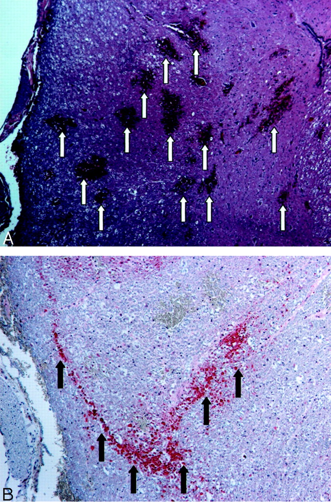
  • Fig 2.
    • Download figure
    • Open in new tab
    • Download powerpoint
    Fig 2.

    Images of animal 4.

    A, Example of hematoxylin and eosin staining obtained at the level of the balloon. Multiple regions of petechial hemorrhage are indicated by arrows. Petechial hemorrhage is a characteristic of spinal cord contusion.

    B, Beta amyloid precursor protein immunostaining of the region shown in A. Amyloid precursor protein is synthesized in neurons in the CNS and peripheral nervous system and is transported to the nerve endings from the cell body. Amyloid precursor protein accumulates when axoplasmic flow is disrupted; it is therefore a sensitive marker of axonal injury. Regions of stain accumulation (arrows) indicate posterior axonal injury.

  • Fig 3.
    • Download figure
    • Open in new tab
    • Download powerpoint
    Fig 3.

    Images of animal 3.

    A, Example of axial view T2-weighted MR image obtained at the level of balloon inflation after the balloon was deflated and removed. The cord displays homogenous signal intensity with no signal intensity change in the dorsal region that shows contrast enhancement in C.

    B, Unenhanced T1-weighted MR image obtained in the axial plane at the level of balloon inflation. No abnormal signal intensity is seen in the spinal cord.

    C, Contrast-enhanced T1-weighted MR image of the region shown in B. A large region of enhancement is located at the dorsal region of the spinal cord (arrow).

    D, Unenhanced sagittal view T1-weighted MR image of the region shown in B. Note that no abnormal signal intensity is seen in the spinal cord.

    E, Focal enhancement is observed after the injection of contrast material (arrow).

    F, Hematoxylin and eosin staining of the spinal cord shows parenchymal hemorrhage in the peripheral white matter (arrows).

    G, Beta amyloid precursor protein immunostaining of the region shown in B highlights well-developed axonal swellings (stain accumulation) in the cross section (arrow).

Tables

  • Figures
    • View popup
    TABLE 1:

    Summary of MR imaging and neuropathological findings

    Animal #Compression (min)Balloon Size (mm)HemorrhageAPPEnhancementT1T2SCO %
    1304NegativeNegativeNegativeNegativeNegative29
    2304NegativePositiveNegativePositiveNegative31
    3307PositivePositivePositiveNegativeNegative79
    4307PositivePositiveNegativeNegativeNegative62
    5307PositivePositivePositivePositiveNegative67
    6307PositiveNegativeNegativePositiveNegative69
    • Note.—APP indicates amyloid precursor protein.

PreviousNext
Back to top

In this issue

American Journal of Neuroradiology: 24 (2)
American Journal of Neuroradiology
Vol. 24, Issue 2
1 Feb 2003
  • Table of Contents
  • Index by author
Advertisement
Print
Download PDF
Email Article

Thank you for your interest in spreading the word on American Journal of Neuroradiology.

NOTE: We only request your email address so that the person you are recommending the page to knows that you wanted them to see it, and that it is not junk mail. We do not capture any email address.

Enter multiple addresses on separate lines or separate them with commas.
Percutaneous Translumbar Spinal Cord Compression Injury in a Dog Model That Uses Angioplasty Balloons: MR Imaging and Histopathologic Findings
(Your Name) has sent you a message from American Journal of Neuroradiology
(Your Name) thought you would like to see the American Journal of Neuroradiology web site.
CAPTCHA
This question is for testing whether or not you are a human visitor and to prevent automated spam submissions.
Cite this article
Phillip D. Purdy, Robert T. Duong, Charles L. White, Donna L. Baer, R. Ross Reichard, G. Lee Pride, Christina Adams, Susan Miller, Christa L. Hladik, Zerrin Yetkin
Percutaneous Translumbar Spinal Cord Compression Injury in a Dog Model That Uses Angioplasty Balloons: MR Imaging and Histopathologic Findings
American Journal of Neuroradiology Feb 2003, 24 (2) 177-184;

Citation Manager Formats

  • BibTeX
  • Bookends
  • EasyBib
  • EndNote (tagged)
  • EndNote 8 (xml)
  • Medlars
  • Mendeley
  • Papers
  • RefWorks Tagged
  • Ref Manager
  • RIS
  • Zotero
0 Responses
Respond to this article
Share
Bookmark this article
Percutaneous Translumbar Spinal Cord Compression Injury in a Dog Model That Uses Angioplasty Balloons: MR Imaging and Histopathologic Findings
Phillip D. Purdy, Robert T. Duong, Charles L. White, Donna L. Baer, R. Ross Reichard, G. Lee Pride, Christina Adams, Susan Miller, Christa L. Hladik, Zerrin Yetkin
American Journal of Neuroradiology Feb 2003, 24 (2) 177-184;
del.icio.us logo Twitter logo Facebook logo Mendeley logo
  • Tweet Widget
  • Facebook Like
  • Google Plus One

Jump to section

  • Article
    • Abstract
    • Methods
    • Results
    • Discussion
    • Conclusion
    • Acknowledgments
    • Footnotes
    • References
  • Figures & Data
  • Info & Metrics
  • Responses
  • References
  • PDF

Related Articles

  • No related articles found.
  • PubMed
  • Google Scholar

Cited By...

  • No citing articles found.
  • Crossref
  • Google Scholar

This article has not yet been cited by articles in journals that are participating in Crossref Cited-by Linking.

More in this TOC Section

  • Optimization of Photon Counting CT Myelography
  • Characteristics of SIH Type I Culprit Lesions
  • Management Outcomes For VO Spine Biopsy
Show more Spine Imaging and Spine Image-Guided Interventions

Similar Articles

Advertisement

Indexed Content

  • Current Issue
  • Accepted Manuscripts
  • Article Preview
  • Past Issues
  • Editorials
  • Editor's Choice
  • Fellows' Journal Club
  • Letters to the Editor
  • Video Articles

Cases

  • Case Collection
  • Archive - Case of the Week
  • Archive - Case of the Month
  • Archive - Classic Case

More from AJNR

  • Trainee Corner
  • Imaging Protocols
  • MRI Safety Corner
  • Book Reviews

Multimedia

  • AJNR Podcasts
  • AJNR Scantastics

Resources

  • Turnaround Time
  • Submit a Manuscript
  • Submit a Video Article
  • Submit an eLetter to the Editor/Response
  • Manuscript Submission Guidelines
  • Statistical Tips
  • Fast Publishing of Accepted Manuscripts
  • Graphical Abstract Preparation
  • Imaging Protocol Submission
  • Evidence-Based Medicine Level Guide
  • Publishing Checklists
  • Author Policies
  • Become a Reviewer/Academy of Reviewers
  • News and Updates

About Us

  • About AJNR
  • Editorial Board
  • Editorial Board Alumni
  • Alerts
  • Permissions
  • Not an AJNR Subscriber? Join Now
  • Advertise with Us
  • Librarian Resources
  • Feedback
  • Terms and Conditions
  • AJNR Editorial Board Alumni

American Society of Neuroradiology

  • Not an ASNR Member? Join Now

© 2025 by the American Society of Neuroradiology All rights, including for text and data mining, AI training, and similar technologies, are reserved.
Print ISSN: 0195-6108 Online ISSN: 1936-959X

Powered by HighWire