
Article Figures & Data
Figures
- Fig 1.
Images from the case of a 31-year-old man with MELAS syndrome, which was documented by a point mutation in the mitochondrial tRNA (Leu). T2-weighted MR image (T2 MRI) (2600/80/1) shows a large focal hyperintensity in the left parietal region, predominantly affecting the gray matter. This area corresponds to elevated choline (Cho), decreased N-acetylaspartate (NAA), and markedly elevated lactate (Lac) on the multisection spectroscopic imaging metabolic maps. In addition, multisection spectroscopic images show globally elevated lactate, which is greatest in the left parietal strokelike lesion, next highest in the remaining gray matter (G.M.) and CSF, and lowest in the white matter. R., right; L., left.
- Fig 2.
Images from the case of a 3-year-old male patient with MELAS-MERRF overlapping syndrome, which was documented by a point mutation. Initial multisection spectroscopic images show no definitive lactate signal intensity. Voxels placed in the right corona radiata (area 1), CSF (area 2), and periventricular white matter (areas 3 and 4) show no clear evidence of lactate doublet. Lipid contamination (*) in this region (1.1–1.4 ppm) may, however, obscure a small lactate peak. R., right; L., left; Cho, choline; Cr, creatine; NAA, N-acetylaspartate; W.M., white matter; T1 MRI, T1-weighted MR image; Lac, lactate.
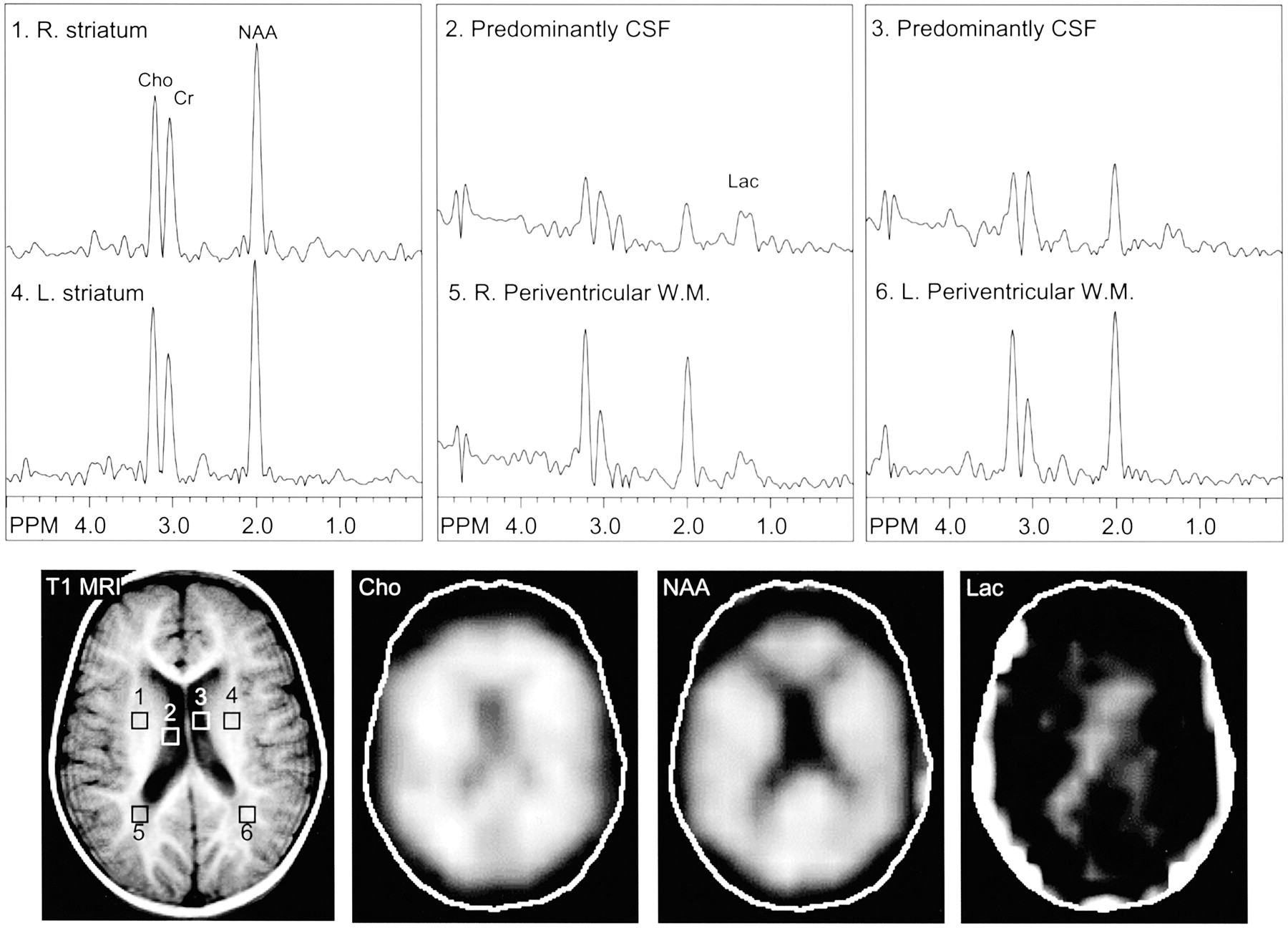
- Fig 3.
Images from the same case of MELAS-MERRF overlapping syndrome shown Figure 2, obtained 1 year later during a subsequent study. MR spectroscopic images show a lactate doublet in the CSF (lateral ventricles, areas 2 and 3) and periventricular white matter (areas 5 and 6), bilaterally. R., right; L., left; Cho, choline; Cr, creatine; NAA, N-acetylaspartate; W.M., white matter; T1 MRI, T1-weighted MR image; Lac, lactate.
- Fig 4.
Images from the case of an 8-year-old female patient with complex I mitochondrial disease, which was diagnosed when the patient was older than 3 years. MR image shows extensive T1-hypointense and T2-hyperintense signal abnormalities in the periventricular regions, particularly in the frontal white matter, in addition to T2 hyperintensity involving the genu of the corpus callosum. MR spectroscopic images show a large lactate peak in the CSF (left lateral ventricle, area 2). A small lactate peak may occur in the frontal white matter lesion (area 1), although spectra are also partially contaminated by lipids (*).W. M., white matter; Lac, lactate; Cho, choline; Cr, creatine; NAA, N-acetylaspartate; G. M., gray matter; T1 MRI, T1-weighted MR image; FLAIR, fluid-attenuated inversion recovery.


Tables
Summary of clinical diagnosis and MR spectroscopy results
Patient No. Sex Age (yr) Clinical Syndrome Supportive Data MR Spectroscopy Findings Lactate Site Interrogated CSF Brain Group 1: well-established diagnosis 1 M 29 Kearns-Sayre Large mtDNA deletion on muscle biopsy N/A − Left mesial frontal lobe (STEAM) 5 M 31 MELAS Point mutation mitochondrial tRNA (Leu) N/A + Small right parietal lesion, more Lac in control left side (STEAM) + + Second MR spectroscopy 3 yr later: gray > white matter stroke-like lesions (MRSI) 7 M 37 MERRF Abundant RRF on muscle biopsy N/A − Left front and occipital, 3 cm single voxel (STEAM) 10 M 7 MELAS Muscle biopsy: RRF, mitochondrial tRNA point mutation, serum Lac 2.9 N/A + Left frontal white matter, single voxel (STEAM) N/A + Second MR spectroscopy 3 mo later: left frontal, smaller after vitamin treatment in 2 days (STEAM) 12 M 1 Elevated Lac Serum Pyr 1.4–2.4*, Lac 1.0–3.2* N/A + Left basal ganglia, single voxel (STEAM) 18 M 3 MELAS/MERRF overlapping syndrome Mitochondrial DNA point mutation − − (MRSI) 1995 normal Lac, Pyr, 1998 Pyr 1.1*, Lac 3.4* + + 2nd MR imaging 11 months later: (MRSI), periventricular white matter 26 F 8 Complex I mitochondrial disease Diagnosed at age 3 yr + + Frontal white matter, corpus callosum lesions (MRSI) 32 M 13 Leigh disease Elevated CSF Lac and Pyr N/A − Post parietal gray and white matter, thalami, single voxel (PRESS) Group 2: possible diagnosis 3 F 27 GI disturbances, “leukodystrophy” pattern white matter abnormality Abnormal ultrastructure of mitochondria on intestinal muscle biopsy N/A − White matter abnormality (STEAM) 4 F 6 Multiple stroke-like episodes Muscle biopsy normal N/A − Deep white matter lesions (STEAM) 8 M 8 Suspected Leigh disease Basal ganglia abnormality, negative CSF Lac N/A − Left high cortical region (STEAM) 9 F 0.5 Developmental delay, poor vision, global cerebral Lac acidosis Diffuse cortical atrophy, decreased white matter volume and hypomyelination N/A + Left frontal cortex, single voxel (STEAM) 11 M 37 “Occipital stroke,” suspected MELAS N/A − Left and right occipital lobes, single voxel (PRESS) 13 F 2 Progressive ataxia, encephalitis-like symptoms + + Basal ganglia (MRSI) 15 M 2 Suspected MELAS, peri-varicella encephalocerebellitis, progressive myoclonus Serum Lac and Pyr, elevated CSF Lac normal, muscle biopsy normal − − (MRSI) 17 M 13 ADEM, 2 episodes Serum Lac normal − − (MRSI) 19 M 1 Lennox-Gastaut with myoclonic epilepsy, cerebral palsy + − (MRSI) 21 M 1 Probable neurodegenerative disease, seizure, mental retardation, failure to thrive Serum Lac 2.6–3.1*, abnormal fatty acid oxidation − − (MRSI) 22 M 3 Developmental delay, seizure, progressive spasticity − − (MRSI) 24 F 17 Progressive spastic paraparesis, ataxia Serum Lac 1.2, Pyr 0.3 − − (MRSI) 28 F 21 Relapsing subacute encephalopathy, suspect Leigh disease History of elevated serum Lac and Pyr − − (MRSI) 29 M 7 Suspected MELAS − − (MRSI) 30 M 12 Movement disorder with seizures + + Left anterior medial temporal lobe T2-hyperintense lesions (MRSI) 31 M 32 Suspected MELAS, mental retardation, mitochondrial encephalopathy − − (MRSI) Group 3: diagnosis excluded by other data 20 M 8 Cerebral palsy w/choreoform movements, probable guanidinoacetate N-methyltransferase deficiency Skin biopsy EM normal − − (MRSI) 23 M 7 Cerebral palsy with spastic diplegia − − (MRSI) 25 F 30 Developmental delay, bipolar syndrome Laboratory results normal − − (MRSI) 27 F 39 Carbohydrate metabolic disease, stroke, endocrine abnormalities, hyperreflexia, poor coordination − − (MRSI) 33 F 20 Acquired lipid myopathy and sensory axonal neuropathy Biochemical ETF-QO deficiency with defined mutation − − (MRSI) Note.—Age indicates patient age in years at time of presentation; Clinical Syndrome, diagnoses made clinically in conjunction with biochemical, genetic, and/or histologic tests (if no final diagnosis available, presenting clinical symptoms listed); M, male; F, female; mtDNA, mitochondrial DNA; N/A, not available; +, presence of lactate; −, absence of lactate; STEAM, stimulated-echo acquisition mode; MELAS, mitochondrial myopathy, encephalopathy, lactic acidosis, and stroke-like episodes; tRNA (Leu), transfer RNA leucine; Lac, lactate; MRSI, MR spectroscopic imaging; MERRF, myoclonus, epilepsy, and ragged red fibers; RRF, ⧫; tRNA, transfer RNA; Pyr, pyruvate;
* , abnormal laboratory value; MELAS/MERRF, overlap of mitochondrial myopathy, encephalopathy, lactic acidosis, and stroke-like episodes with myoclonus, epilepsy, and ragged red fibers; PRESS, point-resolved spectroscopy; GI, gastrointestinal; ADEM, acute disseminated encephalomyelitis; ETF-QO, electron transfer flavoprotein ubiquinone oxidoreductase.
⧫. In general, CSF data were obtained from the MRSI records only.
In this issue


Jump to section
Related Articles
Cited By...
- Large-scale animal model study uncovers altered brain pH and lactate levels as a transdiagnostic endophenotype of neuropsychiatric disorders involving cognitive impairment
- Alterations of Brain Metabolites in Adults With HIV: A Systematic Meta-analysis of Magnetic Resonance Spectroscopy Studies
- Biotin-Responsive Basal Ganglia Disease: Neuroimaging Features before and after Treatment
- Brain imaging in mitochondrial respiratory chain deficiency: combination of brain MRI features as a useful tool for genotype/phenotype correlations
- Cerebral metabolic abnormalities in A3243G mitochondrial DNA mutation carriers
- A guide to diagnosis and treatment of Leigh syndrome