Advertisement

Main menu

  • Home
  • Content
    • Current Issue
    • Accepted Manuscripts
    • Article Preview
    • Past Issue Archive
    • Video Articles
    • AJNR Case Collection
    • Case of the Week Archive
    • Case of the Month Archive
    • Classic Case Archive
  • Special Collections
    • AJNR Awards
    • Low-Field MRI
    • Alzheimer Disease
    • ASNR Foundation Special Collection
    • Photon-Counting CT
    • View All
  • Multimedia
    • AJNR Podcasts
    • AJNR SCANtastic
    • Trainee Corner
    • MRI Safety Corner
    • Imaging Protocols
  • For Authors
    • Submit a Manuscript
    • Submit a Video Article
    • Submit an eLetter to the Editor/Response
    • Manuscript Submission Guidelines
    • Statistical Tips
    • Fast Publishing of Accepted Manuscripts
    • Graphical Abstract Preparation
    • Imaging Protocol Submission
    • Author Policies
  • About Us
    • About AJNR
    • Editorial Board
    • Editorial Board Alumni
  • More
    • Become a Reviewer/Academy of Reviewers
    • Subscribers
    • Permissions
    • Alerts
    • Feedback
    • Advertisers
    • ASNR Home

User menu

  • Alerts
  • Log in

Search

  • Advanced search
American Journal of Neuroradiology

American Journal of Neuroradiology

ASHNR American Society of Functional Neuroradiology ASHNR American Society of Pediatric Neuroradiology ASSR
  • Alerts
  • Log in

Advanced Search

  • Home
  • Content
    • Current Issue
    • Accepted Manuscripts
    • Article Preview
    • Past Issue Archive
    • Video Articles
    • AJNR Case Collection
    • Case of the Week Archive
    • Case of the Month Archive
    • Classic Case Archive
  • Special Collections
    • AJNR Awards
    • Low-Field MRI
    • Alzheimer Disease
    • ASNR Foundation Special Collection
    • Photon-Counting CT
    • View All
  • Multimedia
    • AJNR Podcasts
    • AJNR SCANtastic
    • Trainee Corner
    • MRI Safety Corner
    • Imaging Protocols
  • For Authors
    • Submit a Manuscript
    • Submit a Video Article
    • Submit an eLetter to the Editor/Response
    • Manuscript Submission Guidelines
    • Statistical Tips
    • Fast Publishing of Accepted Manuscripts
    • Graphical Abstract Preparation
    • Imaging Protocol Submission
    • Author Policies
  • About Us
    • About AJNR
    • Editorial Board
    • Editorial Board Alumni
  • More
    • Become a Reviewer/Academy of Reviewers
    • Subscribers
    • Permissions
    • Alerts
    • Feedback
    • Advertisers
    • ASNR Home
  • Follow AJNR on Twitter
  • Visit AJNR on Facebook
  • Follow AJNR on Instagram
  • Join AJNR on LinkedIn
  • RSS Feeds

AJNR Awards, New Junior Editors, and more. Read the latest AJNR updates

OtherBrain

Idiopathic Hypertrophic Cranial Pachymeningitis: Case Report with 7 Years of Imaging Follow-up

Yu Chang Lee, Yao Chung Chueng, Shin Wei Hsu and Chun Chung Lui
American Journal of Neuroradiology January 2003, 24 (1) 119-123;
Yu Chang Lee
  • Find this author on Google Scholar
  • Find this author on PubMed
  • Search for this author on this site
Yao Chung Chueng
  • Find this author on Google Scholar
  • Find this author on PubMed
  • Search for this author on this site
Shin Wei Hsu
  • Find this author on Google Scholar
  • Find this author on PubMed
  • Search for this author on this site
Chun Chung Lui
  • Find this author on Google Scholar
  • Find this author on PubMed
  • Search for this author on this site
  • Article
  • Figures & Data
  • Info & Metrics
  • Responses
  • References
  • PDF
Loading

Article Figures & Data

Figures

  • Tables
  • Fig 1.
    • Download figure
    • Open in new tab
    • Download powerpoint
    Fig 1.

    Contrast-enhanced CT scans show increased enhancement at the right cavernous sinus.

    A, Contrast-enhanced CT scan obtained in 1994 shows that the right tentorium is thickened with homogeneous enhancement due to an inflammatory reaction of the pachymeninges.

    B, Contrast-enhanced CT scan obtained at a more caudal level shows that involvement of the right cavernous sinus correlates with the patient’s symptom of optic neuropathy.
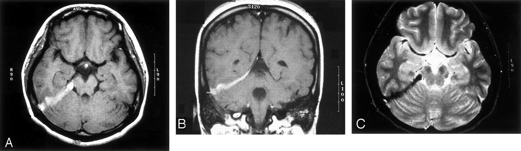
  • Fig 2.
    • Download figure
    • Open in new tab
    • Download powerpoint
    Fig 2.

    MR images obtained in 1995 show the thickened right tentorium. No significant progression of tentorium thickening or cavernous sinus enhancement is noted.

    A, Thickened right tentorium is hypertense on axial T1-weighted image.

    B, Thickened right tentorium is hypertense on coronal T1-weighted image.

    C, Thickened right tentorium is hypointense on coronal T2-weighted image.
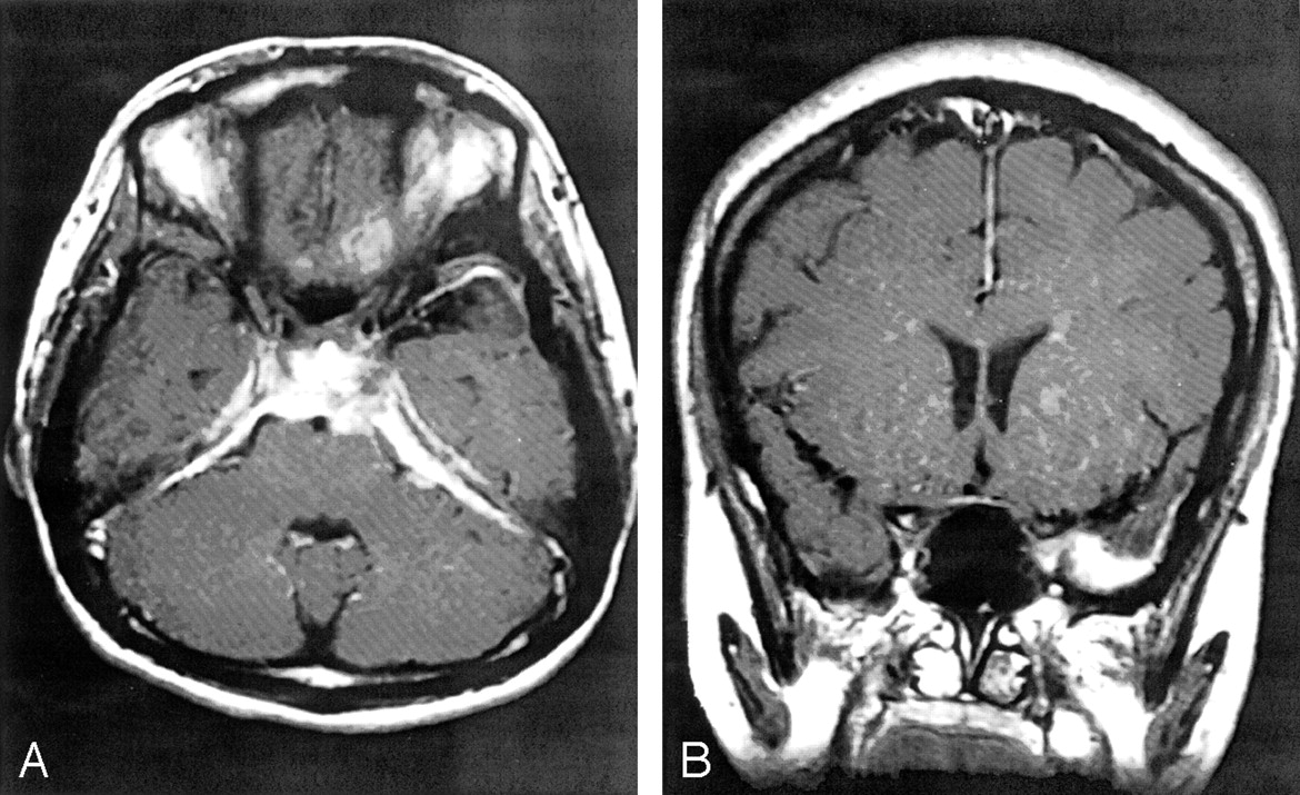
  • Fig 3.
    • Download figure
    • Open in new tab
    • Download powerpoint
    Fig 3.

    MR images show mild increasing thickness and enhancement of the left tentorium.

    A, Axial T1-weighted contrast-enhanced MR image obtained in May of 1998, at the onset of blindness in the right eye and decreased left visual acuity, shows increased right cavernous sinus enhancement and more diffuse involvement of the tentorium bilaterally and of the posterior falx. Prominent dura enhancement can also be seen at the prepontine area, with mild compression of the brain stem.

    B, Coronal T1-weighted contrast-enhanced MR image shows a focus of nodular dural enhancement at left temporal area, with minimal perifocal edema.

  • Fig 4.
    • Download figure
    • Open in new tab
    • Download powerpoint
    Fig 4.

    Coronal T1-weighted contrast-enhanced MR image obtained in June of 1998 shows regression of dural enhancement and the left temporal nodular mass with minimal dural thickening and edema.
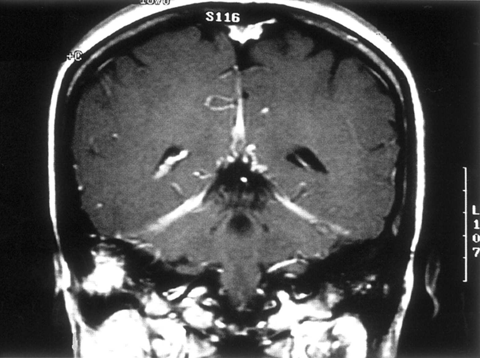
  • Fig 5.
    • Download figure
    • Open in new tab
    • Download powerpoint
    Fig 5.

    Coronal T1-weighted contrast-enhanced MR image obtained in January of 1999 at the onset of right hearing impairment shows increased enhancement of the right mastoid area, although decrease in the tentorium and falx enhancement can be seen.

  • Fig 6.
    • Download figure
    • Open in new tab
    • Download powerpoint
    Fig 6.

    Coronal T1-weighted contrast-enhanced MR image obtained in December of 1999 shows increased intensity of the left mastoid at the onset of left hearing impairment.

  • Fig 7.
    • Download figure
    • Open in new tab
    • Download powerpoint
    Fig 7.

    In July of 2001, the patient suffered from delirium and confusion. CT scan and MR image show enhanced dural mass at the left temporal area, with perifocal edema.

    A, Contrast-enhanced CT scan shows an irregularly enhanced left temporal mass, with compression of the left temporal horn.

    B, Coronal T1-weighted contrast-enhanced MR image shows an enhanced dural-based mass of the left tentorium, with an edematous change of the left temporal lobe. The mass is located in the same area as that revealed in Figure 3B.

  • Fig 8.
    • Download figure
    • Open in new tab
    • Download powerpoint
    Fig 8.

    Cerebral angiogram of the left internal carotid artery shows nonopacification of the straight sinus.
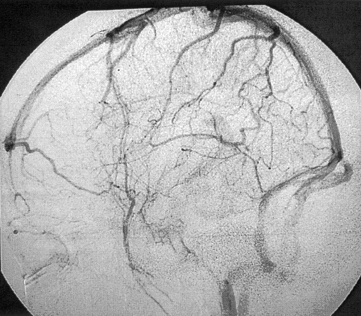
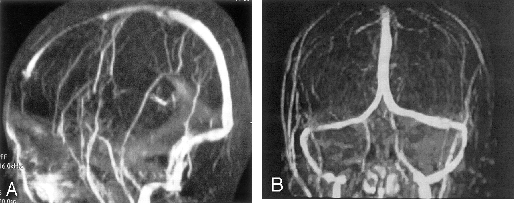
  • Fig 9.
    • Download figure
    • Open in new tab
    • Download powerpoint
    Fig 9.

    Narrowed straight sinus, which correlates with dural sinus thrombosis, was noted on MR venograms.

    A, Sagittal MR venogram shows nonopacification of the straight sinus and indicates thrombosis.

    B, Coronal view.

  • Fig 10.
    • Download figure
    • Open in new tab
    • Download powerpoint
    Fig 10.

    Pathology slide of the meningeal tissue from a left temporal craniotomy shows inflammatory cells with lymphoplasmacytic infiltration, foamy histiocytes, and vascular proliferation (hematoxylin and eosin; original magnification, ×150).

Tables

  • Figures
    • View popup
    TABLE:

    Chronologic imaging findings with clinical correlations

    TimeImaging ModalityImaging FindingsClinical Symptoms
    1994.3.24CTThickened right tentorium: hypodense on unenhanced and with strong enhancementDecreased right vision
    1995.5.6MR imagingRight tentorium thickening: hypointense on T1- and T2-weighted images and homogeneous contrast enhancement
    1998.5.8MR imagingAdditional involvement of left tentorium, posterior falx, and right cavernous sinusRight blindness, decreased left vision
    1998.6.30MR imagingRegressive change of tentorium, falx, and cavernous sinus
    1999.1.11MR imagingRegressive involvement of tentorium but increased enhancement at right mastoidRight hearing loss
    1999.2.8Temporal bone CTRight mastoid cholesteatoma with tegmen and dural plate erosion, left mastoiditis
    1999.12.18MR imagingProgressive enhancement of left mastoidBilateral hearing loss
    2001.4.21MR imagingNo significant interval change of tentorium or mastoid involvement
    2001.7.31CTIll defined enhanced left temporal lobe mass with perifocal edemaDelirium, disorientation
    2001.8.6Cerebral angiographyFaint stain at left temporal, no extra-axial tumor
    2001.8.7MR imagingEnhanced left temporal dural mass with mass effect of temporal lobe, poor opacified straight sinus
PreviousNext
Back to top

In this issue

American Journal of Neuroradiology: 24 (1)
American Journal of Neuroradiology
Vol. 24, Issue 1
1 Jan 2003
  • Table of Contents
  • Index by author
Advertisement
Print
Download PDF
Email Article

Thank you for your interest in spreading the word on American Journal of Neuroradiology.

NOTE: We only request your email address so that the person you are recommending the page to knows that you wanted them to see it, and that it is not junk mail. We do not capture any email address.

Enter multiple addresses on separate lines or separate them with commas.
Idiopathic Hypertrophic Cranial Pachymeningitis: Case Report with 7 Years of Imaging Follow-up
(Your Name) has sent you a message from American Journal of Neuroradiology
(Your Name) thought you would like to see the American Journal of Neuroradiology web site.
CAPTCHA
This question is for testing whether or not you are a human visitor and to prevent automated spam submissions.
Cite this article
Yu Chang Lee, Yao Chung Chueng, Shin Wei Hsu, Chun Chung Lui
Idiopathic Hypertrophic Cranial Pachymeningitis: Case Report with 7 Years of Imaging Follow-up
American Journal of Neuroradiology Jan 2003, 24 (1) 119-123;

Citation Manager Formats

  • BibTeX
  • Bookends
  • EasyBib
  • EndNote (tagged)
  • EndNote 8 (xml)
  • Medlars
  • Mendeley
  • Papers
  • RefWorks Tagged
  • Ref Manager
  • RIS
  • Zotero
0 Responses
Respond to this article
Share
Bookmark this article
Idiopathic Hypertrophic Cranial Pachymeningitis: Case Report with 7 Years of Imaging Follow-up
Yu Chang Lee, Yao Chung Chueng, Shin Wei Hsu, Chun Chung Lui
American Journal of Neuroradiology Jan 2003, 24 (1) 119-123;
del.icio.us logo Twitter logo Facebook logo Mendeley logo
  • Tweet Widget
  • Facebook Like
  • Google Plus One

Jump to section

  • Article
    • Abstract
    • Case Report
    • Discussion
    • Footnotes
    • References
  • Figures & Data
  • Info & Metrics
  • Responses
  • References
  • PDF

Related Articles

  • No related articles found.
  • PubMed
  • Google Scholar

Cited By...

  • Idiopathic hypertrophic pachymeningitis in a patient with a history of diffuse large B cell lymphoma
  • Hypertrophic pachymeningoencephalitis associated with temporal giant cell arteritis
  • Imaging Features of Meningeal Inflammatory Myofibroblastic Tumor
  • Imaging Lesions of the Cavernous Sinus
  • Idiopathic hypertrophic pachymeningitis
  • Crossref
  • Google Scholar

This article has not yet been cited by articles in journals that are participating in Crossref Cited-by Linking.

More in this TOC Section

  • Predictors of Reperfusion in Patients with Acute Ischemic Stroke
  • Enhanced Axonal Metabolism during Early Natalizumab Treatment in Relapsing-Remitting Multiple Sclerosis
  • SWI or T2*: Which MRI Sequence to Use in the Detection of Cerebral Microbleeds? The Karolinska Imaging Dementia Study
Show more Brain

Similar Articles

Advertisement

Indexed Content

  • Current Issue
  • Accepted Manuscripts
  • Article Preview
  • Past Issues
  • Editorials
  • Editor's Choice
  • Fellows' Journal Club
  • Letters to the Editor
  • Video Articles

Cases

  • Case Collection
  • Archive - Case of the Week
  • Archive - Case of the Month
  • Archive - Classic Case

More from AJNR

  • Trainee Corner
  • Imaging Protocols
  • MRI Safety Corner
  • Book Reviews

Multimedia

  • AJNR Podcasts
  • AJNR Scantastics

Resources

  • Turnaround Time
  • Submit a Manuscript
  • Submit a Video Article
  • Submit an eLetter to the Editor/Response
  • Manuscript Submission Guidelines
  • Statistical Tips
  • Fast Publishing of Accepted Manuscripts
  • Graphical Abstract Preparation
  • Imaging Protocol Submission
  • Evidence-Based Medicine Level Guide
  • Publishing Checklists
  • Author Policies
  • Become a Reviewer/Academy of Reviewers
  • News and Updates

About Us

  • About AJNR
  • Editorial Board
  • Editorial Board Alumni
  • Alerts
  • Permissions
  • Not an AJNR Subscriber? Join Now
  • Advertise with Us
  • Librarian Resources
  • Feedback
  • Terms and Conditions
  • AJNR Editorial Board Alumni

American Society of Neuroradiology

  • Not an ASNR Member? Join Now

© 2025 by the American Society of Neuroradiology All rights, including for text and data mining, AI training, and similar technologies, are reserved.
Print ISSN: 0195-6108 Online ISSN: 1936-959X

Powered by HighWire