
Article Figures & Data
Figures
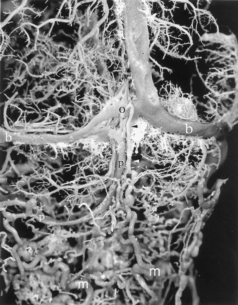
- Fig 1.
Posterior view of a corrosion cast shows an occipital emissary vein (o) draining into the deep cervical veins and a left occipital sinus (p). Black arrowhead and white arrowhead, extracranial connection of the occipital emissary vein; b, transverse sinus; m, deep cervical vein.
- Fig 2.
Left jugular bulb region. The ACC (asterisk) and its connections with surrounding veins are shown. The proximal portions of both transverse sinuses and confluens sinuum have been removed for better visualization. Double arrowhead, inferior petrooccipital vein; arrow, basilar plexus; double arrow, branch to prevertebral venous plexus; r, middle meningeal veins; d, cavernous sinus; a, superior jugular bulb; e, inferior petrosal sinus; c, sigmoid sinus; g, posterior condylar vein; h, lateral condylar vein; f, anterior condylar vein; j, vertebral artery venous plexus; k, anastomosis between anterior internal vertebral venous plexus and vertebral artery venous plexus; i, anterior internal vertebral venous plexus; m, deep cervical vein; b, transverse sinus; l, internal carotid artery venous plexus of Rektorzik; s, emissary vein of the foramen ovale; v, pterygoid plexus; t, intervertebral veins, including inter atlanto-occipital vein.
A, Posterior view.
B, Anterior view.
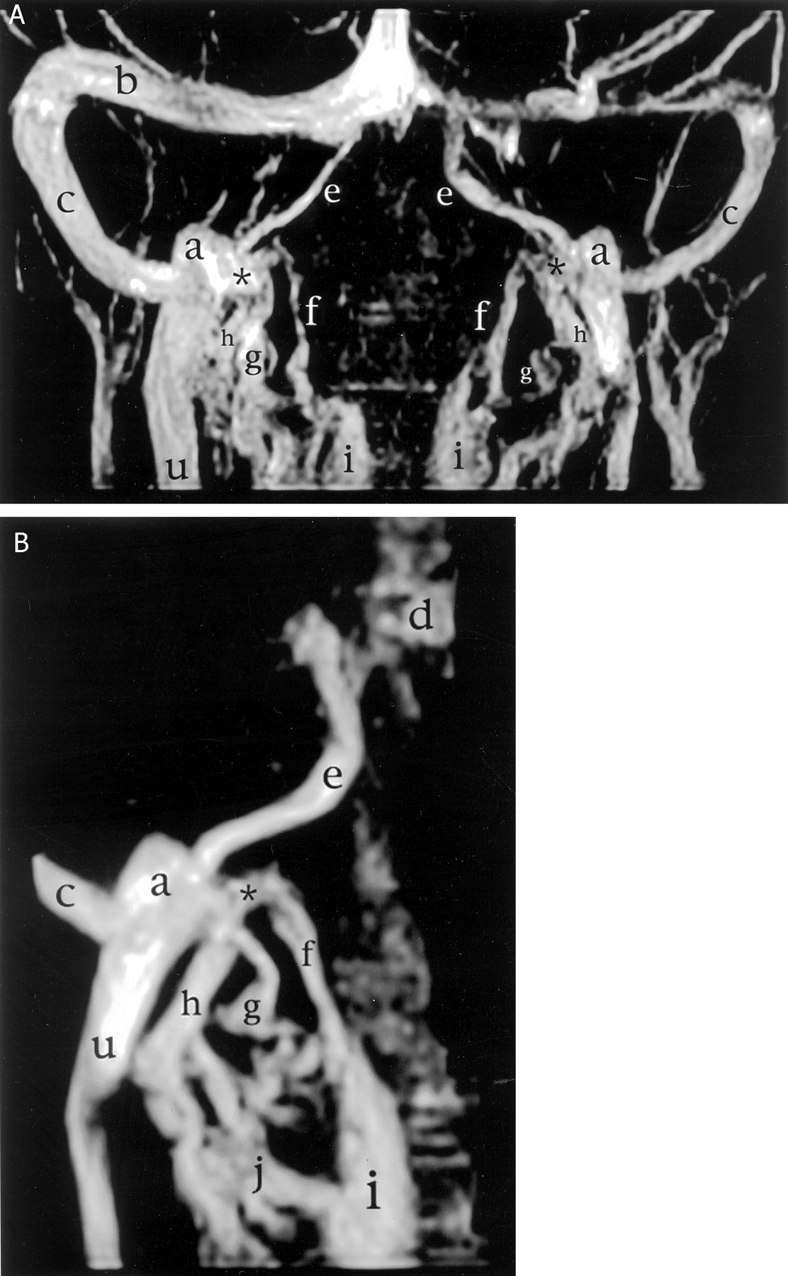
- Fig 3.
MR venograms of the craniocervical region (2D time-of-flight gradient echo images with magnetization transfer; 40/7; field of view, 15 cm; 18; flip angle, 35 degrees; resolution, 220/256; section thickness, 1.5 mm; section overlap, 0.5 mm; matrix, 256 × 256). The ACC (asterisk) and the anterior (f), posterior (g), and lateral (h) condylar veins are shown. b, transverse sinus; c, sigmoid sinus; e, inferior petrosal sinus; a, superior jugular bulb; u, internal jugular vein; i, anterior internal vertebral venous plexus; d, cavernous sinus; j, vertebral artery venous plexus.
A, Overview.
B, Focused on the right superior jugular bulb.
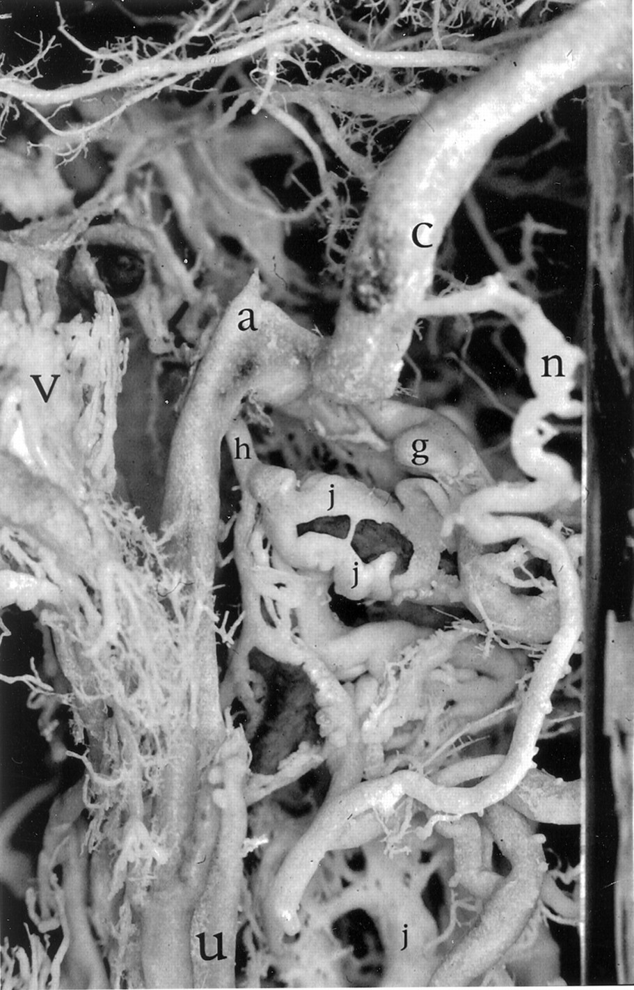
- Fig 4.
Left lateral view of the sigmoid sinus/jugular bulb junction. The mastoid emissary vein (n) and lateral (h) and posterior (g) condylar veins are shown. The plexiform nature of the vertebral artery venous plexus (j), shown here around the horizontal portion of V3, is well seen. c, sigmoid sinus; a, superior jugular bulb; v, pterygoid plexus; u, internal jugular vein.
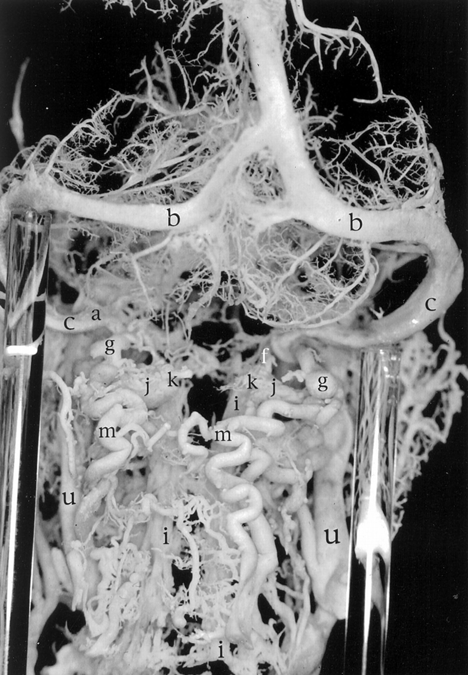
- Fig 5.
Posterior overview of the craniocervical junction in a venous corrosion cast. Note prominent deep cervical veins bilaterally (m). The left posterior condylar vein (g) is clearly seen to drain into both the horizontal portion of the left vertebral artery venous plexus at the C1 level (j) and the left deep cervical vein (m). b, transverse sinus; c, sigmoid sinus; a, superior jugular bulb; f, anterior condylar vein; k, anastomosis between anterior internal vertebral venous plexus and vertebral artery venous plexus; i, anterior internal vertebral venous plexus; u, internal jugular vein.
- Fig 6.
Right lateral view of the craniocervical junction in a venous corrosion cast. Note the presence of a prominent mastoid emissary vein (n) connecting to a deep cervical vein (m). The carotid artery venous plexus is clearly visible (l). b, transverse sinus; d, cavernous sinus; c, sigmoid sinus; a, superior jugular bulb; v, pterygoid plexus; g, posterior condylar vein; h, lateral condylar vein; u, internal jugular vein.
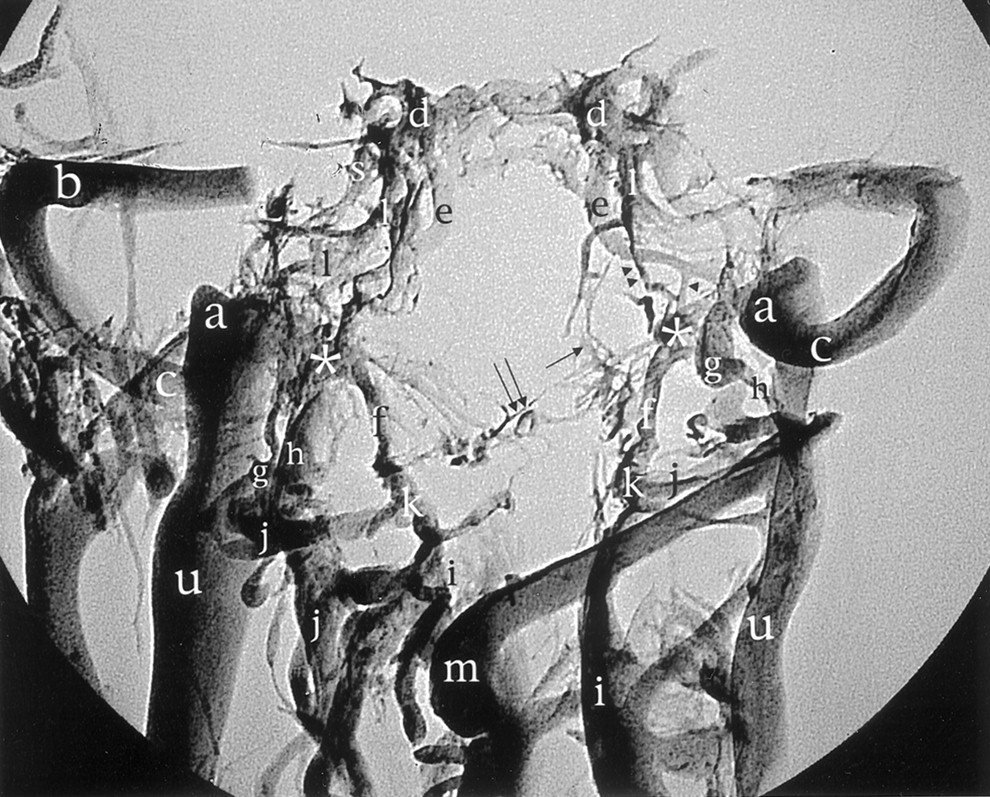
- Fig 7.
Standard radiographic picture of the corrosion cast from Figure 2 in the anteroposterior projection shows the ACC (asterisk) and its relation with surrounding veins. The proximal portions of the transverse sinus (b), confluens sinuum, and straight and superior longitudinal sinuses have been removed for better visualization. Arrowhead, branch between the inferior petrosal sinus and the ACC; double arrowhead, inferior petrooccipital vein; arrow, basilar plexus; double arrow, branch to prevertebral venous plexus; d, cavernous sinus; e, inferior petrosal sinus; l, internal carotid artery venous plexus of Rektorzik; a, superior jugular bulb; g, posterior condylar vein; c, sigmoid sinus; h, lateral condylar vein; k, anastomosis between anterior internal vertebral venous plexus and vertebral artery venous plexus; j, vertebral artery venous plexus; u, internal jugular vein; i, anterior internal vertebral venous plexus; m, deep cervical vein.
- Fig 8.
Schematic representation of anterior view of the ACC and its connections. Note the six main contributions, from the anterior condylar vein (f), the lateral condylar vein (h), the internal jugular vein (u), the inferior petrosal sinus (e), the venous plexus of Rektorzik (double arrowhead), and the prevertebral venous plexus (double arrow). d, cavernous sinus; s, emissary vein of the foramen ovale; a, superior jugular bulb; c, sigmoid sinus; g, posterior condylar vein; k, anastomosis between anterior internal vertebral venous plexus and vertebral artery venous plexus; j, vertebral artery venous plexus; m, deep cervical vein; i, anterior internal vertebral venous plexus.


In this issue


Jump to section
Related Articles
Cited By...
- Posterior condylar canal dural arteriovenous fistula: anatomical, symptomatological, and therapeutic considerations in comparison with hypoglossal canal dural arteriovenous fistula
- Cerebral venous anatomy: implications for the neurointerventionalist
- Cerebral venous anatomy: implications for the neurointerventionalist
- Endovascular treatment strategy, technique, and outcomes for dural arteriovenous fistulas of the marginal sinus region
- Dural Arteriovenous Fistulas of the Foramen Magnum Region: Clinical Features and Angioarchitectural Phenotypes
- Selective embolization of the mastoid emissary vein for pulsatile tinnitus treatment: when is it indicated?
- The Occipital Emissary Vein: A Possible Marker for Pseudotumor Cerebri
- Transvenous coil embolization with intra-operative cone beam CT assistance in the treatment of hypoglossal canal dural arteriovenous fistulae
- Unique percutaneous direct puncture technique for occlusion of a hypoglossal canal dural arteriovenous fistula
- Jugular Anomalies in Multiple Sclerosis Are Associated with Increased Collateral Venous Flow
- Anterior condylar confluence dural arteriovenous fistula: a rare cause of hoarseness
- Republished: Endovascular treatment of posterior condylar canal dural arteriovenous fistula
- Nonaneurysmal Perimesencephalic Hemorrhage Is Associated with Deep Cerebral Venous Drainage Anomalies: A Systematic Literature Review and Meta-Analysis
- Endovascular treatment of posterior condylar canal dural arteriovenous fistula
- Acute subarachnoid hemorrhage in posterior condylar canal dural arteriovenous fistula: imaging features with endovascular management
- Acute subarachnoid hemorrhage in posterior condylar canal dural arteriovenous fistula: imaging features with endovascular management
- Onyx embolization of anterior condylar confluence dural arteriovenous fistula
- Intracranial Nonjugular Venous Pathways: A Possible Compensatory Drainage Mechanism
- Onyx embolization of anterior condylar confluence dural arteriovenous fistula
- Extracranial Venous Drainage Patterns in Patients with Multiple Sclerosis and Healthy Controls
- Incidence of Extrinsic Compression of the Internal Jugular Vein in Unselected Patients Undergoing CT Angiography
- The perfect crime? CCSVI not leaving a trace in MS
- Venous structures at the craniocervical junction: anatomical variations evaluated by multidetector row CT
- Intraosseous Cranial Dural Arteriovenous Fistula Treated with Transvenous Embolization