Advertisement

Main menu

  • Home
  • Content
    • Current Issue
    • Accepted Manuscripts
    • Article Preview
    • Past Issue Archive
    • Video Articles
    • AJNR Case Collection
    • Case of the Week Archive
    • Case of the Month Archive
    • Classic Case Archive
  • Special Collections
    • AJNR Awards
    • Low-Field MRI
    • Alzheimer Disease
    • ASNR Foundation Special Collection
    • Photon-Counting CT
    • View All
  • Multimedia
    • AJNR Podcasts
    • AJNR SCANtastic
    • Trainee Corner
    • MRI Safety Corner
    • Imaging Protocols
  • For Authors
    • Submit a Manuscript
    • Submit a Video Article
    • Submit an eLetter to the Editor/Response
    • Manuscript Submission Guidelines
    • Statistical Tips
    • Fast Publishing of Accepted Manuscripts
    • Graphical Abstract Preparation
    • Imaging Protocol Submission
    • Author Policies
  • About Us
    • About AJNR
    • Editorial Board
    • Editorial Board Alumni
  • More
    • Become a Reviewer/Academy of Reviewers
    • Subscribers
    • Permissions
    • Alerts
    • Feedback
    • Advertisers
    • ASNR Home

User menu

  • Alerts
  • Log in

Search

  • Advanced search
American Journal of Neuroradiology

American Journal of Neuroradiology

ASHNR American Society of Functional Neuroradiology ASHNR American Society of Pediatric Neuroradiology ASSR
  • Alerts
  • Log in

Advanced Search

  • Home
  • Content
    • Current Issue
    • Accepted Manuscripts
    • Article Preview
    • Past Issue Archive
    • Video Articles
    • AJNR Case Collection
    • Case of the Week Archive
    • Case of the Month Archive
    • Classic Case Archive
  • Special Collections
    • AJNR Awards
    • Low-Field MRI
    • Alzheimer Disease
    • ASNR Foundation Special Collection
    • Photon-Counting CT
    • View All
  • Multimedia
    • AJNR Podcasts
    • AJNR SCANtastic
    • Trainee Corner
    • MRI Safety Corner
    • Imaging Protocols
  • For Authors
    • Submit a Manuscript
    • Submit a Video Article
    • Submit an eLetter to the Editor/Response
    • Manuscript Submission Guidelines
    • Statistical Tips
    • Fast Publishing of Accepted Manuscripts
    • Graphical Abstract Preparation
    • Imaging Protocol Submission
    • Author Policies
  • About Us
    • About AJNR
    • Editorial Board
    • Editorial Board Alumni
  • More
    • Become a Reviewer/Academy of Reviewers
    • Subscribers
    • Permissions
    • Alerts
    • Feedback
    • Advertisers
    • ASNR Home
  • Follow AJNR on Twitter
  • Visit AJNR on Facebook
  • Follow AJNR on Instagram
  • Join AJNR on LinkedIn
  • RSS Feeds

AJNR Awards, New Junior Editors, and more. Read the latest AJNR updates

Research ArticlePediatric Neuroimaging

Neonatal Alloimmune Thrombocytopenia: Antenatal and Postnatal Imaging Findings in the Pediatric Brain

Sonia T. Dale and Lee T. Coleman
American Journal of Neuroradiology October 2002, 23 (9) 1457-1465;
Sonia T. Dale
  • Find this author on Google Scholar
  • Find this author on PubMed
  • Search for this author on this site
Lee T. Coleman
  • Find this author on Google Scholar
  • Find this author on PubMed
  • Search for this author on this site
  • Article
  • Figures & Data
  • Info & Metrics
  • Responses
  • References
  • PDF
Loading

Article Figures & Data

Figures

  • Tables
  • Fig 1.
    • Download figure
    • Open in new tab
    • Download powerpoint
    Fig 1.

    Case 3.

    A, Cranial sonogram obtained on day 2 of life shows a large right occipital porencephalic cyst containing echogenic clot.

    B and C, Axial view unenhanced cranial CT scans obtained on day 3 of life show large porencephalic cysts within the right occipital and temporal lobes. The occipital cyst communicates with the right lateral ventricle.
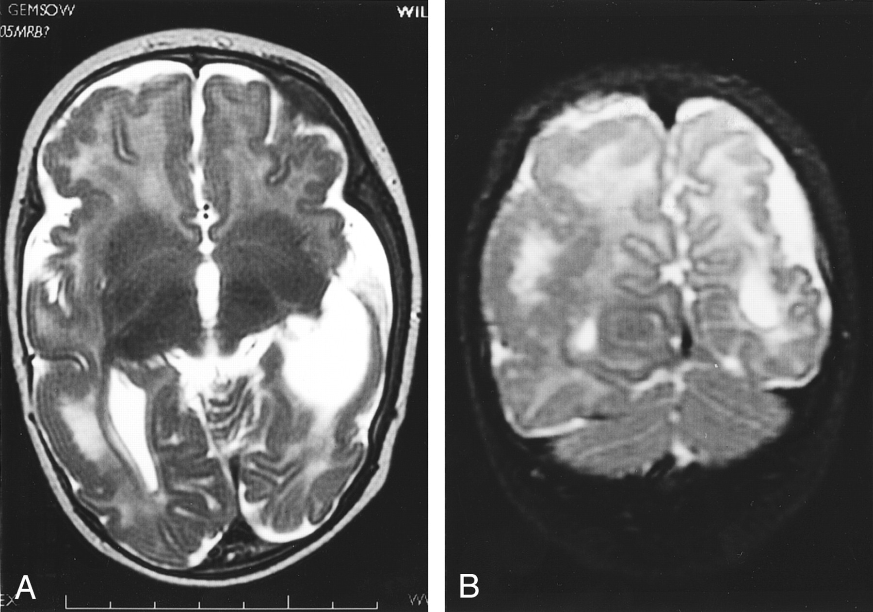
  • Fig 2.
    • Download figure
    • Open in new tab
    • Download powerpoint
    Fig 2.

    Case 1.

    A, Unenhanced axial view cranial CT scan shows large right frontotemporal and left temporal porencephalic cysts with enlargement and remodeling of the middle cranial fossae.

    B, Coronal view fluid-attenuated inversion recovery image (10002/147 [TR/TE]; inversion time, 2200 ms), obtained when the patient was 8 years old, shows large bilateral porencephalic cysts. There is absence of any surrounding high signal intensity gliosis. There is ex vacuo dilation of the right lateral ventricle.

    C, Coronal T2-weighted image (6200/101 [TR/TE]) shows bilateral porencephalic cysts. Communication of the right cyst with the ventricular system after fenestration surgery can be seen.

  • Fig 3.
    • Download figure
    • Open in new tab
    • Download powerpoint
    Fig 3.

    Case 4.

    A and B, Axial view unenhanced cranial CT scans, obtained on day 3 of life, show very large bilateral areas of porencephaly with marked loss of cerebral parenchyma. The cysts contain clots of varying ages and attenuation. Both cysts communicate with the ventricles.

  • Fig 4.
    • Download figure
    • Open in new tab
    • Download powerpoint
    Fig 4.

    Case 2.

    A, Sagittal view cranial sonogram, obtained on day 3 of life, shows increased bulk and echogenicity of the choroid plexus within the lateral ventricle, compatible with choroid plexus hemorrhage, as well as ventricular dilation.

    B, Unenhanced axial view cranial CT scan, obtained on day 4 of life, shows abnormal enlargement and variable attenuation of the extraaxial spaces, compatible with extraaxial hemorrhagic collections. Hyperattenuated blood can be seen along the tentorium cerebelli, compatible with more acute hemorrhage. A left temporal porencephalic cyst is also present.

    C, Coronal view gradient echo image (625/14.9), obtained on day 8 of life, shows hemosiderin deposition (very low signal intensity) within the choroid plexus of both lateral ventricles, along the tentorium cerebelli, and over the cerebellar surface. Loculated, mixed signal intensity and bilateral extraaxial collections compatible with hemorrhagic collections of varying ages can be seen. The lateral ventricles are dilated.

  • Fig 5.
    • Download figure
    • Open in new tab
    • Download powerpoint
    Fig 5.

    Case 6.

    A and B, Fetal MR images obtained at 21 weeks’ gestation. Axial and coronal view T2-weighted images show a large left-sided extraaxial hemorrhage of variable signal intensity, with mass effect on the underlying cerebral hemisphere.

    C and D, Fetal MR images obtained at 23 weeks’ gestation. Axial and coronal view T2-weighted images (26480/100) show the mixed signal intensity extraaxial hemorrhage with more clarity. Signal intensity change within the left parietal lobe can be seen, with breech of the overlying cortex and adjacent wall of the lateral ventricle representing an area of evolving porencephaly.

    E and F, Fetal MR images obtained at 33 weeks’ gestation. Axial and coronal view T2-weighted images (42597/95.7) show further evolution and resolution of the left extraaxial hemorrhage. There is left temporoparietal porencephaly with ventricular communication. High signal intensity abnormality is now noted within the white matter of the right temporal lobe.

    G and H, Cranial MR images obtained when the patient was 5 months of age. Axial and coronal view T2-weighted images (3420/168) show left temporoparietal porencephaly with ventricular communication and loss of cerebral substance. Ventriculomegaly can be seen. Persisting signal intensity abnormality can be seen within the white matter of the right temporal and parietal lobes. A new finding is of a neuronal migrational disorder with extensive polymicrogyria and areas of bandlike heterotopia with an almost “double cortex” appearance.

  • Fig 6.
    • Download figure
    • Open in new tab
    • Download powerpoint
    Fig 6.

    Case 6.

    A and B, Axial and coronal view T2-weighted images (3420/168), obtained when the patient was 5 months of age, show left parietal and temporal porencephaly in association with a neuronal migrational disorder with extensive areas of polymicrogyria.

Tables

  • Figures
    • View popup
    TABLE 1:

    Summary of clinical details for patients with neonatal alloimmune thrombocytopenia

    CaseSexGestation at DeliveryMode of DeliveryPlatelet Count(Cord Blood Sample)Normal = 150–400 × 109Early Clinical Features
    1F41 /40SVD12 × 109Generalized petechiae, bruising
    2F38 /40Elective LUSCS12 × 109Generalized petechiae, purpura
    3F37 /40SVD13 × 109Generalized petechiae, bruising
    4M38 /40Emergency LUSCS20 × 109Bruising, large head circumference, intraretinal hemorrhages, small optic discs
    5M40 /40Forceps28 × 109Generalized petechiae
    6F33 /40Elective LUSCS83 × 109 (Antenatal)Microcephaly, seizures
    • Note.—F indicates female; M, male; SVD, spontaneous vaginal delivery; LUSCS, lower uterine segment caesarian section.

    • View popup
    TABLE 2:

    Summary of imaging findings associated with neonatal alloimmune thrombocytopenia

    Case No.Imaging Modality and AgeImaging FindingsClinical Outcome
    1US, 1 moLarge left temporal and right frontotemporal porencephalic cystsSeizures, cerebral palsy, left hemiplegia, developmental delay, microcephaly
    CT, 1–6 yrBilateral porencephalic cysts, right lateral ventricular enlargement
    MR imaging, 8 yrMultilocular left temporal porencephalic cyst, right frontotemporal porencephaly with ventricular communication (after fenestration surgery), right lateral ventricular enlargement
    2US, days 1 and 3Bilateral choroid plexus hemorrhages, thick echogenic extraaxial spaces, left temporal porencephalic cyst with ventricular communication, ventriculomegalySeizures, developmental delay
    CT, day 4Choroid plexus hemorrhages, acute on chronic bilateral extraaxial hemorrhages, left temporal porencephaly
    MR imaging, day 8Choroid plexus hemorrhages (hemosiderin), hemosiderin over cerebellar sulci, mixed signal loculated extraaxial collections, left temporal porencephalic cyst with ventricular communication, lateral ventriculomegaly with colpocephaly
    3US, day 2Large right temporal porencephalic cyst with clot, right occipital lobe porencephalic cyst with clot communicating with lateral ventricleSeizures, developmental delay, microcephaly
    CT, day 3Right-sided porencephalic cysts with clot, small acute parenchymal hemorrhage in right frontal white matter, dilated right lateral ventricle
    4US, day 3Large left frontotemporal porencephalic cyst with clot communicating with lateral ventricle, large right parietotemporal and occipital porencephalic cyst with clot and ventricular communication, ventriculomegaly (colpocephaly)Died during neonatal period
    CT, day 3Bilateral large porencephalic cysts with clot and ventricular communication, ventriculomegaly
    5CT, 4 moLarge right occipitotemporal porencephalic cyst communicating with right lateral ventricle, mild right lateral ventricular dilatationSeizures, blindness, developmental delay
    MR imaging, 2 yrLarge right temporal porencephalic cyst with ventricular communication, ventriculoperitoneal shunt, reduced periventricular/periatrial white matter with increased fluid-attenuated inversion recovery/T2 signal (gliosis)
    6Fetal MR imaging, 21/40Left extraaxial hemorrhage with mass effect, signal change left parietal lobe with cortical breech, porencephaly?Microcephaly, seizures
    Resolving extraaxial hemorrhage, extensive parietotemporal porencephaly with hemosiderin lining cyst, ex vacuo dilation right lateral ventricle, high signal right temporal lobe, delayed maturation with delayed sulcation
    MR imaging, 5 moLeft parietotemporal porencephaly, ventriculomegaly, neuronal migrational disorder with extensive polymicrogyria and periventricular heterotopia with almost double cortex appearance, abnormal signal right temporal and parietal white matter
PreviousNext
Back to top

In this issue

American Journal of Neuroradiology: 23 (9)
American Journal of Neuroradiology
Vol. 23, Issue 9
1 Oct 2002
  • Table of Contents
  • Index by author
Advertisement
Print
Download PDF
Email Article

Thank you for your interest in spreading the word on American Journal of Neuroradiology.

NOTE: We only request your email address so that the person you are recommending the page to knows that you wanted them to see it, and that it is not junk mail. We do not capture any email address.

Enter multiple addresses on separate lines or separate them with commas.
Neonatal Alloimmune Thrombocytopenia: Antenatal and Postnatal Imaging Findings in the Pediatric Brain
(Your Name) has sent you a message from American Journal of Neuroradiology
(Your Name) thought you would like to see the American Journal of Neuroradiology web site.
CAPTCHA
This question is for testing whether or not you are a human visitor and to prevent automated spam submissions.
Cite this article
Sonia T. Dale, Lee T. Coleman
Neonatal Alloimmune Thrombocytopenia: Antenatal and Postnatal Imaging Findings in the Pediatric Brain
American Journal of Neuroradiology Oct 2002, 23 (9) 1457-1465;

Citation Manager Formats

  • BibTeX
  • Bookends
  • EasyBib
  • EndNote (tagged)
  • EndNote 8 (xml)
  • Medlars
  • Mendeley
  • Papers
  • RefWorks Tagged
  • Ref Manager
  • RIS
  • Zotero
0 Responses
Respond to this article
Share
Bookmark this article
Neonatal Alloimmune Thrombocytopenia: Antenatal and Postnatal Imaging Findings in the Pediatric Brain
Sonia T. Dale, Lee T. Coleman
American Journal of Neuroradiology Oct 2002, 23 (9) 1457-1465;
del.icio.us logo Twitter logo Facebook logo Mendeley logo
  • Tweet Widget
  • Facebook Like
  • Google Plus One

Jump to section

  • Article
    • Abstract
    • Methods
    • Results
    • Discussion
    • Conclusion
    • References
  • Figures & Data
  • Info & Metrics
  • Responses
  • References
  • PDF

Related Articles

  • No related articles found.
  • PubMed
  • Google Scholar

Cited By...

  • Haematological analysis of calves with bovine neonatal pancytopenia
  • The role of fetal magnetic resonance imaging
  • Severe bilateral intracranial haemorrhage due to alloimmune thrombozytopenia in a premature infant
  • Crossref
  • Google Scholar

This article has not yet been cited by articles in journals that are participating in Crossref Cited-by Linking.

More in this TOC Section

  • Frontal Paraventricular Cysts
  • Sodium MRI in Pediatric Brain Tumors
  • FRACTURE MR in Congenital Vertebral Anomalies
Show more Pediatric Neuroimaging

Similar Articles

Advertisement

Indexed Content

  • Current Issue
  • Accepted Manuscripts
  • Article Preview
  • Past Issues
  • Editorials
  • Editor's Choice
  • Fellows' Journal Club
  • Letters to the Editor
  • Video Articles

Cases

  • Case Collection
  • Archive - Case of the Week
  • Archive - Case of the Month
  • Archive - Classic Case

More from AJNR

  • Trainee Corner
  • Imaging Protocols
  • MRI Safety Corner
  • Book Reviews

Multimedia

  • AJNR Podcasts
  • AJNR Scantastics

Resources

  • Turnaround Time
  • Submit a Manuscript
  • Submit a Video Article
  • Submit an eLetter to the Editor/Response
  • Manuscript Submission Guidelines
  • Statistical Tips
  • Fast Publishing of Accepted Manuscripts
  • Graphical Abstract Preparation
  • Imaging Protocol Submission
  • Evidence-Based Medicine Level Guide
  • Publishing Checklists
  • Author Policies
  • Become a Reviewer/Academy of Reviewers
  • News and Updates

About Us

  • About AJNR
  • Editorial Board
  • Editorial Board Alumni
  • Alerts
  • Permissions
  • Not an AJNR Subscriber? Join Now
  • Advertise with Us
  • Librarian Resources
  • Feedback
  • Terms and Conditions
  • AJNR Editorial Board Alumni

American Society of Neuroradiology

  • Not an ASNR Member? Join Now

© 2025 by the American Society of Neuroradiology All rights, including for text and data mining, AI training, and similar technologies, are reserved.
Print ISSN: 0195-6108 Online ISSN: 1936-959X

Powered by HighWire