
Article Figures & Data
Figures
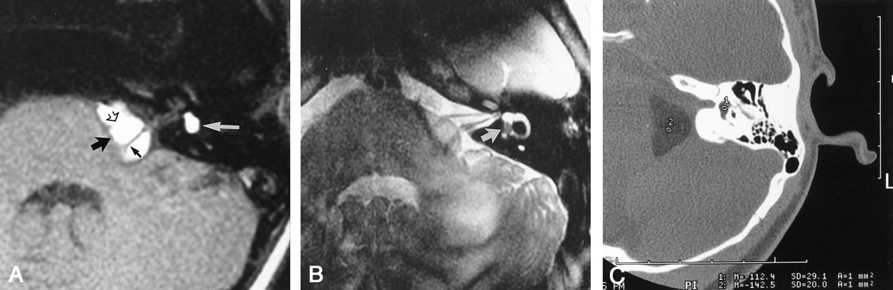
- Fig 1.
Patient 1.
A, T1-weighted axial image (400/15/2 [TR/TE/NEX]), obtained through the left internal auditory canal, shows a hyperintense mass in the cerebellopontine angle (thick black arrow) and vestibule (white arrow). The seventh (open arrow) and eighth (small black arrow) cranial nerves can be seen coursing through the cerebellopontine angle component.
B, T2-weighted fast spin-echo axial image (4000/90/4), obtained through the left internal auditory canal, clearly shows the intravestibular location of the mass (arrow). C, Axial CT scan, obtained through the left internal auditory canal, shows the hypoattenuated cerebellopontine angle and vestibular masses. Hounsfield attenuation units are −112 for the vestibular component (1) and −142 for the cerebellopontine angle component (2), consistent with fat attenuation (lipoma).
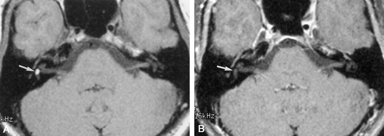
- Fig 2.
Patient 2.
A, Axial T1-weighted image (400/14/2) shows a hyperintense mass in the right vestibule (arrow).
B, Axial T2-weighted fast spin-echo image (4000/102/4) shows the intravestibular mass to be hypointense relative to the fluid-filled membranous labyrinth (arrow).
C, Coronal T2-weighted fast spin-echo image (4000/102/4) shows the intravestibular mass (large arrow), the horizontal semicircular canal (small arrow), and the basal turn of the cochlea (open arrow).
D, Axial T1-weighted image (500/20/4) with fat saturation reveals saturation of the intravestibular mass (arrow), consistent with a lipoma.
- Fig 3.
Patient 3. (Images courtesy of Nancy J. Fischbein, MD.)
A, Axial T1-weighted image (533/12/4) shows a hyperintense left cerebellopontine angle (curved white arrow) and an intravestibular (straight white arrow) mass. The eighth cranial nerve can be seen coursing through the posterior portion of the cerebellopontine angle component (black arrow).
B, Axial T1-weighted image (533/12/4) with fat saturation displays saturation of the cerebellopontine angle (black arrow) and vestibular (white arrow) lipomas.
- Fig 4.
Patient 4. (Images courtesy of Joel M. Schwartz, MD.)
A, Axial T1-weighted image (400/14/2) reveals a hyperintense mass in the right vestibule (arrow).
B, Right vestibular mass saturates on this T1-weighted axial fat-saturated image (500/20/4) (arrow), confirming a lipoma.
- Fig 5.
Four-week embryo. (Reprinted with permission from: Gilchrist F. A survey of embryology. New York: McGraw-Hill; 1968:295–297.)
- Fig 6.
Five- to 6-week embryo. (Reprinted with permission from: Gilchrist F. A survey of embryology. New York: McGraw-Hill; 1968:295–297.)


Tables
Clinical data and lipoma location
Patient Age (y)/Sex Side Location Symptoms 1 10/F Left Vestibule and CPA Progressive high-frequency hearing loss 2 15/F Right Vestibule Congenital high-frequency hearing loss 3 32/F Left Vestibule and CPA Congenital profound high-frequency hearing loss 4 40/F Right Vestibule New-onset high-frequency hearing loss 5 26/M Right Vestibule and CPA Chronic mild sensorineural hearing loss Note.—CPA indicates cerebellopontine angle.


