Advertisement

Main menu

  • Home
  • Content
    • Current Issue
    • Accepted Manuscripts
    • Article Preview
    • Past Issue Archive
    • Video Articles
    • AJNR Case Collection
    • Case of the Week Archive
    • Case of the Month Archive
    • Classic Case Archive
  • Special Collections
    • AJNR Awards
    • Low-Field MRI
    • Alzheimer Disease
    • ASNR Foundation Special Collection
    • Photon-Counting CT
    • View All
  • Multimedia
    • AJNR Podcasts
    • AJNR SCANtastic
    • Trainee Corner
    • MRI Safety Corner
    • Imaging Protocols
  • For Authors
    • Submit a Manuscript
    • Submit a Video Article
    • Submit an eLetter to the Editor/Response
    • Manuscript Submission Guidelines
    • Statistical Tips
    • Fast Publishing of Accepted Manuscripts
    • Graphical Abstract Preparation
    • Imaging Protocol Submission
    • Author Policies
  • About Us
    • About AJNR
    • Editorial Board
    • Editorial Board Alumni
  • More
    • Become a Reviewer/Academy of Reviewers
    • Subscribers
    • Permissions
    • Alerts
    • Feedback
    • Advertisers
    • ASNR Home

User menu

  • Alerts
  • Log in

Search

  • Advanced search
American Journal of Neuroradiology

American Journal of Neuroradiology

ASHNR American Society of Functional Neuroradiology ASHNR American Society of Pediatric Neuroradiology ASSR
  • Alerts
  • Log in

Advanced Search

  • Home
  • Content
    • Current Issue
    • Accepted Manuscripts
    • Article Preview
    • Past Issue Archive
    • Video Articles
    • AJNR Case Collection
    • Case of the Week Archive
    • Case of the Month Archive
    • Classic Case Archive
  • Special Collections
    • AJNR Awards
    • Low-Field MRI
    • Alzheimer Disease
    • ASNR Foundation Special Collection
    • Photon-Counting CT
    • View All
  • Multimedia
    • AJNR Podcasts
    • AJNR SCANtastic
    • Trainee Corner
    • MRI Safety Corner
    • Imaging Protocols
  • For Authors
    • Submit a Manuscript
    • Submit a Video Article
    • Submit an eLetter to the Editor/Response
    • Manuscript Submission Guidelines
    • Statistical Tips
    • Fast Publishing of Accepted Manuscripts
    • Graphical Abstract Preparation
    • Imaging Protocol Submission
    • Author Policies
  • About Us
    • About AJNR
    • Editorial Board
    • Editorial Board Alumni
  • More
    • Become a Reviewer/Academy of Reviewers
    • Subscribers
    • Permissions
    • Alerts
    • Feedback
    • Advertisers
    • ASNR Home
  • Follow AJNR on Twitter
  • Visit AJNR on Facebook
  • Follow AJNR on Instagram
  • Join AJNR on LinkedIn
  • RSS Feeds

AJNR Awards, New Junior Editors, and more. Read the latest AJNR updates

OtherNeurointervention

Risks of Tumor Embolization in the Presence of an Unrecognized Patent Foramen Ovale: Case Report

Michael Bruce Horowitz, Ricardo Carrau, Donald Crammond and Emanuel Kanal
American Journal of Neuroradiology June 2002, 23 (6) 982-984;
Michael Bruce Horowitz
aDepartment of Neurosurgery, University of Pittsburgh Medical Center, Presbyterian University Hospital, PA
  • Find this author on Google Scholar
  • Find this author on PubMed
  • Search for this author on this site
Ricardo Carrau
aDepartment of Neurosurgery, University of Pittsburgh Medical Center, Presbyterian University Hospital, PA
  • Find this author on Google Scholar
  • Find this author on PubMed
  • Search for this author on this site
Donald Crammond
aDepartment of Neurosurgery, University of Pittsburgh Medical Center, Presbyterian University Hospital, PA
  • Find this author on Google Scholar
  • Find this author on PubMed
  • Search for this author on this site
Emanuel Kanal
aDepartment of Neurosurgery, University of Pittsburgh Medical Center, Presbyterian University Hospital, PA
  • Find this author on Google Scholar
  • Find this author on PubMed
  • Search for this author on this site
  • Article
  • Figures & Data
  • Info & Metrics
  • Responses
  • References
  • PDF
Loading

Abstract

Summary: A patent foramen ovale (PFO) is a fairly common septal defect in the general population. Paradoxical embolization through a PFO is a known cause of stroke. Preprocedural recognition of a PFO in a patient undergoing particulate arterial embolization can help reduce the risk of cerebral infarction.

A patent foramen ovale (PFO) is a common cardiac anomaly. The incidence of PFOs in autopsy studies are reported to be 12–35%, and clinical reports of patients younger than 55 years state that unexplained stroke due to PFO occurs in 12–41% of the population studied (1, 2). Large defects, those 0.6–1.0 cm, occurred in 6% of the autopsied group (1). When particulate emboli are injected into the arterial system to embolize a lesion that contains an arteriovenous shunt such as a tumor, arteriovenous malformation, or arteriovenous fistula, the interventionalist faces the risk of causing paradoxical infarction in the presence of a PFO. Knowing that such a cardiac abnormality is present can permit the physician to alter the treatment plan.

Case Report

A 14-year-old right-handed boy presented with chronic nasal congestion. MR imaging revealed a contrast-enhancing mass in the nasopharynx consistent with a juvenile nasopharyngeal angiofibroma (JNA). Prior to surgical resection, preoperative embolization was requested to reduce intraoperative blood loss.

In view of the patient’s age and inability to cooperate with the embolization procedure, he was placed under general endotracheal anesthesia. Neurophysiologic monitoring of bilateral somatosensory evoked potentials (SSEP) and electroencephalography (EEG) were performed. Arterial access was obtained via the right femoral artery by using a 6F sheath. A 6F guiding catheter was advanced into the right common carotid artery, and arteriographic series were performed. These studies demonstrated supply to the mass via the right internal maxillary (RIMax) artery, right ascending pharyngeal artery, and a branch from the anterior genu (C3 segment) of the right cavernous internal carotid artery. The guiding catheter was advanced into the right external carotid artery, and the RImax and right ascending pharyngeal arteries were sequentially catheterized by using a 0.018-inch microcatheter. Angiograms demonstrated no clear connections to the intracranial circulation. Methohexital (4 mg) was injected into each vessel prior to embolization, and no SSEP or EEG changes were seen. The mass was next embolized by using 300–500-μm Embospheres (Biosphere Medical, Rockland, MA) until early reflux was seen. The vessels were then permanently occluded with platinum coils. At the end of the procedure, the mass obtained its vascular supply from the cavernous carotid alone.

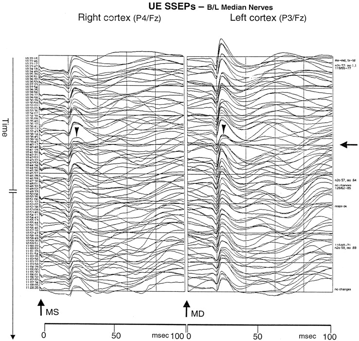
During the initial embolization of the RIMax artery, the neurophysiologist reported left hemispheric changes in the SSEPs and then right hemisphere changes. In both hemispheres, SSEPs were reduced by approximately 50%, with the left hemisphere slightly worse than the right. These changes then stabilized and improved slightly throughout the remainder of the procedure (Fig 1). Because no changes in arterial blood pressure or anesthetic agent could explain the neurophysiologic alterations and because the left hemisphere was affected during RIMax arterial embolization, we believed that the SSEP changes might have been technical. The procedure was completed. No supply arose from the left carotid system.

Fig 1.
  • Download figure
  • Open in new tab
  • Download powerpoint
Fig 1.

SSEP tracing demonstrates changes in recordings during the embolization. Upper extremity (UE) SSEP were obtained from stimulating the left (MS) and right (MD) median nerves at the wrists and by recording evoked potentials from the scalp. The arrow on the far right indicates the time at which the SSEP waveforms began to deviate. The arrowheads indicate the wave change with a 60% loss of amplitude in the P30 response in each hemisphere.

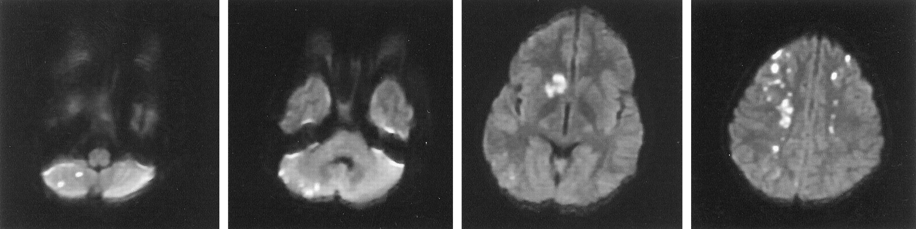
The patient awoke from anesthesia and was extubated. Motor examination findings were normal, yet the patient followed commands poorly and seemed confused. Later in the evening, the patient was incontinent of urine and failed to act appropriately with his mother. He was nearly mute and moderately combative. Electrolyte and oxygen saturation levels were normal. The following morning, cerebral MR imaging revealed diffuse patchy infarcts involving the caudate nuclei, deep white matter, and cerebellar hemisphere (Fig 2). A transthoracic echocardiogram with a bubble study demonstrated a right-to-left cardiac shunt consistent with a patent foramen ovale. A chest radiograph did not show signs of pulmonary arteriovenous fistulas. Over the next 3 days, the patient’s neurologic function returned to his baseline. Detailed neuropsychiatric testing was not performed.

Fig 2.
  • Download figure
  • Open in new tab
  • Download powerpoint
Fig 2.

Diffusion-weighted MR image shows multiple infarctions secondary to paradoxical emboli.

Discussion

A PFO is a persistent valvelike connection between the right and left atria that, in most individuals, is closed by fibrous adhesions between the septum primum and the septum secundum during the 1st month of life (2). Autopsy studies have demonstrated a prevalence of PFO of 17–35% (2). Many studies have linked PFO to paradoxical cerebral emboli (3–16).

When particulate emboli are intentionally injected into the arterial circulation to treat a lesion that contains arteriovenous shunts such as vascular tumors and vascular malformations, the likelihood of paradoxical embolization increases when a patient also has a PFO. In the case described here, the patient did not have a cardiac murmur, and his PFO manifested itself only during coughing. The circuit of the anesthesia machine induced 2.5 mmHg of positive end-expiratory pressure. This, combined with the particle size, (300–500 μm) may have been enough to promote the paradoxical events. In retrospect, neurophysiologic monitoring enabled the early detection of the events; however, we did not recognize their importance.

How has this event changed our practice? We have routinely used neurophysiologic monitoring with embolizations in patients under general anesthesia and will continue to do so. External carotid circulation procedures should never be considered low risk in view of the numerous, dangerous external carotid–to–internal carotid anastomoses that may not be apparent during superselective arteriography. The information provided by brainstem auditory evoked potentials and EEG and SSEP results, although not a substitute for a detailed neurologic examination, can help alert the physician to unexpected embolic events or changes in cerebral perfusion. We have always performed our particulate embolization procedures in awake, sedated patients when possible, and will continue to do so, to perform neurologic examinations throughout the procedure. Nevertheless, some individuals cannot tolerate an awake procedure. All patients undergoing nonemergent particulate embolization now undergo pretreatment echocardiography. Those who have a PFO or other atrial septal defect do not undergo embolization, or they are treated by using acrylic agents or larger particles that are less likely to migrate far along the venous system and back to the heart. We suspect that, in view of the relatively high prevalence of intracardiac shunts in the general population, this issue will present itself again soon.

References

  1. ↵
    Kerut EK, Norfleet WT, Plotnick GD, Giles TD. Patent foramen ovale: a review of associated conditions and the impact of physiological size. J Am Coll Cardiol 2001;38:613–623
    CrossRefPubMed
  2. ↵
    Franke A, Hanrath P. The role of atrial septal abnormalities in cryptogenic stroke-still questionable? Eur Heart J 2001;22:198–200
    CrossRefPubMed
  3. ↵
    McGraw D, Harper R. Patent foramen ovale and cryptogenic cerebral infarction. Intern Med J 2001;31:42–47
    CrossRefPubMed
  4. Hayashida K, Fukuchi K, Inubushi M, Fukushima K, Imakita S, Kimura K. Embolic distribution through patent foramen ovale demonstrated by (99m) Tc-MAA SPECT after Valsalva radionucleotide venography. J Nucl Med 2001;42:859–863
    Abstract/FREE Full Text
  5. Noser EA, Felberg RA, Alexandrov AV. Thrombolytic therapy in an adolescent ischemic stroke. J Child Neurol 2001;16:286–288
    CrossRefPubMed
  6. Chant H, McCollum C. Stroke in young adults: the role of paradoxical embolism. Thromb Haemost 2001;85:22–29
    PubMed
  7. Vahedi K, Amarenco P. Cardiac causes of stroke. Curr Treat Options Neurol 2000;4:305–318
  8. De Castro S, Cartoni D, Fiorelli M, et al. Morphological and functional characteristics of patent foramen ovale and their embolic implications. Stroke 2000;31:2407–2413
    Abstract/FREE Full Text
  9. De Castro S, Cartoni D, Fiorelli M, et al. Patent foramen ovale and its embolic implications. Am J Cardiol 2000;86:51G–52G
    PubMed
  10. Beattie JR, Cohen DJ, Manning WJ, Douglas PS. Role of routine transthoracic echocardiography in evaluation and management of stroke. J Intern Med 1998;24:281–291
  11. Steiner MM, De Tullio MR, Rundek T, et al. Patent foramen ovale size and embolic brain imaging findings among patients with ischemic stroke. Stroke 1998;29:944–948
    Abstract/FREE Full Text
  12. Kulpeksa J, Jobson B, Byrd RP, Roy TM. Paradoxical embolism and a patent foramen ovale. Tenn Med 1998;91:143–145
    PubMed
  13. Vanoverbeke H, Vanoverbeke J, Van der Tempel G, Verheul G. Paradoxical embolism through patent foramen ovale. Acta Chir Belg 1997;97:299–301
    PubMed
  14. Christenson B, Malm J, Carlberg B, et al. Epidemiology and etiology of ischemic stroke in young adults aged 18 to 44 in northern Sweden. Stroke 1997;28:1702–1709
    Abstract/FREE Full Text
  15. Rauh G, Fischereder M, Florentin AS. Transesophageal echocardiography in patients with focal cerebral ischemia of unknown cause. Stroke 1996;27:691–694
    Abstract/FREE Full Text
  • Received February 1, 2002.
  • Accepted after revision March 4, 2002.
  • Copyright © American Society of Neuroradiology
View Abstract
PreviousNext
Back to top

In this issue

American Journal of Neuroradiology: 23 (6)
American Journal of Neuroradiology
Vol. 23, Issue 6
1 Jun 2002
  • Table of Contents
  • Index by author
Advertisement
Print
Download PDF
Email Article

Thank you for your interest in spreading the word on American Journal of Neuroradiology.

NOTE: We only request your email address so that the person you are recommending the page to knows that you wanted them to see it, and that it is not junk mail. We do not capture any email address.

Enter multiple addresses on separate lines or separate them with commas.
Risks of Tumor Embolization in the Presence of an Unrecognized Patent Foramen Ovale: Case Report
(Your Name) has sent you a message from American Journal of Neuroradiology
(Your Name) thought you would like to see the American Journal of Neuroradiology web site.
CAPTCHA
This question is for testing whether or not you are a human visitor and to prevent automated spam submissions.
Cite this article
Michael Bruce Horowitz, Ricardo Carrau, Donald Crammond, Emanuel Kanal
Risks of Tumor Embolization in the Presence of an Unrecognized Patent Foramen Ovale: Case Report
American Journal of Neuroradiology Jun 2002, 23 (6) 982-984;

Citation Manager Formats

  • BibTeX
  • Bookends
  • EasyBib
  • EndNote (tagged)
  • EndNote 8 (xml)
  • Medlars
  • Mendeley
  • Papers
  • RefWorks Tagged
  • Ref Manager
  • RIS
  • Zotero
0 Responses
Respond to this article
Share
Bookmark this article
Risks of Tumor Embolization in the Presence of an Unrecognized Patent Foramen Ovale: Case Report
Michael Bruce Horowitz, Ricardo Carrau, Donald Crammond, Emanuel Kanal
American Journal of Neuroradiology Jun 2002, 23 (6) 982-984;
del.icio.us logo Twitter logo Facebook logo Mendeley logo
  • Tweet Widget
  • Facebook Like
  • Google Plus One

Jump to section

  • Article
    • Abstract
    • Case Report
    • Discussion
    • References
  • Figures & Data
  • Info & Metrics
  • Responses
  • References
  • PDF

Related Articles

  • No related articles found.
  • PubMed
  • Google Scholar

Cited By...

  • Paradoxical embolus to the brain from embolization of a carotid body tumor
  • A case of brain embolism during catheter embolisation of head arteriovenous malformation. What is the mechanism of stroke?
  • A case of brain embolism during catheter embolisation of head arteriovenous malformation. What is the mechanism of stroke?
  • Crossref
  • Google Scholar

This article has not yet been cited by articles in journals that are participating in Crossref Cited-by Linking.

More in this TOC Section

  • Rescue Reentry in Carotid Near-Occlusion
  • Contour Neurovascular System: Five Year Follow Up
  • Effect of SARS-CoV2 on Endovascular Thrombectomy
Show more Neurointervention

Similar Articles

Advertisement

Indexed Content

  • Current Issue
  • Accepted Manuscripts
  • Article Preview
  • Past Issues
  • Editorials
  • Editor's Choice
  • Fellows' Journal Club
  • Letters to the Editor
  • Video Articles

Cases

  • Case Collection
  • Archive - Case of the Week
  • Archive - Case of the Month
  • Archive - Classic Case

More from AJNR

  • Trainee Corner
  • Imaging Protocols
  • MRI Safety Corner
  • Book Reviews

Multimedia

  • AJNR Podcasts
  • AJNR Scantastics

Resources

  • Turnaround Time
  • Submit a Manuscript
  • Submit a Video Article
  • Submit an eLetter to the Editor/Response
  • Manuscript Submission Guidelines
  • Statistical Tips
  • Fast Publishing of Accepted Manuscripts
  • Graphical Abstract Preparation
  • Imaging Protocol Submission
  • Evidence-Based Medicine Level Guide
  • Publishing Checklists
  • Author Policies
  • Become a Reviewer/Academy of Reviewers
  • News and Updates

About Us

  • About AJNR
  • Editorial Board
  • Editorial Board Alumni
  • Alerts
  • Permissions
  • Not an AJNR Subscriber? Join Now
  • Advertise with Us
  • Librarian Resources
  • Feedback
  • Terms and Conditions
  • AJNR Editorial Board Alumni

American Society of Neuroradiology

  • Not an ASNR Member? Join Now

© 2025 by the American Society of Neuroradiology All rights, including for text and data mining, AI training, and similar technologies, are reserved.
Print ISSN: 0195-6108 Online ISSN: 1936-959X

Powered by HighWire