
Article Figures & Data
Figures
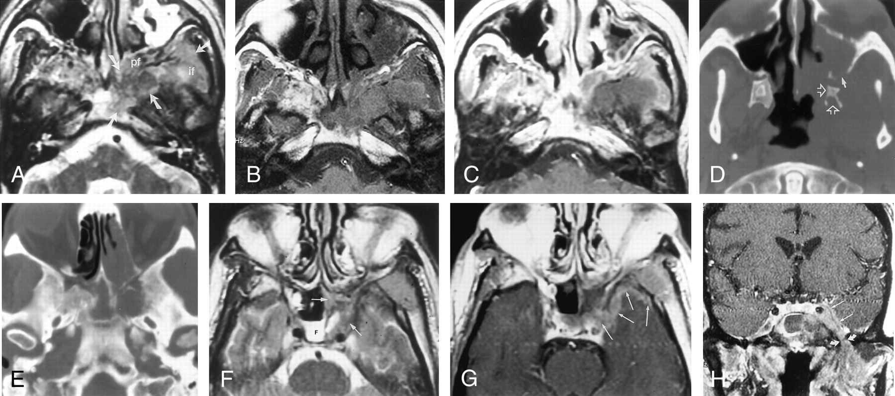
- Fig 1.
Images from the case of a 53-year-old man who developed B-cell lymphoma in the paranasal sinuses, with extension to the skull base after lung transplantation.
A, Axial view fast spin-echo T2-weighted (4000/80[TR/TE]) MR image, obtained at the base of the skull, shows abnormal hypointense tissue (arrows) involving the clivus, pterygopalatine fossa (pf), and infratemporal fossa (if).
B, Axial view unenhanced T1-weighted (500/9) MR image, obtained at same level as that shown in A, shows that the abnormal tissue is slightly hyperintense to muscle.
C, Corresponding contrast-enhanced axial view T1-weighted (600/20) MR image shows no central or solid enhancement of the lesion.
D, Axial view CT scan, obtained at the level of the pterygoid and superior maxillary sinus, shows osseous erosion of the posterior left maxillary sinus wall (solid arrow) and abnormality of the left pterygoid bone (open arrows).
E, Axial view CT scan, obtained at the level of the skull base, shows erosion of the inferior sphenoid sinus/basisphenoid.
F, Axial view fast spin-echo T2-weighted (2500/85) MR image shows fluid (F) in the right sphenoid sinus and hypointense tissue (arrows) in the left sphenoid sinus.
G, Axial view contrast-enhanced T1-weighted (600/20) MR image, obtained at the same level as that shown in F, shows peripheral enhancement of the tissue in the left sphenoid sinus and enhancing tissue in and along the lateral dural margin of the left cavernous sinus and the anterior aspect of the left middle cranial fossa (arrows).
H, Coronal view contrast-enhanced T1-weighted (700/14) MR image shows abnormal tissue along the ventral margin of the cavernous sinus (straight arrows), just anterior to Meckel’s cave and extending through the foramen ovale (curved arrows) into the masticator space.


