Advertisement

Main menu

  • Home
  • Content
    • Current Issue
    • Accepted Manuscripts
    • Article Preview
    • Past Issue Archive
    • Video Articles
    • AJNR Case Collection
    • Case of the Week Archive
    • Case of the Month Archive
    • Classic Case Archive
  • Special Collections
    • AJNR Awards
    • Low-Field MRI
    • Alzheimer Disease
    • ASNR Foundation Special Collection
    • Photon-Counting CT
    • View All
  • Multimedia
    • AJNR Podcasts
    • AJNR SCANtastic
    • Trainee Corner
    • MRI Safety Corner
    • Imaging Protocols
  • For Authors
    • Submit a Manuscript
    • Submit a Video Article
    • Submit an eLetter to the Editor/Response
    • Manuscript Submission Guidelines
    • Statistical Tips
    • Fast Publishing of Accepted Manuscripts
    • Graphical Abstract Preparation
    • Imaging Protocol Submission
    • Author Policies
  • About Us
    • About AJNR
    • Editorial Board
    • Editorial Board Alumni
  • More
    • Become a Reviewer/Academy of Reviewers
    • Subscribers
    • Permissions
    • Alerts
    • Feedback
    • Advertisers
    • ASNR Home

User menu

  • Alerts
  • Log in

Search

  • Advanced search
American Journal of Neuroradiology

American Journal of Neuroradiology

ASHNR American Society of Functional Neuroradiology ASHNR American Society of Pediatric Neuroradiology ASSR
  • Alerts
  • Log in

Advanced Search

  • Home
  • Content
    • Current Issue
    • Accepted Manuscripts
    • Article Preview
    • Past Issue Archive
    • Video Articles
    • AJNR Case Collection
    • Case of the Week Archive
    • Case of the Month Archive
    • Classic Case Archive
  • Special Collections
    • AJNR Awards
    • Low-Field MRI
    • Alzheimer Disease
    • ASNR Foundation Special Collection
    • Photon-Counting CT
    • View All
  • Multimedia
    • AJNR Podcasts
    • AJNR SCANtastic
    • Trainee Corner
    • MRI Safety Corner
    • Imaging Protocols
  • For Authors
    • Submit a Manuscript
    • Submit a Video Article
    • Submit an eLetter to the Editor/Response
    • Manuscript Submission Guidelines
    • Statistical Tips
    • Fast Publishing of Accepted Manuscripts
    • Graphical Abstract Preparation
    • Imaging Protocol Submission
    • Author Policies
  • About Us
    • About AJNR
    • Editorial Board
    • Editorial Board Alumni
  • More
    • Become a Reviewer/Academy of Reviewers
    • Subscribers
    • Permissions
    • Alerts
    • Feedback
    • Advertisers
    • ASNR Home
  • Follow AJNR on Twitter
  • Visit AJNR on Facebook
  • Follow AJNR on Instagram
  • Join AJNR on LinkedIn
  • RSS Feeds

AJNR Awards, New Junior Editors, and more. Read the latest AJNR updates

Research ArticleSpine Imaging and Spine Image-Guided Interventions

Laron Syndrome Abnormalities: Spinal Stenosis, Os Odontoideum, Degenerative Changes of the Atlanto-odontoid Joint, and Small Oropharynx

Liora Kornreich, Gadi Horev, Michael Schwarz, Boaz Karmazyn and Zvi Laron
American Journal of Neuroradiology April 2002, 23 (4) 625-631;
Liora Kornreich
  • Find this author on Google Scholar
  • Find this author on PubMed
  • Search for this author on this site
Gadi Horev
  • Find this author on Google Scholar
  • Find this author on PubMed
  • Search for this author on this site
Michael Schwarz
  • Find this author on Google Scholar
  • Find this author on PubMed
  • Search for this author on this site
Boaz Karmazyn
  • Find this author on Google Scholar
  • Find this author on PubMed
  • Search for this author on this site
Zvi Laron
  • Find this author on Google Scholar
  • Find this author on PubMed
  • Search for this author on this site
  • Article
  • Figures & Data
  • Info & Metrics
  • Responses
  • References
  • PDF
Loading

Article Figures & Data

Figures

  • Tables
  • Fig 1.
    • Download figure
    • Open in new tab
    • Download powerpoint
    Fig 1.

    Patient 1. Sagittal T2-weighted images (4000/160/16 [TR/TE/NEX], 4-mm thickness) show stenosis of the cervical spine. Note bulging disks at multiple levels, causing compression of the thecal sac.

  • Fig 2.
    • Download figure
    • Open in new tab
    • Download powerpoint
    Fig 2.

    Patient 11. Sagittal T1-weighted MR image (450/25/2, 3.2-mm thickness) of the cervical spine shows that the spinal canal is of normal width. Dens is intact. Arrows indicate the level of measurement of soft-palate thickness.
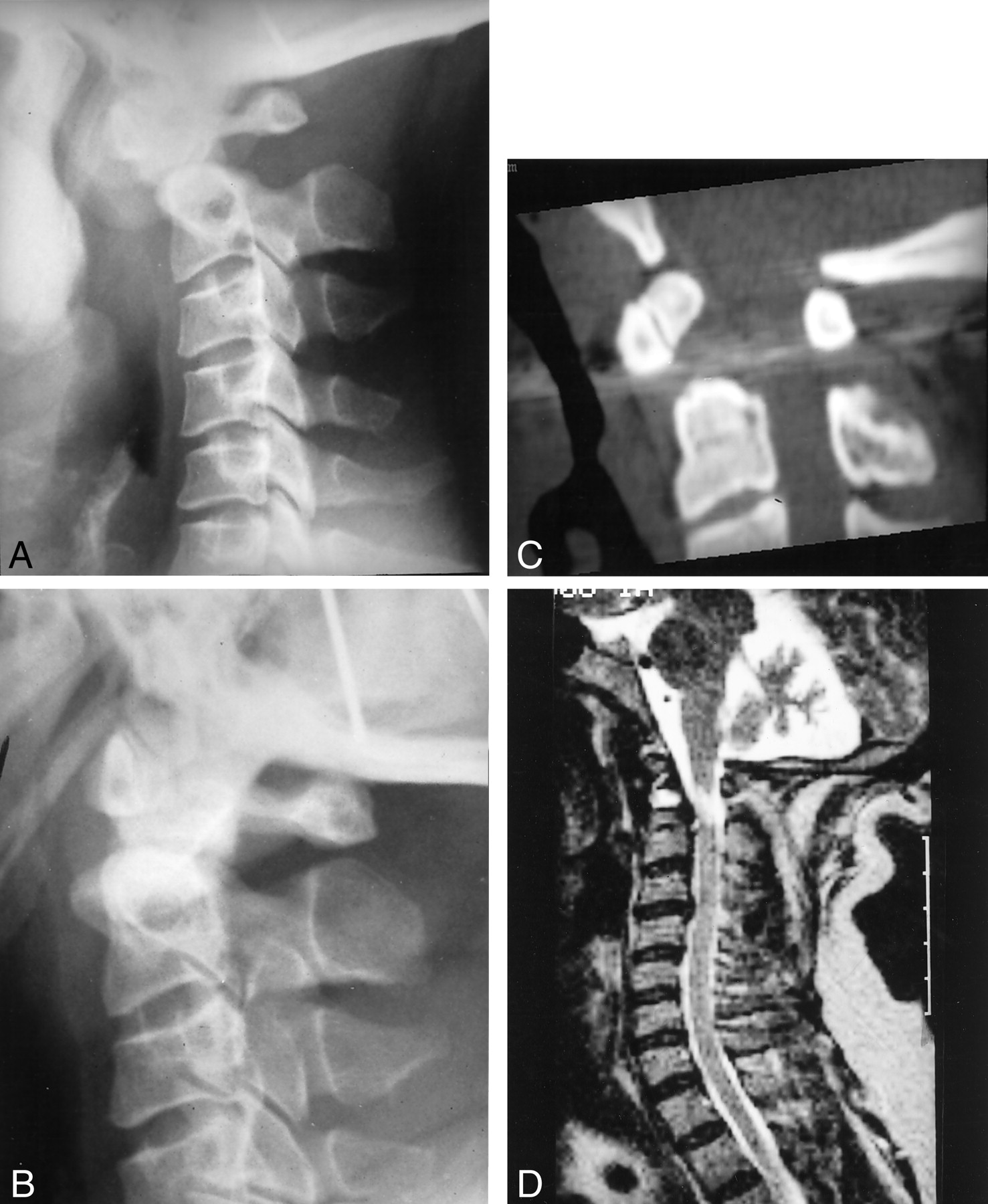
  • Fig 3.
    • Download figure
    • Open in new tab
    • Download powerpoint
    Fig 3.

    Patient 3.

    A and B, Lateral flexion (A) and extension (B) radiographs of the cervical spine. Dens is not visualized. Atlantoaxial instability is demonstrated. Note the small vertebral bodies and the narrow cervical canal.

    C, Sagittal reconstruction of CT data obtained at C1-C2 shows a gap between the base of the dens and the small bone above it, compatible with os odontoideum.

    D, Sagittal T2-weighted MR image (4600/160/1) shows stenosis of the cervial spine. A focus of myelomalacia is seen at the C1-C2 level. High signal intensity from the region of the disrupted dens probably represents reactive tissue.

  • Fig 4.
    • Download figure
    • Open in new tab
    • Download powerpoint
    Fig 4.

    Patient 9.

    A, Lateral radiograph of the cervical spine shows the dens, which appears thickened and sclerotic.

    B, Sagittal reconstruction of CT data obtained at C1-C2 shows no malformation of the dens. Note the cortical thickening and the narrowing of the atlantoaxial joint, possibly compatible with degenerative changes.

    C and D, Sagittal T1-weighted (C; 500/20/2, 4-mm thickness) and T2-weighted (D; 4000/160/1, 4-mm thickness) MR images show a thin dens. Note the mild compression of the subarachnoid space at the C1-C2 level owing to cortical thickening and sclerosis of the dens.

  • Fig 5.
    • Download figure
    • Open in new tab
    • Download powerpoint
    Fig 5.

    Axial T1-weighted (500/20/2, 5-mm thickness) MR images of the oropharynx.

    A, MR image in control subject for comparison. Arrows indicate the anteroposterior and mediolateral diameters.

    B, MR image in patient 5. Oropharynx is strikingly small. Note the abundance of adipose tissue (arrows) between the muscles.

Tables

  • Figures
    • View popup
    TABLE 1:

    Pertinent clinical data of 11 patients with Laron syndrom

    Patient No.Sex/Age (y)Height ft, in (cm)Weight lb (kg)
    Untreated patients
        1M/364, 7 (139.0)172 (78)
        2M/413, 10, (115.7)75 (34)
        3M/464, 10 (129.0)88 (40)
        4M/464, 0(121.0)72 (33)
        5M/684, 8 (142.0)138 (63)
        6F/393, 10 (118.0)90 (41)
        7F/424, 3 (129.3)134 (61)
        8F/433, 8 (112.0)66 (30)
        9F/443, 8 (112.3)90 (41)
        10F/484, 1 (123.6)103 (47)
    IFG-1–treated patient*
        11F/93, 9 (115.0)74 (34)
    • * Since age 3 years.

    • View popup
    TABLE 2:

    Cervical spine measurements and additional findings in nine patients with Laron syndrom

    Patitent No.*AP Canal DiameterVertebral Body DiameterRatio†Normal Ratio†Additional Findings
    Untreated patients
        Men0.98 ± 0.11 (n = 98) 95% CI 0.97–0.99
            110.013.50.74Disk bulge C5-C6-C7, compression of SAS
            210.013.00.77None
            310.515.00.70Disk bulge C3-C4
        Women1.12 ± 0.14 (n = 94) 95% CI 1.09–1.15
            69.510.50.90Disk bulge C5-C6
            710.011.50.87Disk bulge C3-C4, C5-C6
            810.012.00.80None
            911.010.01.10None
            109.013.00.70Disk bulge C3-C4, C5-C6
    IGF-1–treated patient     1113.010.01.301.32 ± 0.17None
    • Note.—AP indicates anteroposterior; CI, confidence interval; SAS, subarachnoid space.

    • * Patient 4 underwent only conventional radiography, and patient 5 underwent MR imaging of only the head; thus, they are not included.

    • † Ratio between the anteroposterior diameter of the canal and the vertebral body.

    • View popup
    TABLE 3:

    Findings at the atlanto-odontoid joint in 10 untreated patients with Laron syndrom

    Patient No.Imaging StudiesOs OdontoideumMyelomalaciaDegenerative ChangesCompression of SAS
        1MR, LC, OM––++
        2MR, LC, OM, F/E++–+
        3MR, CT, LC, OM, F/E++–+
        4LC, OM, F/E+NA–NA
        5MR, CT, LC, OM––++
        6MR, LC, OM––––
        7MR, CT, LC––++
        8MR, LC––++
        9MR, CT––++
        10MR, LC, OM––++
    • Note.—CT indicates CT at C1-C2; F/E, flexion and extension radiographs of cervical spine; LC, lateral radiograph of cervical spine; OM, open mouth dens view; +, present; −, absent; NA, not available; SAS, subarachnoid space.

    • View popup
    TABLE 4:

    Oropharynx measurements in nine patients with Laron syndrome compared with the normal mea

    Patient No.Trasverse Diameter (mm)AP Diameter (mm)Soft-Palate Thickness (mm)
    Untreated patients
        1117.011
        2104.08
        374.09
        574.020
        6101.213
        744.010
        975.010
        1046.013
    Patient mean ± SD7.14 ± 2.64.45 ± 1.812.28 ± 3.7
    Control mean ± SD12.9 ± 4.05.8 ± 2.49.88 ± 1.2
    P value<.005NSNS
    IGF-1–treated patient 11456
    • Note.—AP indicates anteroposterior; NS, difference between patient and control means was not statistically significant; SD, standard deviation. Patient 4 did not undergo MR imaging, and in patient 8 measurements were not diagnostic.

PreviousNext
Back to top

In this issue

American Journal of Neuroradiology: 23 (4)
American Journal of Neuroradiology
Vol. 23, Issue 4
1 Apr 2002
  • Table of Contents
  • Index by author
Advertisement
Print
Download PDF
Email Article

Thank you for your interest in spreading the word on American Journal of Neuroradiology.

NOTE: We only request your email address so that the person you are recommending the page to knows that you wanted them to see it, and that it is not junk mail. We do not capture any email address.

Enter multiple addresses on separate lines or separate them with commas.
Laron Syndrome Abnormalities: Spinal Stenosis, Os Odontoideum, Degenerative Changes of the Atlanto-odontoid Joint, and Small Oropharynx
(Your Name) has sent you a message from American Journal of Neuroradiology
(Your Name) thought you would like to see the American Journal of Neuroradiology web site.
CAPTCHA
This question is for testing whether or not you are a human visitor and to prevent automated spam submissions.
Cite this article
Liora Kornreich, Gadi Horev, Michael Schwarz, Boaz Karmazyn, Zvi Laron
Laron Syndrome Abnormalities: Spinal Stenosis, Os Odontoideum, Degenerative Changes of the Atlanto-odontoid Joint, and Small Oropharynx
American Journal of Neuroradiology Apr 2002, 23 (4) 625-631;

Citation Manager Formats

  • BibTeX
  • Bookends
  • EasyBib
  • EndNote (tagged)
  • EndNote 8 (xml)
  • Medlars
  • Mendeley
  • Papers
  • RefWorks Tagged
  • Ref Manager
  • RIS
  • Zotero
0 Responses
Respond to this article
Share
Bookmark this article
Laron Syndrome Abnormalities: Spinal Stenosis, Os Odontoideum, Degenerative Changes of the Atlanto-odontoid Joint, and Small Oropharynx
Liora Kornreich, Gadi Horev, Michael Schwarz, Boaz Karmazyn, Zvi Laron
American Journal of Neuroradiology Apr 2002, 23 (4) 625-631;
del.icio.us logo Twitter logo Facebook logo Mendeley logo
  • Tweet Widget
  • Facebook Like
  • Google Plus One

Jump to section

  • Article
    • Abstract
    • Methods
    • Results
    • Discussion
    • Conclusion
    • Acknowledgments
    • References
  • Figures & Data
  • Info & Metrics
  • Responses
  • References
  • PDF

Related Articles

  • No related articles found.
  • PubMed
  • Google Scholar

Cited By...

  • No citing articles found.
  • Crossref
  • Google Scholar

This article has not yet been cited by articles in journals that are participating in Crossref Cited-by Linking.

More in this TOC Section

  • Optimization of Photon Counting CT Myelography
  • Characteristics of SIH Type I Culprit Lesions
  • Management Outcomes For VO Spine Biopsy
Show more Spine Imaging and Spine Image-Guided Interventions

Similar Articles

Advertisement

Indexed Content

  • Current Issue
  • Accepted Manuscripts
  • Article Preview
  • Past Issues
  • Editorials
  • Editor's Choice
  • Fellows' Journal Club
  • Letters to the Editor
  • Video Articles

Cases

  • Case Collection
  • Archive - Case of the Week
  • Archive - Case of the Month
  • Archive - Classic Case

More from AJNR

  • Trainee Corner
  • Imaging Protocols
  • MRI Safety Corner
  • Book Reviews

Multimedia

  • AJNR Podcasts
  • AJNR Scantastics

Resources

  • Turnaround Time
  • Submit a Manuscript
  • Submit a Video Article
  • Submit an eLetter to the Editor/Response
  • Manuscript Submission Guidelines
  • Statistical Tips
  • Fast Publishing of Accepted Manuscripts
  • Graphical Abstract Preparation
  • Imaging Protocol Submission
  • Evidence-Based Medicine Level Guide
  • Publishing Checklists
  • Author Policies
  • Become a Reviewer/Academy of Reviewers
  • News and Updates

About Us

  • About AJNR
  • Editorial Board
  • Editorial Board Alumni
  • Alerts
  • Permissions
  • Not an AJNR Subscriber? Join Now
  • Advertise with Us
  • Librarian Resources
  • Feedback
  • Terms and Conditions
  • AJNR Editorial Board Alumni

American Society of Neuroradiology

  • Not an ASNR Member? Join Now

© 2025 by the American Society of Neuroradiology All rights, including for text and data mining, AI training, and similar technologies, are reserved.
Print ISSN: 0195-6108 Online ISSN: 1936-959X

Powered by HighWire