Advertisement

Main menu

  • Home
  • Content
    • Current Issue
    • Accepted Manuscripts
    • Article Preview
    • Past Issue Archive
    • Video Articles
    • AJNR Case Collection
    • Case of the Week Archive
    • Case of the Month Archive
    • Classic Case Archive
  • Special Collections
    • AJNR Awards
    • Low-Field MRI
    • Alzheimer Disease
    • ASNR Foundation Special Collection
    • Photon-Counting CT
    • View All
  • Multimedia
    • AJNR Podcasts
    • AJNR SCANtastic
    • Trainee Corner
    • MRI Safety Corner
    • Imaging Protocols
  • For Authors
    • Submit a Manuscript
    • Submit a Video Article
    • Submit an eLetter to the Editor/Response
    • Manuscript Submission Guidelines
    • Statistical Tips
    • Fast Publishing of Accepted Manuscripts
    • Graphical Abstract Preparation
    • Imaging Protocol Submission
    • Author Policies
  • About Us
    • About AJNR
    • Editorial Board
    • Editorial Board Alumni
  • More
    • Become a Reviewer/Academy of Reviewers
    • Subscribers
    • Permissions
    • Alerts
    • Feedback
    • Advertisers
    • ASNR Home

User menu

  • Alerts
  • Log in

Search

  • Advanced search
American Journal of Neuroradiology

American Journal of Neuroradiology

ASHNR American Society of Functional Neuroradiology ASHNR American Society of Pediatric Neuroradiology ASSR
  • Alerts
  • Log in

Advanced Search

  • Home
  • Content
    • Current Issue
    • Accepted Manuscripts
    • Article Preview
    • Past Issue Archive
    • Video Articles
    • AJNR Case Collection
    • Case of the Week Archive
    • Case of the Month Archive
    • Classic Case Archive
  • Special Collections
    • AJNR Awards
    • Low-Field MRI
    • Alzheimer Disease
    • ASNR Foundation Special Collection
    • Photon-Counting CT
    • View All
  • Multimedia
    • AJNR Podcasts
    • AJNR SCANtastic
    • Trainee Corner
    • MRI Safety Corner
    • Imaging Protocols
  • For Authors
    • Submit a Manuscript
    • Submit a Video Article
    • Submit an eLetter to the Editor/Response
    • Manuscript Submission Guidelines
    • Statistical Tips
    • Fast Publishing of Accepted Manuscripts
    • Graphical Abstract Preparation
    • Imaging Protocol Submission
    • Author Policies
  • About Us
    • About AJNR
    • Editorial Board
    • Editorial Board Alumni
  • More
    • Become a Reviewer/Academy of Reviewers
    • Subscribers
    • Permissions
    • Alerts
    • Feedback
    • Advertisers
    • ASNR Home
  • Follow AJNR on Twitter
  • Visit AJNR on Facebook
  • Follow AJNR on Instagram
  • Join AJNR on LinkedIn
  • RSS Feeds

AJNR Awards, New Junior Editors, and more. Read the latest AJNR updates

OtherSpine Imaging and Spine Image-Guided Interventions

Real-Time CT-Guided Spinal Biopsy with a Disposable Stereotactic Device: A Technical Note

Donald Chakeres, Wayne Slone, Greg Christoforidis and Eric Bourekas
American Journal of Neuroradiology April 2002, 23 (4) 605-608;
Donald Chakeres
  • Find this author on Google Scholar
  • Find this author on PubMed
  • Search for this author on this site
Wayne Slone
  • Find this author on Google Scholar
  • Find this author on PubMed
  • Search for this author on this site
Greg Christoforidis
  • Find this author on Google Scholar
  • Find this author on PubMed
  • Search for this author on this site
Eric Bourekas
  • Find this author on Google Scholar
  • Find this author on PubMed
  • Search for this author on this site
  • Article
  • Figures & Data
  • Info & Metrics
  • Responses
  • References
  • PDF
Loading

Article Figures & Data

Figures

  • Tables
  • Fig 1.
    • Download figure
    • Open in new tab
    • Download powerpoint
    Fig 1.

    Device components.

    A, Nonsterile skin entry localizer is composed of two image-conspicuous lines (barium impregnated silicon) in a truncated inverted V-shaped triangular configuration. The millimeter distance measured between the two V limbs of the pattern is equal to the actual image section plane location on the printed millimeter scale. The pattern has a thicker image-conspicuous band paralleling the radiologic right V limb that is used as a right-left reference. The radiologic right V limb is the 0-mm reference. A series of offset lines at 10-mm increments are printed on the pattern. The intersection of the chosen vector with the pattern is defined as a specific point on the pattern by the two coordinates (slice select, offset). In this example, the skin entry point located at slice select 53 mm and offset 8 mm is marked with a pen.

    B, Sterile skin entry-point localizer. The center of this pattern is centered over the predefined marked skin entry point.

    C, View of the upper vector point components parallel to the skin. The device comes with an incorporated sterile adhesive drape and a circular opening with an attached base ring to support the upper level pattern platform.

    D, View of the upper vector point localizer seen from above. The upper vector point localizer has a clear diaphragm pattern and printed scale that is used in a manner identical to that of the skin point localizer.

  • Fig 2.
    • Download figure
    • Open in new tab
    • Download powerpoint
    Fig 2.

    Diaphragm device—assisted procedure.

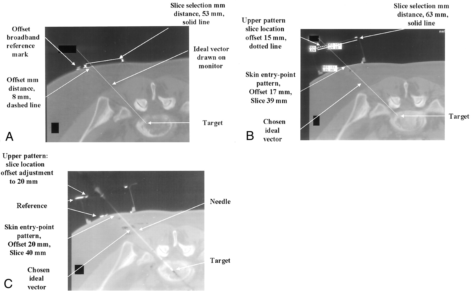
    A, A 3-mm axial CT image with a 19° angle gantry tilt. This is the planning image for the procedure, with the goal to biopsy the L5-S1 level. An ideal vector is drawn from the target to the skin. Nonsterile skin entry-point pattern is seen as two hyperattenuating dots. The distance measured is 53 mm and defines the entry-point slice select coordinate. The second coordinate offset value is 8 mm. The skin entry-point localizer is punctured at this point and lines are drawn parallel to the slice on the patient’s skin.

    B, First poststerile CT image with the multilevel device placed over the target. The sterile pattern localizer is seen as two small hyperattenuating dots on the skin. The ideal targeting vector is drawn on the monitor, and its intersection point with the upper level pattern component is measured. The slice location is 63 mm and the offset 15 mm. The diaphragm is punctured at this point, and the needle is placed at the predefined skin entry point. After one adjustment, the needle is advance the measured depth to the target.

    C, CT image shows the needle entering the target along the predefined vector. The upper diaphragm was sliced, allowing the probe to pass through the diaphragm to reach the target. Despite the complex double oblique vector, the entire needle is visible, demonstrating the accuracy of the device.

Tables

  • Figures
    • View popup
    TABLE

    Comparison of the freehand technique with the stereotactic device technique

    VariableFreehandStereotactic Device
    No. of patients88
    Skin entry-point verification (min)17.54
    Sterile setup time (min)24.913
    Needle aligned with vector (min)27.618.8
    Needle in target (min)36.925.3
    Total no. of images147.9
    Distance of skin entry point (mm)4.50
    Distance off target (mm)4.81.6
    Depth to target (mm)79.873.1
PreviousNext
Back to top

In this issue

American Journal of Neuroradiology: 23 (4)
American Journal of Neuroradiology
Vol. 23, Issue 4
1 Apr 2002
  • Table of Contents
  • Index by author
Advertisement
Print
Download PDF
Email Article

Thank you for your interest in spreading the word on American Journal of Neuroradiology.

NOTE: We only request your email address so that the person you are recommending the page to knows that you wanted them to see it, and that it is not junk mail. We do not capture any email address.

Enter multiple addresses on separate lines or separate them with commas.
Real-Time CT-Guided Spinal Biopsy with a Disposable Stereotactic Device: A Technical Note
(Your Name) has sent you a message from American Journal of Neuroradiology
(Your Name) thought you would like to see the American Journal of Neuroradiology web site.
CAPTCHA
This question is for testing whether or not you are a human visitor and to prevent automated spam submissions.
Cite this article
Donald Chakeres, Wayne Slone, Greg Christoforidis, Eric Bourekas
Real-Time CT-Guided Spinal Biopsy with a Disposable Stereotactic Device: A Technical Note
American Journal of Neuroradiology Apr 2002, 23 (4) 605-608;

Citation Manager Formats

  • BibTeX
  • Bookends
  • EasyBib
  • EndNote (tagged)
  • EndNote 8 (xml)
  • Medlars
  • Mendeley
  • Papers
  • RefWorks Tagged
  • Ref Manager
  • RIS
  • Zotero
0 Responses
Respond to this article
Share
Bookmark this article
Real-Time CT-Guided Spinal Biopsy with a Disposable Stereotactic Device: A Technical Note
Donald Chakeres, Wayne Slone, Greg Christoforidis, Eric Bourekas
American Journal of Neuroradiology Apr 2002, 23 (4) 605-608;
del.icio.us logo Twitter logo Facebook logo Mendeley logo
  • Tweet Widget
  • Facebook Like
  • Google Plus One

Jump to section

  • Article
    • Abstract
    • Technique
    • Discussion
    • References
  • Figures & Data
  • Info & Metrics
  • Responses
  • References
  • PDF

Related Articles

  • No related articles found.
  • PubMed
  • Google Scholar

Cited By...

  • No citing articles found.
  • Crossref
  • Google Scholar

This article has not yet been cited by articles in journals that are participating in Crossref Cited-by Linking.

More in this TOC Section

  • Optimization of Photon Counting CT Myelography
  • Characteristics of SIH Type I Culprit Lesions
  • Management Outcomes For VO Spine Biopsy
Show more Spine Imaging and Spine Image-Guided Interventions

Similar Articles

Advertisement

Indexed Content

  • Current Issue
  • Accepted Manuscripts
  • Article Preview
  • Past Issues
  • Editorials
  • Editor's Choice
  • Fellows' Journal Club
  • Letters to the Editor
  • Video Articles

Cases

  • Case Collection
  • Archive - Case of the Week
  • Archive - Case of the Month
  • Archive - Classic Case

More from AJNR

  • Trainee Corner
  • Imaging Protocols
  • MRI Safety Corner
  • Book Reviews

Multimedia

  • AJNR Podcasts
  • AJNR Scantastics

Resources

  • Turnaround Time
  • Submit a Manuscript
  • Submit a Video Article
  • Submit an eLetter to the Editor/Response
  • Manuscript Submission Guidelines
  • Statistical Tips
  • Fast Publishing of Accepted Manuscripts
  • Graphical Abstract Preparation
  • Imaging Protocol Submission
  • Evidence-Based Medicine Level Guide
  • Publishing Checklists
  • Author Policies
  • Become a Reviewer/Academy of Reviewers
  • News and Updates

About Us

  • About AJNR
  • Editorial Board
  • Editorial Board Alumni
  • Alerts
  • Permissions
  • Not an AJNR Subscriber? Join Now
  • Advertise with Us
  • Librarian Resources
  • Feedback
  • Terms and Conditions
  • AJNR Editorial Board Alumni

American Society of Neuroradiology

  • Not an ASNR Member? Join Now

© 2025 by the American Society of Neuroradiology All rights, including for text and data mining, AI training, and similar technologies, are reserved.
Print ISSN: 0195-6108 Online ISSN: 1936-959X

Powered by HighWire