
Article Figures & Data
Figures
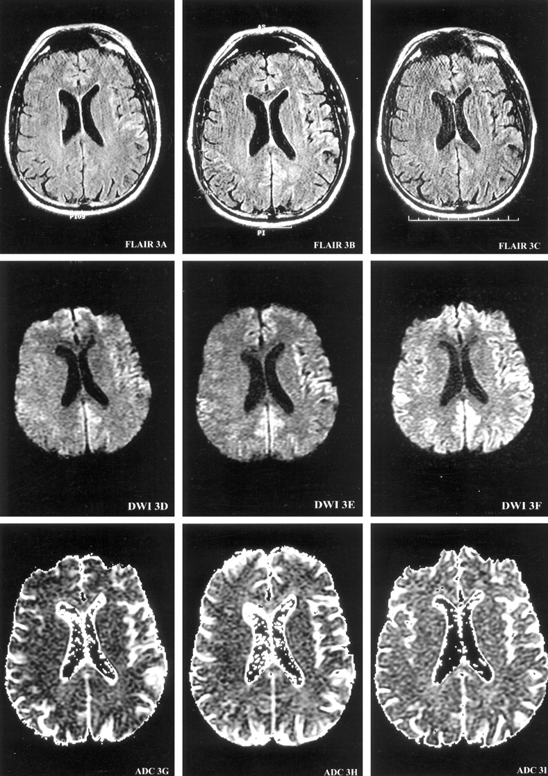
- Fig 1.
Serial MR images demonstrate the evolution of ribbon-like cortical signal intensity abnormalities. MR images are presented at each of three brain axial levels (1–3), at each of three time points from the onset of symptoms (left columns, 4 months; middle columns, 5.5 months; right columns, 6 months) for FLAIR (top row), DW imaging (middle row), and ADC (bottom row) studies. At 4 months from onset of symptoms, DW images demonstrate gyriform increased signal intensity predominantly in the right temporal cortex (DWI1D) with decreased ADC signal intensity consistent with restricted diffusion (ADC1G). At 5.5 months from onset, the hyperintense signals on DW images involve more cortical gyri, extending into the left temporoparietal cortex (DWI3E). At 6 months from onset, DW images (DWI1F, DWI2F, and DWI3F) show ribbon-like areas of hyperintensity involving the right temporoparietooccipital cortex, as well as the left frontotempoparietal cortex, extending into the parafalcine occipital region.
Images on this page were obtained at level 1.
(continued) Images on this page were obtained at level 2.
(continued) Images on this page were obtained at level 3.
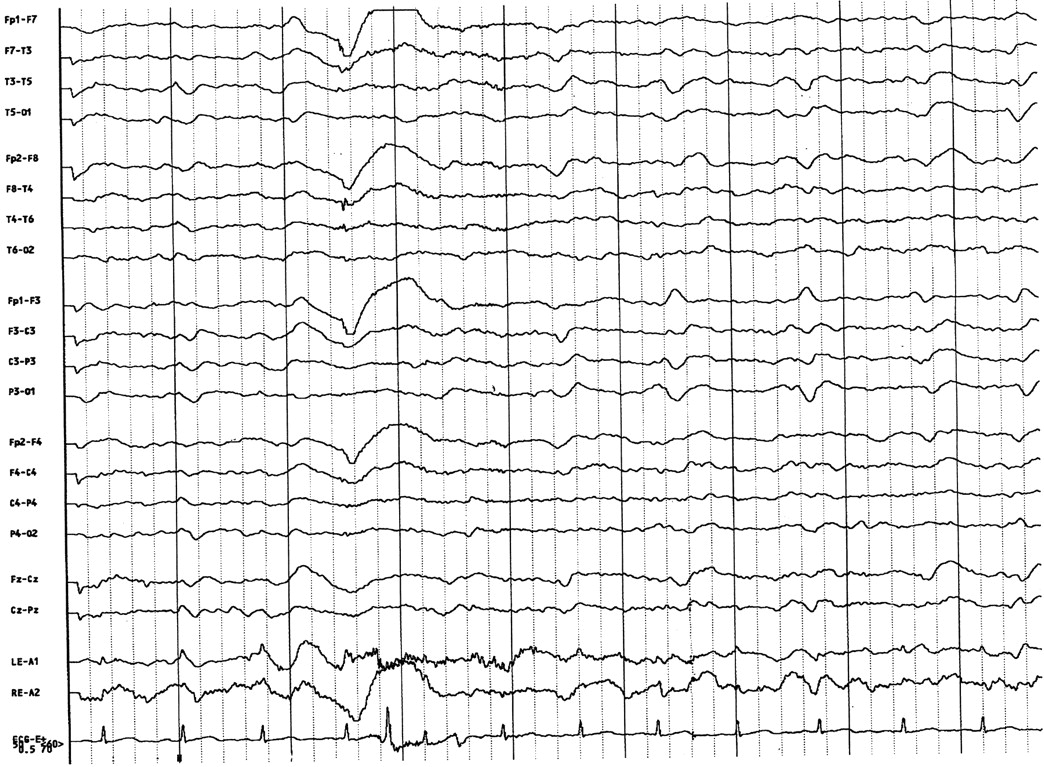
- Fig 2.
A 21-channel EEG with the patient awake demonstrates periodic sharp-wave complex discharges, which are of greater amplitude over the left frontotemporal region and are superimposed on diffusely disorganized and slow background.
- Fig 3.
Photomicrograph of brain biopsy specimen obtained from the cortex of the left frontal lobe demonstrates prominent neuronal loss. Astrocytosis is not conspicuous. There are characteristic spongiform changes (**) that tend to be cell-associated (hematoxylin-eosin stain, original magnification ×400).


Tables
- TABLE
Review of pathologically proved CJD cases with positive DW imaging findings reported in the literature
Report Patient Age (y)/Sex Time of Study after Onset of Symptoms (mo) Location of Hyperintensity on DW Image MR Imaging (T2/FLAIR) Abnormalities Others Findings Demaerel et al 1997 (6) 68/F 1 Cortex (+) T2 (+) EEG Bahn et al 1997 (7) 61/F 4 Basal ganglia/cortex (+/−) FLAIR> T2 (−) EEG Samman et al 1999 (8) 68/F 8 Basal ganglia/cortex (+) T2 (−) EEG (−) CSF Demaerel et al 1999 (9) 68/F 0.75, 1.75 Basal ganglia/cortex (+/−) T2 NP 65/M 2 Basal ganglia/cortex (−) NP 59/F 1, 1.5 Basal ganglia/cortex (−) NP Bahn et al 1999 (10) 61/F 7 Basal ganglia (+) FLAIR > T2 (+) CSF 53/F 1 Basal ganglia (+) T2 NP 64/F 2 Cortex (+/−) FLAIR (+) CSF Yee et al 1999 (11) 69/M 1.5 Cortex (−) (+) EEG Kropp et al 2000 (12) 68/F 9 Basal ganglia/cortex (+/−) (+) CSF 70/F 7 Basal ganglia (+/−) (+) CSF 58/M 5 Basal ganglia (+/−) (+) CSF 48/M 3 Basal ganglia/cortex (+/−) (+) CSF 68/F 1 Basal ganglia (+/−) (+) CSF Romi et al 2000 (13) 54/F 8 Basal ganglia/co rtex (−) (−) EEG Current case 63/M 4, 5.5, 6 Cortex (+/−) FLAIR (+) EEG (−) CSF Note.—CSF markers used were 14-3-3 protein or neuron-specific enolase. (+) indicates positive findings; (+/−), subtle findings; (−), negative findings; NP, no other studies were performed.


