Advertisement

Main menu

  • Home
  • Content
    • Current Issue
    • Accepted Manuscripts
    • Article Preview
    • Past Issue Archive
    • Video Articles
    • AJNR Case Collection
    • Case of the Week Archive
    • Case of the Month Archive
    • Classic Case Archive
  • Special Collections
    • AJNR Awards
    • Low-Field MRI
    • Alzheimer Disease
    • ASNR Foundation Special Collection
    • Photon-Counting CT
    • View All
  • Multimedia
    • AJNR Podcasts
    • AJNR SCANtastic
    • Trainee Corner
    • MRI Safety Corner
    • Imaging Protocols
  • For Authors
    • Submit a Manuscript
    • Submit a Video Article
    • Submit an eLetter to the Editor/Response
    • Manuscript Submission Guidelines
    • Statistical Tips
    • Fast Publishing of Accepted Manuscripts
    • Graphical Abstract Preparation
    • Imaging Protocol Submission
    • Author Policies
  • About Us
    • About AJNR
    • Editorial Board
    • Editorial Board Alumni
  • More
    • Become a Reviewer/Academy of Reviewers
    • Subscribers
    • Permissions
    • Alerts
    • Feedback
    • Advertisers
    • ASNR Home

User menu

  • Alerts
  • Log in

Search

  • Advanced search
American Journal of Neuroradiology

American Journal of Neuroradiology

ASHNR American Society of Functional Neuroradiology ASHNR American Society of Pediatric Neuroradiology ASSR
  • Alerts
  • Log in

Advanced Search

  • Home
  • Content
    • Current Issue
    • Accepted Manuscripts
    • Article Preview
    • Past Issue Archive
    • Video Articles
    • AJNR Case Collection
    • Case of the Week Archive
    • Case of the Month Archive
    • Classic Case Archive
  • Special Collections
    • AJNR Awards
    • Low-Field MRI
    • Alzheimer Disease
    • ASNR Foundation Special Collection
    • Photon-Counting CT
    • View All
  • Multimedia
    • AJNR Podcasts
    • AJNR SCANtastic
    • Trainee Corner
    • MRI Safety Corner
    • Imaging Protocols
  • For Authors
    • Submit a Manuscript
    • Submit a Video Article
    • Submit an eLetter to the Editor/Response
    • Manuscript Submission Guidelines
    • Statistical Tips
    • Fast Publishing of Accepted Manuscripts
    • Graphical Abstract Preparation
    • Imaging Protocol Submission
    • Author Policies
  • About Us
    • About AJNR
    • Editorial Board
    • Editorial Board Alumni
  • More
    • Become a Reviewer/Academy of Reviewers
    • Subscribers
    • Permissions
    • Alerts
    • Feedback
    • Advertisers
    • ASNR Home
  • Follow AJNR on Twitter
  • Visit AJNR on Facebook
  • Follow AJNR on Instagram
  • Join AJNR on LinkedIn
  • RSS Feeds

AJNR Awards, New Junior Editors, and more. Read the latest AJNR updates

Research ArticleBRAIN

Diffusion Tensor MR Imaging of High-Grade Cerebral Gliomas

Saurabh Sinha, Mark E. Bastin, Ian R. Whittle and Joanna M. Wardlaw
American Journal of Neuroradiology April 2002, 23 (4) 520-527;
Saurabh Sinha
  • Find this author on Google Scholar
  • Find this author on PubMed
  • Search for this author on this site
Mark E. Bastin
  • Find this author on Google Scholar
  • Find this author on PubMed
  • Search for this author on this site
Ian R. Whittle
  • Find this author on Google Scholar
  • Find this author on PubMed
  • Search for this author on this site
Joanna M. Wardlaw
  • Find this author on Google Scholar
  • Find this author on PubMed
  • Search for this author on this site
  • Article
  • Figures & Data
  • Info & Metrics
  • Responses
  • References
  • PDF
Loading

Article Figures & Data

Figures

  • Tables
  • Fig 1.
    • Download figure
    • Open in new tab
    • Download powerpoint
    Fig 1.

    Images in patient 4.

    A and B, Contrast-enhanced T1-weighted volume images. In B, the solid arrow indicates a region of an enhancing tumor margin; dashed arrow, enhancing tumor core; dotted arrow, nonenhancing tumor core.

    C and D, T2-weighted EP images. Arrow in D indicates a region of edematous brain.

    E, 〈D〉 parametric map.

    F, FA parametric map.
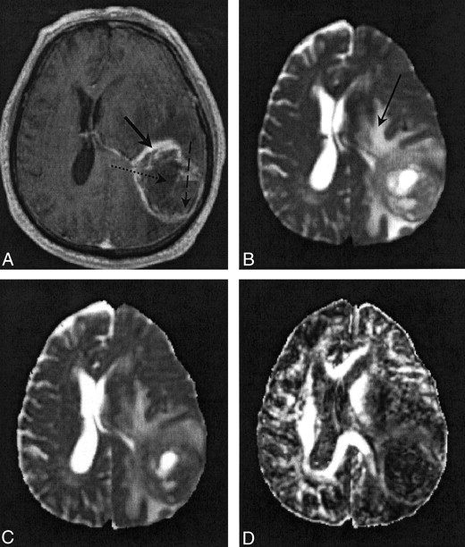
  • Fig 2.
    • Download figure
    • Open in new tab
    • Download powerpoint
    Fig 2.

    Images in patient 5.

    A, Contrast-enhanced T1-weighted volume image. The solid arrow indicates a region of enhancing tumor margin; dashed arrow, enhancing tumor core; dotted arrow, nonenhancing tumor core.

    B, T2-weighted EP images. Arrow indicates a region of edematous brain.

    C and D, Note the high 〈D〉 and low FA values in the nonenhancing tumor core and edematous brain on the 〈D〉 parametric map (C) and FA parametric map (D).

  • Fig 3.
    • Download figure
    • Open in new tab
    • Download powerpoint
    Fig 3.

    Images in patient 3. The thick arrow indicates a region of enhancing tumor margin; the thin arrow in B indicates edematous brain.

    A, Contrast-enhanced T1-weighted volume image.

    B, T2-weighted EP images.

    C, 〈D〉 parametric map. Note the diffuse nature of the edema.

    D, FA parametric map.

Tables

  • Figures
    • View popup
    TABLE 1:

    TABLE 1: 〈D〉 values in the neoplasm, surrounding edematous brain, and normal white matter in nine patients with high-grade glioma

    Patient/Age (y)/SexMean 〈D〉 Value (× 10−6 mm2/s)
    Enhancing Tumor MarginEnhancing Tumor CoreNonenhancing Tumor CoreEdematous BrainNormal White Matter
    IpsilateralContralateral
    1/61/F*958.69 ± 64.28NANA1127.17 ± 155.62725.53 ± 16.08720.90 ± 37.18
    2/43/M*†948.56 ± 52.35NANA923.87 ± 60.71747.34 ± 1.43731.76 ± 11.34
    3/56/F*‡928.34 ± 50.88NANA1221.99 ± 261.25NANA
    4/65/M1238.54 ± 158.931500.83 ± 275.191861.26 ± 292.521494.35 ± 210.91717.50 ± 23.28704.44 ± 19.58
    5/71/M1195.37 ± 206.511181.63 ± 294.641890.93 ± 275.341477.25 ± 279.53753.10 ± 50.43774.64 ± 63.37
    6/50/M1289.65 ± 227.581571.22 ± 108.361784.93 ± 301.461575.23 ± 215.07745.16 ± 25.87735.33 ± 29.34
    7/42/M1208.40 ± 170.221256.69 ± 213.051264.92 ± 395.201143.00 ± 326.96739.16 ± 21.58720.83 ± 18.43
    8/55/F1185.35 ± 262.881031.14 ± 326.10NA1674.29 ± 253.11725.90 ± 27.08715.58 ± 34.79
    9/74/M§1309.58 ± 142.091356.45 ± 225.761957.34 ± 424.161565.69 ± 219.01NA768.67 ± 9.05
    Mean1229.80 ± 206.801308.67 ± 292.501825.38 ± 404.061411.23 ± 322.31733.51 ± 31.59730.00 ± 37.69
    • Note.—All values are reported as the mean ± the standard deviation. Normal white matter was the centrum semiovale. NA indicates not applicable.

    • * This patient had a multifocal glioma.

    • † This patient had a low-grade glioma with a malignant core.

    • ‡ T2-weighted EP images depicted abnormal signal intensity throughout the ipsilateral and contralateral centrum semiovale.

    • § T2-weighted EP images depicted abnormal signal intensity throughout the ipsilateral centrum semiovale.

    • View popup
    TABLE 2:

    TABLE 2: FA values in the neoplasm, surrounding edematous brain, and normal white matter in nine patients with high-grade glioma

    PatientFA Value
    Enhancing Tumor MarginEnhancing Tumor CoreNonenhancing Tumor CoreEdematous BrainNormal White Matter
    IpsilateralContralateral
    1*0.20 ± 0.06NANA0.21 ± 0.070.51 ± 0.020.42 ± 0.07
    2*†0.22 ± 0.04NANA0.23 ± 0.080.50 ± 0.040.47 ± 0.11
    3*‡0.18 ± 0.06NANA0.17 ± 0.07NANA
    40.16 ± 0.070.10 ± 0.030.07 ± 0.010.14 ± 0.050.43 ± 0.100.48 ± 0.06
    50.15 ± 0.050.14 ± 0.060.08 ± 0.020.18 ± 0.090.47 ± 0.070.52 ± 0.07
    60.13 ± 0.030.08 ± 0.030.07 ± 0.010.15 ± 0.050.45 ± 0.090.51 ± 0.05
    70.18 ± 0.050.15 ± 0.070.10 ± 0.030.25 ± 0.100.44 ± 0.100.46 ± 0.09
    80.20 ± 0.050.14 ± 0.04NA0.15 ± 0.060.48 ± 0.080.48 ± 0.08
    9§0.18 ± 0.060.13 ± 0.050.10 ± 0.030.17 ± 0.06NA0.46 ± 0.02
    Mean0.16 ± 0.060.13 ± 0.060.09 ± 0.030.17 ± 0.080.46 ± 0.080.48 ± 0.08
    • Note.—All values are reported as the mean ± the standard deviation. Normal white matter was the centrum semiovale. NA indicates not applicable.

    • * This patient had a multifocal glioma.

    • † This patient had a low-grade glioma with a malignant core.

    • ‡ T2-weighted EP images depicted abnormal signal intensity throughout the ipsilateral and contralateral centrum semiovale.

    • § T2-weighted EP images depicted abnormal signal intensity throughout the ipsilateral centrum semiovale.

    • View popup
    TABLE 3:

    TABLE 3: Statistical analysis of differences in 〈D〉 and FA values in nine patients with high-grade glioma

    ComparisonPatients with a Statistically Significant Difference*
    〈D〉 ValueFA
    Edematous brain versus enhancing tumor margin1–91, 4–9
    Ipsilateral white matter versus enhancing tumor margin1, 2, 4–91, 2, 4–9
    Ipsilateral white matter versus edematous brain1, 2, 4–91, 2, 4–9
    • Note.—In patient 3, T2-weighted EP images depicted abnormal signal intensity throughout the ipsilateral and contralateral centrum semiovale. In patient 9, T2-weighted EP images depicted abnormal signal intensity throughout ipsilateral centrum semiovale. Data in the contralateral centrum semiovale were used in this analysis.

    • * P < .05, post hoc Scheffé test.

PreviousNext
Back to top

In this issue

American Journal of Neuroradiology: 23 (4)
American Journal of Neuroradiology
Vol. 23, Issue 4
1 Apr 2002
  • Table of Contents
  • Index by author
Advertisement
Print
Download PDF
Email Article

Thank you for your interest in spreading the word on American Journal of Neuroradiology.

NOTE: We only request your email address so that the person you are recommending the page to knows that you wanted them to see it, and that it is not junk mail. We do not capture any email address.

Enter multiple addresses on separate lines or separate them with commas.
Diffusion Tensor MR Imaging of High-Grade Cerebral Gliomas
(Your Name) has sent you a message from American Journal of Neuroradiology
(Your Name) thought you would like to see the American Journal of Neuroradiology web site.
CAPTCHA
This question is for testing whether or not you are a human visitor and to prevent automated spam submissions.
Cite this article
Saurabh Sinha, Mark E. Bastin, Ian R. Whittle, Joanna M. Wardlaw
Diffusion Tensor MR Imaging of High-Grade Cerebral Gliomas
American Journal of Neuroradiology Apr 2002, 23 (4) 520-527;

Citation Manager Formats

  • BibTeX
  • Bookends
  • EasyBib
  • EndNote (tagged)
  • EndNote 8 (xml)
  • Medlars
  • Mendeley
  • Papers
  • RefWorks Tagged
  • Ref Manager
  • RIS
  • Zotero
0 Responses
Respond to this article
Share
Bookmark this article
Diffusion Tensor MR Imaging of High-Grade Cerebral Gliomas
Saurabh Sinha, Mark E. Bastin, Ian R. Whittle, Joanna M. Wardlaw
American Journal of Neuroradiology Apr 2002, 23 (4) 520-527;
del.icio.us logo Twitter logo Facebook logo Mendeley logo
  • Tweet Widget
  • Facebook Like
  • Google Plus One

Jump to section

  • Article
    • Abstract
    • Methods
    • Results
    • Discussion
    • Conclusion
    • Acknowledgments
    • Footnotes
    • References
  • Figures & Data
  • Info & Metrics
  • Responses
  • References
  • PDF

Related Articles

  • No related articles found.
  • PubMed
  • Google Scholar

Cited By...

  • Memory recovery is related to default mode network impairment and neurite density during brain tumours treatment
  • Utility of Diffusion Tensor Imaging in Evaluation of the Peritumoral Region in Patients with Primary and Metastatic Brain Tumors
  • Differentiation of Brain Abscesses from Necrotic Glioblastomas and Cystic Metastatic Brain Tumors with Diffusion Tensor Imaging
  • Diffusion Tensor MR Imaging of Cerebral Gliomas: Evaluating Fractional Anisotropy Characteristics
  • Apparent Diffusion and Fractional Anisotropy of Diffuse Intrinsic Brain Stem Gliomas
  • Diffusion Tensor Imaging in Glioblastoma Multiforme and Brain Metastases: The Role of p, q, L, and Fractional Anisotropy
  • Advances Toward an Understanding of Brainstem Gliomas
  • Enhanced visualization and quantification of magnetic resonance diffusion tensor imaging using the p:q tensor decomposition
  • Differentiation between solitary brain metastasis and high-grade glioma by diffusion tensor imaging
  • Temporal evolution of water diffusion parameters is different in grey and white matter in human ischaemic stroke
  • Effects of dexamethasone on peritumoural oedematous brain: a DT-MRI study
  • Differentiation of Toxoplasmosis and Lymphoma in AIDS Patients by Using Apparent Diffusion Coefficients
  • Crossref
  • Google Scholar

This article has not yet been cited by articles in journals that are participating in Crossref Cited-by Linking.

More in this TOC Section

  • Optimal MRI Sequence for Identifying Occlusion Location in Acute Stroke: Which Value of Time-Resolved Contrast-Enhanced MRA?
  • Evaluating the Effects of White Matter Multiple Sclerosis Lesions on the Volume Estimation of 6 Brain Tissue Segmentation Methods
  • Quiet PROPELLER MRI Techniques Match the Quality of Conventional PROPELLER Brain Imaging Techniques
Show more Brain

Similar Articles

Advertisement

Indexed Content

  • Current Issue
  • Accepted Manuscripts
  • Article Preview
  • Past Issues
  • Editorials
  • Editor's Choice
  • Fellows' Journal Club
  • Letters to the Editor
  • Video Articles

Cases

  • Case Collection
  • Archive - Case of the Week
  • Archive - Case of the Month
  • Archive - Classic Case

More from AJNR

  • Trainee Corner
  • Imaging Protocols
  • MRI Safety Corner
  • Book Reviews

Multimedia

  • AJNR Podcasts
  • AJNR Scantastics

Resources

  • Turnaround Time
  • Submit a Manuscript
  • Submit a Video Article
  • Submit an eLetter to the Editor/Response
  • Manuscript Submission Guidelines
  • Statistical Tips
  • Fast Publishing of Accepted Manuscripts
  • Graphical Abstract Preparation
  • Imaging Protocol Submission
  • Evidence-Based Medicine Level Guide
  • Publishing Checklists
  • Author Policies
  • Become a Reviewer/Academy of Reviewers
  • News and Updates

About Us

  • About AJNR
  • Editorial Board
  • Editorial Board Alumni
  • Alerts
  • Permissions
  • Not an AJNR Subscriber? Join Now
  • Advertise with Us
  • Librarian Resources
  • Feedback
  • Terms and Conditions
  • AJNR Editorial Board Alumni

American Society of Neuroradiology

  • Not an ASNR Member? Join Now

© 2025 by the American Society of Neuroradiology All rights, including for text and data mining, AI training, and similar technologies, are reserved.
Print ISSN: 0195-6108 Online ISSN: 1936-959X

Powered by HighWire