
Article Figures & Data
Figures
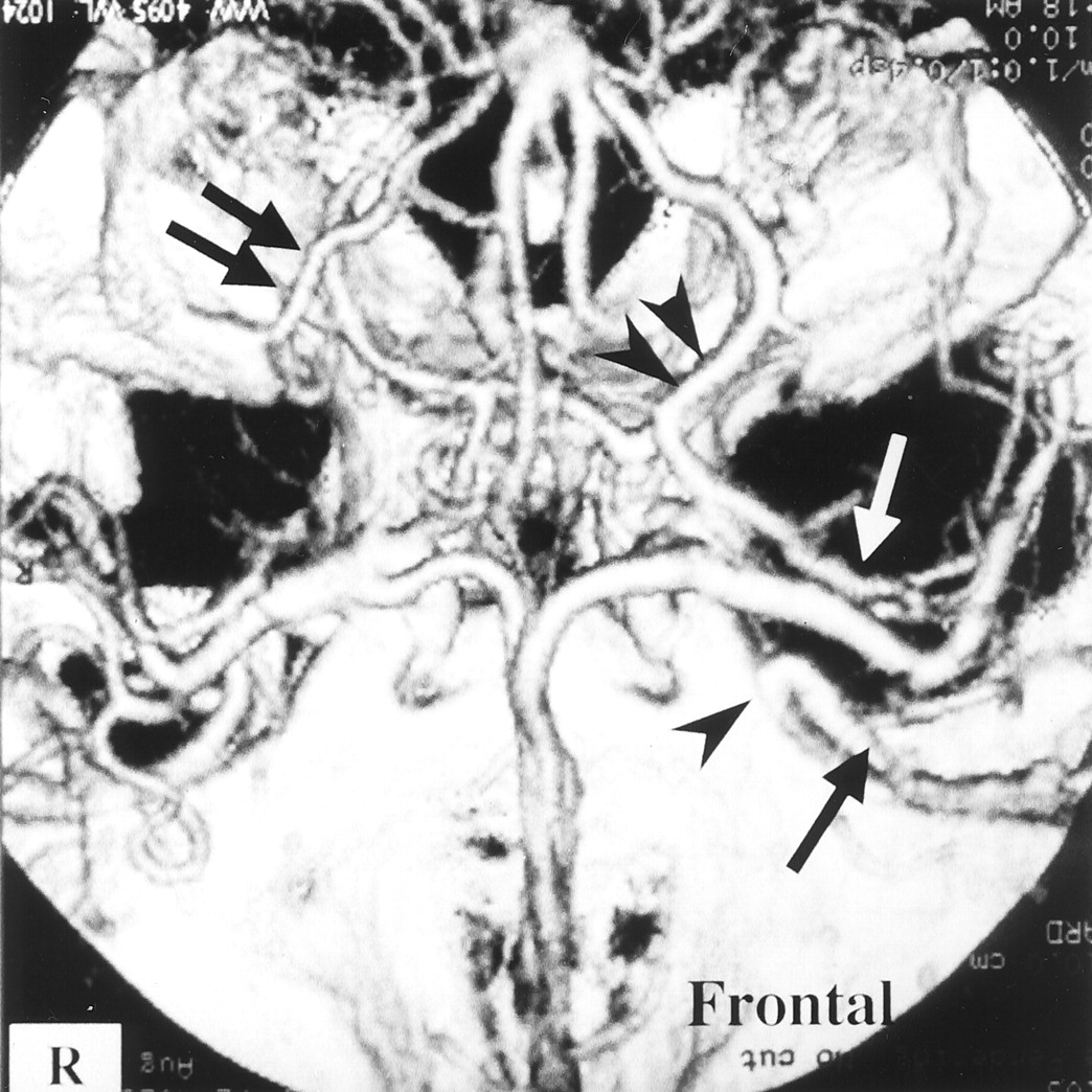
- Fig 1.
Patient with a left internal carotid artery aneurysm.
3D-CTA (superior view) shows drainage of well-developed SSVs (black arrow) into the SPS at the inner portion of the sphenoid wing (black arrowhead) on the left side. A well-developed DMCV (white arrow) connecting to the BVR (double black arrowheads) is also visualized on the left side. DMCV and BVR development (double black arrows) on the right side is poor compared with that on the left side.
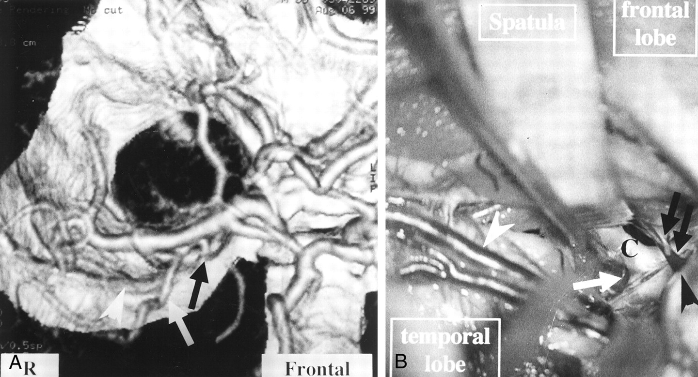
- Fig 2.
Patient with a right middle cerebral artery aneurysm.
A, 3D-CTA (right anterior view) shows the connecting vein (white arrowhead) between the BVR (white arrow) and the SPS. The connecting vein lies in contact with the aneurysm (black arrowhead). A black arrow and double black arrows indicate the SSVs and right middle cerebral artery, respectively.
B, A microscopic operative photograph shows that the right sylvian fissure is opened by the pterional approach. Spatulas (Spat.) retract the right frontal and right temporal lobes. The connecting vein between the BVR and the SPS (white arrow) adheres strongly to the middle cerebral artery (black double arrows) and the aneurysm (black arrow). A black arrowhead indicates the SSVs.
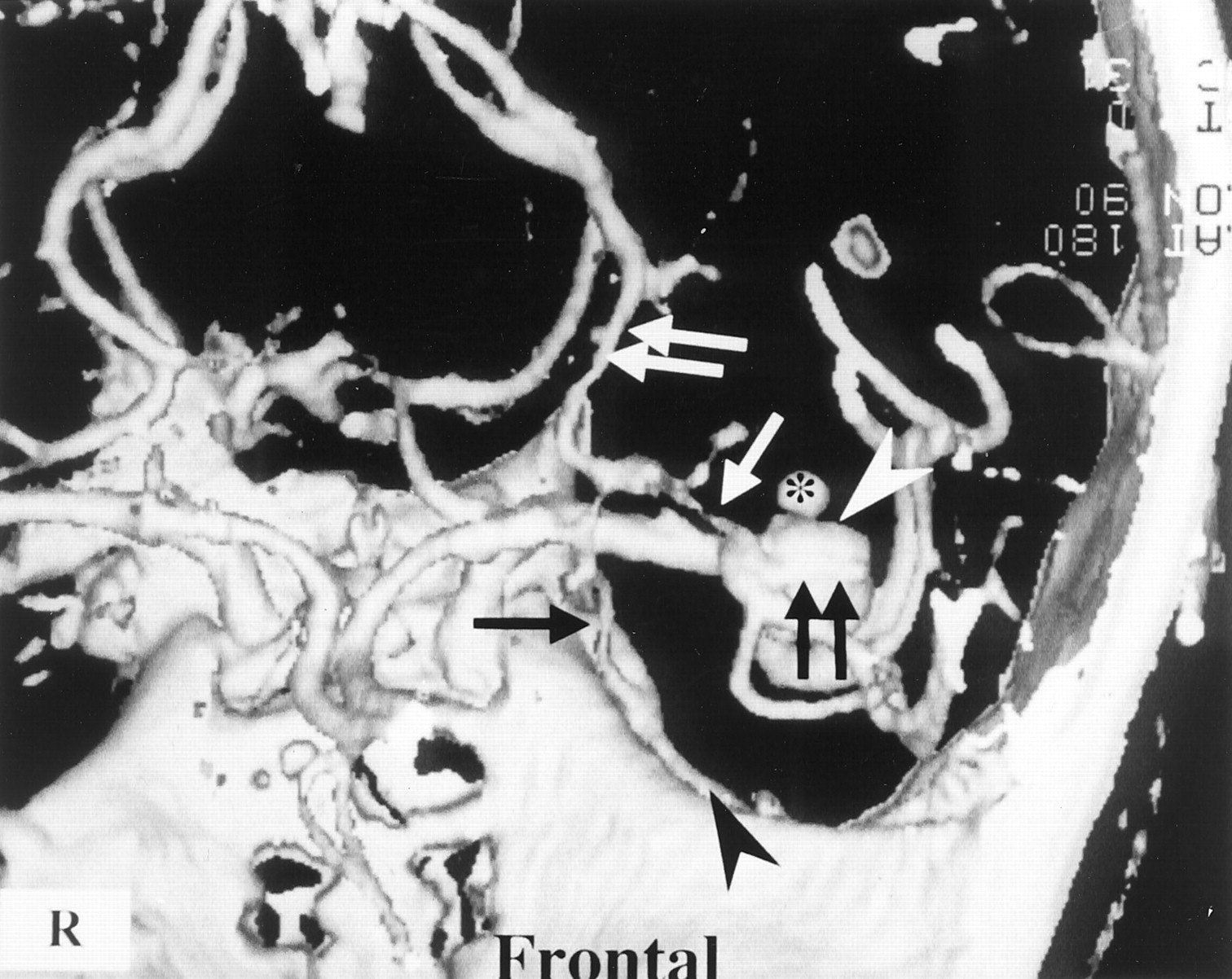
- Fig 3.
Patient with a left middle cerebral artery aneurysm. 3D-CTA (superior view) suggests strong adhesion (double black arrows) of an insular vein (white arrow) to the cerebral aneurysm (white arrowhead). In this case, a connecting vein (black arrow) between the BVR (double white arrows) and the SPS, SSVs (black arrowhead), and an aneurysmal bleb (*) are also revealed.
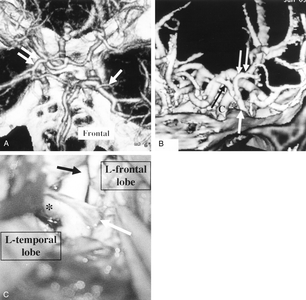
- Fig 4.
Patient with a left internal carotid artery aneurysm.
A, 3D-CTA (superior view) shows the left DMCV (arrow) emptying into the SPS and poor BVR development on the left side compared with that on the right side (double arrows).
B, 3D-CTA (left anterior view) reveals that the anterior cerebral vein (black arrow) and DMCV (double white arrows) join and empty into the SPS (white arrow). 3D-CTA also shows these veins in front of a left internal carotid artery aneurysm (*) but not in contact with it. C indicates the proximal portion of the left internal carotid artery.
C, A microscopic operative view shows that the left frontal lobe and the left temporal lobe are retracted to expose the left internal carotid artery aneurysm (*). A black arrow indicates the proximal portion of the left internal carotid artery. The connecting vein (white arrow) depicted in B was smoothly detached from the aneurysm and the internal carotid artery, but application of the clip was disturbed by this vein.
- Fig 5.
Patient with an AcomA aneurysm.
A, 3D-CTA (superior view) shows the insular vein (white arrow) emptying into the SPS. A black arrowhead and double black arrows indicate the AcomA aneurysm and the SSVs.
B and C, Via the pterional approach to the AcomA aneurysm, dissection of the aneurysm was attempted rostral (B) and caudal (C) to the bridging veins from the insular vein to the SPS (microscopic operative views). The white arrowhead, white arrow, black arrow, and SW indicate the bridging veins, the left anterior cerebral artery, the right anterior cerebral artery, and the sphenoid wing, respectively.
Retraction of the right frontal lobe was restricted, because the right frontal lobe was anchored to these bridging veins. Therefore, the aneurysmal neck was not completely explored. Because 3D-CTA depicted poor venous drainage from the insular cortex to the BVR, as shown in A, we could not sacrifice these veins and halted clipping of the aneurysm with this approach.
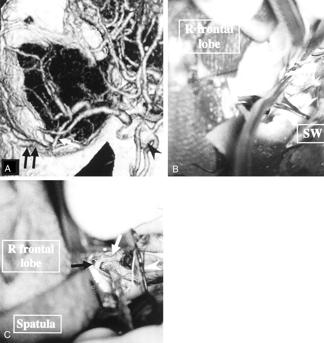
- Fig 6.
Patient with a right internal carotid artery aneurysm.
A, 3D-CTA (superior view) reveals the fronto-orbital vein (white arrow) emptying into the SPS at the inner portion of the sphenoid wing (black arrow) A white arrowhead indicates the SSVs.
B, A microscopic operative photograph shows that the right frontal lobe was retracted by the spatula to expose the right internal carotid artery (C). The aneurysm (white arrow) was barely clipped, although the bridging vein between the fronto-orbital vein and the SPS (black arrows) limited retraction of the frontal lobe. The black arrowhead indicates the draining point of this bridging vein into the SPS. The white arrowhead indicates the SSVs.
- Fig 7.
Patient with a ruptured right middle cerebral artery aneurysm.
3D-CTA (inferior view) shows a connecting vein (arrow) between the BVR (double arrows) and the SPS contiguous to the right middle cerebral artery aneurysm (white arrowhead), but it also depicts a gap between the aneurysm and the connecting vein. A black arrow indicates the SSVs.
- Fig 8.
Diagram of the cerebral veins around the sylvian fissure (inferior view).
A, Representative venous-draining system is revealed. ACV, BVR, DMCV, FOV, IVs, SSVs, and UV indicate the anterior cerebral vein, basal vein of Rosenthal, deep middle cerebral vein, fronto-orbital vein, insular veins, superficial sylvian veins, and uncal vein, respectively.
B, Bridging veins from the IVs, DMCV, FOV, and BVR to the SPS are indicated by solid arrows. The draining veins from the SSVs to the SPS or cavernous sinus are revealed by broken arrows. The final draining points are depicted at the midportion of the sphenoid wing in 11 patients (A), at the inner portion of the sphenoid wing in four (B), and the cavernous sinus in two (C).


