Advertisement

Main menu

  • Home
  • Content
    • Current Issue
    • Accepted Manuscripts
    • Article Preview
    • Past Issue Archive
    • Video Articles
    • AJNR Case Collection
    • Case of the Week Archive
    • Case of the Month Archive
    • Classic Case Archive
  • Special Collections
    • AJNR Awards
    • Low-Field MRI
    • Alzheimer Disease
    • ASNR Foundation Special Collection
    • Photon-Counting CT
    • View All
  • Multimedia
    • AJNR Podcasts
    • AJNR SCANtastic
    • Trainee Corner
    • MRI Safety Corner
    • Imaging Protocols
  • For Authors
    • Submit a Manuscript
    • Submit a Video Article
    • Submit an eLetter to the Editor/Response
    • Manuscript Submission Guidelines
    • Statistical Tips
    • Fast Publishing of Accepted Manuscripts
    • Graphical Abstract Preparation
    • Imaging Protocol Submission
    • Author Policies
  • About Us
    • About AJNR
    • Editorial Board
    • Editorial Board Alumni
  • More
    • Become a Reviewer/Academy of Reviewers
    • Subscribers
    • Permissions
    • Alerts
    • Feedback
    • Advertisers
    • ASNR Home

User menu

  • Alerts
  • Log in

Search

  • Advanced search
American Journal of Neuroradiology

American Journal of Neuroradiology

ASHNR American Society of Functional Neuroradiology ASHNR American Society of Pediatric Neuroradiology ASSR
  • Alerts
  • Log in

Advanced Search

  • Home
  • Content
    • Current Issue
    • Accepted Manuscripts
    • Article Preview
    • Past Issue Archive
    • Video Articles
    • AJNR Case Collection
    • Case of the Week Archive
    • Case of the Month Archive
    • Classic Case Archive
  • Special Collections
    • AJNR Awards
    • Low-Field MRI
    • Alzheimer Disease
    • ASNR Foundation Special Collection
    • Photon-Counting CT
    • View All
  • Multimedia
    • AJNR Podcasts
    • AJNR SCANtastic
    • Trainee Corner
    • MRI Safety Corner
    • Imaging Protocols
  • For Authors
    • Submit a Manuscript
    • Submit a Video Article
    • Submit an eLetter to the Editor/Response
    • Manuscript Submission Guidelines
    • Statistical Tips
    • Fast Publishing of Accepted Manuscripts
    • Graphical Abstract Preparation
    • Imaging Protocol Submission
    • Author Policies
  • About Us
    • About AJNR
    • Editorial Board
    • Editorial Board Alumni
  • More
    • Become a Reviewer/Academy of Reviewers
    • Subscribers
    • Permissions
    • Alerts
    • Feedback
    • Advertisers
    • ASNR Home
  • Follow AJNR on Twitter
  • Visit AJNR on Facebook
  • Follow AJNR on Instagram
  • Join AJNR on LinkedIn
  • RSS Feeds

AJNR Awards, New Junior Editors, and more. Read the latest AJNR updates

Research ArticleNeurointervention

Cyanoacrylate Embolization of Experimental Aneurysms

Jean Raymond, France Berthelet, Anne-Cécile Desfaits, Igor Salazkin and Daniel Roy
American Journal of Neuroradiology January 2002, 23 (1) 129-138;
Jean Raymond
  • Find this author on Google Scholar
  • Find this author on PubMed
  • Search for this author on this site
France Berthelet
  • Find this author on Google Scholar
  • Find this author on PubMed
  • Search for this author on this site
Anne-Cécile Desfaits
  • Find this author on Google Scholar
  • Find this author on PubMed
  • Search for this author on this site
Igor Salazkin
  • Find this author on Google Scholar
  • Find this author on PubMed
  • Search for this author on this site
Daniel Roy
  • Find this author on Google Scholar
  • Find this author on PubMed
  • Search for this author on this site
  • Article
  • Figures & Data
  • Info & Metrics
  • Responses
  • References
  • PDF
Loading

Article Figures & Data

Figures

  • Tables
  • Fig 1.
    • Download figure
    • Open in new tab
    • Download powerpoint
    Fig 1.

    Axial photomicrographs show histologic findings 3 months after embolization. h indicates cyanoacrylate; c, coils; ca, carotid artery; n, neck; ra, recurrent aneurysm; vp, venous pouch; t, vascularized fibrous tissue. (HPS, original magnification ×20).

    A, Lateral wall aneurysm, obtained after embolization with cyanoacrylate-coated sponge. Aneurysm remains obliterated by a thick and fibrous neointima.

    B, Lateral wall aneurysm, obtained after embolization with sponge alone. Aneurysm shows large recurrence and hypertrophy of the venous pouch.

    C, Bifurcation aneurysm, obtained after embolization with coils alone, shows recurrence around the mass of coils. Neointima is thin and incomplete at the neck. Coils are embedded in vascularized fibrous tissue.

    D, Bifurcation aneurysm, obtained after embolization with coils and cyanoacrylate, shows no recurrence. Note aneurysm filled with cyanoacrylate.
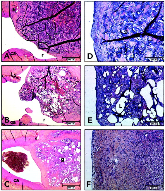
  • Fig 2.
    • Download figure
    • Open in new tab
    • Download powerpoint
    Fig 2.

    Histologic findings 3 weeks after embolization. ca indicates carotid artery; g, sponge; r, recurrence; S, suture; L, iodized oil. (HPS; magnification ×20 [A–C], ×100 [D–F]).

    A and D, Axial section of lateral wall aneurysm and carotid arteries, obtained 3 weeks after embolization with gelatin sponge. Note recurrence and thin neointima covering the uncoated sponge.

    B and E, Axial section of lateral wall aneurysm and carotid arteries, obtained 3 weeks after embolization with iodized oil. Note recurrences and thin neointima covering the iodized oil-coated sponges. Higher magnification reveals that the neointima is composed of a few layers of spindle cells embedded in a collagenous matrix and covered by an endothelium in aneurysms treated with iodized oil-coated sponges.

    C and F, Axial section of lateral wall aneurysms and carotid arteries, obtained 3 weeks after embolization with cyanoacrylate-coated sponges. Aneurysms treated with cyanoacrylate-coated sponges show a thicker neointima and complete obliteration of the neck.

  • Fig 3.
    • Download figure
    • Open in new tab
    • Download powerpoint
    Fig 3.

    Endovascular cyanoacrylate embolization.

    A and B, Spot radiographs obtained during endovascular cyanoacrylate delivery. The cyanoacrylate deposition is limited to the aneurysm at the beginning.

    C, Spot radiograph obtained after endovascular cyanoacrylate delivery. The cyanoacrylate spills through the neck at the end of the injection.

    D, Carotid arteriogram shows that spilling of cyanoacrylate caused severe stenosis of the bifurcation.

  • Fig 4.
    • Download figure
    • Open in new tab
    • Download powerpoint
    Fig 4.

    Endovascular cyanoacrylate embolization using coil protection.

    A and B, Radiographs obtained before (A) and after (B) carotid injection of contrast agent show how cyanoacrylate deposition can be limited by a single coil. Cyanoacrylate is seen as radiopaque material within and surrounding loops of coils. Note complete obliteration of the aneurysm without compromising the bifurcation.

Tables

  • Figures
    • View popup
    TABLE 1:

    Angiographic scores and neointima thickness in lateral wall and bifurcation aneurysms treated with coils

    ParameterLateral Wall Aneurysms (n = 6)Bifurcation Aneurysms (n = 6)
    Initial angiographic score0.67 ± 1.030.40 ± 0.89
    Score at 3 wks1.50 ± 0.550.75 ± 0.96
    Evolution of score at 3 wks0.83 ± 0.750.75 ± 0.96
    Score at 3 mo1.17 ± 0.753.00 ± 0.71*
    Evolution of score at 3 mo0.50 ± 1.052.60 ± 0.55†
    Neointima at 3 mo (μm)170.00 ± 42.4329.80 ± 24.35‡
    • Note.—Data are the mean ± SD.

    • * This score was significantly worse in bifurcation than in lateral wall aneurysms (P = .004).

    • † This score was significantly worse in bifurcation than in lateral wall aneurysms (P = .003).

    • ‡ This measurement was significantly thinner in bifurcation than in lateral wall aneurysms (P = .0004).

    • View popup
    TABLE 2:

    Angiographic scores and neointima thickness in lateral wall aneurysms treated with gelatin sponges

    ParameterUncoated SpongeIodized Oil-Coated SpongeCyanoacrylate-Coated Sponge
    Initial angiographic score1.68 ± 0.78 (21)2.40 ± 0.55 (5)2.19 ± 0.68 (16)
    Score at 3 wks2.68 ± 1.00 (21)2.40 ± 0.55 (5)1.59 ± 1.33* (16)
    Evolution of score at 3 wks1.00 ± 0.87 (21)0.00 ± 1.0 (5)−0.52 ± 1.29† (16)
    Score at 3 mo3.43 ± 0.79 (6)—1.50 ± 1.64 (6)
    Evolution of score at 3 mo1.86 ± 0.69 (6)—−0.67 ± 1.51† (6)
    Neointima at 3 wks (μm)99.15 ± 112.35 (21)185.75 ± 198.14 (5)575.53 ± 328.27‡ (16)
    Neointima at 3 mo (μm)124.43 ± 120.02 (6)—429.50 ± 183.03‡ (6)
    • Note.—Data are the mean ± SD. Numbers in parentheses are numbers of aneurysms.

    • * This score was significantly better in aneurysms treated with cyanoacrylate-coated sponges than in those treated with uncoated sponges (P < .05).

    • † This score was significantly better in aneurysms treated with cyanoacrylate-coated sponges than in those treated with uncoated sponges (P < .05).

    • ‡ This measurement was significantly thicker in aneurysms treated with cyanoacrylate-coated sponges than in those treated with uncoated sponges (P < .05).

    • View popup
    TABLE 3:

    Angiographic scores and neointima thickness in bifurcation aneurysms treated with coils alone or with cyanoacrylate

    ParameterCoils Alone (n = 6)Cyanoacrylate and Coils (n = 6)
    Initial angiographic score0.40 ± 0.891.00 ± 1.16
    Score at 3 wks0.75 ± 0.960.00 ± 0.00
    Evolution of score at 3 wks0.75 ± 0.96−1.00 ± 1.16
    Score at 3 mo3.00 ± 0.710.50 ± 1.00*
    Evolution of score at 3 mo2.60 ± 0.55−0.50 ± 1.92†
    Neointima at 3 mo (μm)29.80 ± 24.36300.00 ± 50.09‡
    • Note.—Data are the mean ± SD.

    • * This score was significantly better in aneurysms treated with cyanoacrylate over a coil than in those treated with coils alone (P = .001).

    • † This score is significantly better in aneurysms treated with cyanoacrylate over a coil than in those treated with coils alone (P = .01).

    • ‡ This measurement at the neck of aneurysms treated with cyanoacrylate behind a coil is significantly thicker than that in aneurysms treated with coils alone (P < .05).

PreviousNext
Back to top

In this issue

American Journal of Neuroradiology: 23 (1)
American Journal of Neuroradiology
Vol. 23, Issue 1
1 Jan 2002
  • Table of Contents
  • Index by author
Advertisement
Print
Download PDF
Email Article

Thank you for your interest in spreading the word on American Journal of Neuroradiology.

NOTE: We only request your email address so that the person you are recommending the page to knows that you wanted them to see it, and that it is not junk mail. We do not capture any email address.

Enter multiple addresses on separate lines or separate them with commas.
Cyanoacrylate Embolization of Experimental Aneurysms
(Your Name) has sent you a message from American Journal of Neuroradiology
(Your Name) thought you would like to see the American Journal of Neuroradiology web site.
CAPTCHA
This question is for testing whether or not you are a human visitor and to prevent automated spam submissions.
Cite this article
Jean Raymond, France Berthelet, Anne-Cécile Desfaits, Igor Salazkin, Daniel Roy
Cyanoacrylate Embolization of Experimental Aneurysms
American Journal of Neuroradiology Jan 2002, 23 (1) 129-138;

Citation Manager Formats

  • BibTeX
  • Bookends
  • EasyBib
  • EndNote (tagged)
  • EndNote 8 (xml)
  • Medlars
  • Mendeley
  • Papers
  • RefWorks Tagged
  • Ref Manager
  • RIS
  • Zotero
0 Responses
Respond to this article
Share
Bookmark this article
Cyanoacrylate Embolization of Experimental Aneurysms
Jean Raymond, France Berthelet, Anne-Cécile Desfaits, Igor Salazkin, Daniel Roy
American Journal of Neuroradiology Jan 2002, 23 (1) 129-138;
del.icio.us logo Twitter logo Facebook logo Mendeley logo
  • Tweet Widget
  • Facebook Like
  • Google Plus One

Jump to section

  • Article
    • Abstract
    • Methods
    • Results
    • Discussion
    • Conclusion
    • Acknowledgments
    • Footnotes
    • References
  • Figures & Data
  • Info & Metrics
  • Responses
  • References
  • PDF

Related Articles

  • No related articles found.
  • PubMed
  • Google Scholar

Cited By...

  • Treatment of experimental aneurysms with a new liquid embolic agent and a retrievable stent: proof of concept and feasibility study
  • Preliminary in vivo evaluation of a novel intrasaccular cerebral aneurysm occlusion device
  • Stent-Assisted Coiling of Bifurcation Aneurysms May Improve Endovascular Treatment: A Critical Evaluation in an Experimental Model
  • Intra-procedural aneurysm rupture treated with n-butyl cyanoacrylate embolization: technical note
  • A New Canine Carotid Artery Bifurcation Aneurysm Model for the Evaluation of Neurovascular Devices
  • In Vivo Experimental Intracranial Aneurysm Models: A Systematic Review
  • Safety and Effectiveness of Radioactive Coil Embolization of Aneurysms: Effects of Radiation on Recanalization, Clot Organization, Neointima Formation, and Surrounding Nerves in Experimental Models
  • Role of the Endothelial Lining in Recurrences After Coil Embolization: Prevention of Recanalization by Endothelial Denudation
  • Alginate for Endovascular Treatment of Aneurysms and Local Growth Factor Delivery
  • Role of the Endothelial Lining in Persistence of Residual Lesions and Growth of Recurrences After Endovascular Treatment of Experimental Aneurysms
  • Crossref
  • Google Scholar

This article has not yet been cited by articles in journals that are participating in Crossref Cited-by Linking.

More in this TOC Section

  • Rescue Reentry in Carotid Near-Occlusion
  • Contour Neurovascular System: Five Year Follow Up
  • Effect of SARS-CoV2 on Endovascular Thrombectomy
Show more Neurointervention

Similar Articles

Advertisement

Indexed Content

  • Current Issue
  • Accepted Manuscripts
  • Article Preview
  • Past Issues
  • Editorials
  • Editor's Choice
  • Fellows' Journal Club
  • Letters to the Editor
  • Video Articles

Cases

  • Case Collection
  • Archive - Case of the Week
  • Archive - Case of the Month
  • Archive - Classic Case

More from AJNR

  • Trainee Corner
  • Imaging Protocols
  • MRI Safety Corner
  • Book Reviews

Multimedia

  • AJNR Podcasts
  • AJNR Scantastics

Resources

  • Turnaround Time
  • Submit a Manuscript
  • Submit a Video Article
  • Submit an eLetter to the Editor/Response
  • Manuscript Submission Guidelines
  • Statistical Tips
  • Fast Publishing of Accepted Manuscripts
  • Graphical Abstract Preparation
  • Imaging Protocol Submission
  • Evidence-Based Medicine Level Guide
  • Publishing Checklists
  • Author Policies
  • Become a Reviewer/Academy of Reviewers
  • News and Updates

About Us

  • About AJNR
  • Editorial Board
  • Editorial Board Alumni
  • Alerts
  • Permissions
  • Not an AJNR Subscriber? Join Now
  • Advertise with Us
  • Librarian Resources
  • Feedback
  • Terms and Conditions
  • AJNR Editorial Board Alumni

American Society of Neuroradiology

  • Not an ASNR Member? Join Now

© 2025 by the American Society of Neuroradiology All rights, including for text and data mining, AI training, and similar technologies, are reserved.
Print ISSN: 0195-6108 Online ISSN: 1936-959X

Powered by HighWire