Advertisement

Main menu

  • Home
  • Content
    • Current Issue
    • Accepted Manuscripts
    • Article Preview
    • Past Issue Archive
    • Video Articles
    • AJNR Case Collection
    • Case of the Week Archive
    • Case of the Month Archive
    • Classic Case Archive
  • Special Collections
    • AJNR Awards
    • Low-Field MRI
    • Alzheimer Disease
    • ASNR Foundation Special Collection
    • Photon-Counting CT
    • View All
  • Multimedia
    • AJNR Podcasts
    • AJNR SCANtastic
    • Trainee Corner
    • MRI Safety Corner
    • Imaging Protocols
  • For Authors
    • Submit a Manuscript
    • Submit a Video Article
    • Submit an eLetter to the Editor/Response
    • Manuscript Submission Guidelines
    • Statistical Tips
    • Fast Publishing of Accepted Manuscripts
    • Graphical Abstract Preparation
    • Imaging Protocol Submission
    • Author Policies
  • About Us
    • About AJNR
    • Editorial Board
    • Editorial Board Alumni
  • More
    • Become a Reviewer/Academy of Reviewers
    • Subscribers
    • Permissions
    • Alerts
    • Feedback
    • Advertisers
    • ASNR Home

User menu

  • Alerts
  • Log in

Search

  • Advanced search
American Journal of Neuroradiology

American Journal of Neuroradiology

ASHNR American Society of Functional Neuroradiology ASHNR American Society of Pediatric Neuroradiology ASSR
  • Alerts
  • Log in

Advanced Search

  • Home
  • Content
    • Current Issue
    • Accepted Manuscripts
    • Article Preview
    • Past Issue Archive
    • Video Articles
    • AJNR Case Collection
    • Case of the Week Archive
    • Case of the Month Archive
    • Classic Case Archive
  • Special Collections
    • AJNR Awards
    • Low-Field MRI
    • Alzheimer Disease
    • ASNR Foundation Special Collection
    • Photon-Counting CT
    • View All
  • Multimedia
    • AJNR Podcasts
    • AJNR SCANtastic
    • Trainee Corner
    • MRI Safety Corner
    • Imaging Protocols
  • For Authors
    • Submit a Manuscript
    • Submit a Video Article
    • Submit an eLetter to the Editor/Response
    • Manuscript Submission Guidelines
    • Statistical Tips
    • Fast Publishing of Accepted Manuscripts
    • Graphical Abstract Preparation
    • Imaging Protocol Submission
    • Author Policies
  • About Us
    • About AJNR
    • Editorial Board
    • Editorial Board Alumni
  • More
    • Become a Reviewer/Academy of Reviewers
    • Subscribers
    • Permissions
    • Alerts
    • Feedback
    • Advertisers
    • ASNR Home
  • Follow AJNR on Twitter
  • Visit AJNR on Facebook
  • Follow AJNR on Instagram
  • Join AJNR on LinkedIn
  • RSS Feeds

AJNR Awards, New Junior Editors, and more. Read the latest AJNR updates

Research ArticleBrain

Diffusion-weighted MR Imaging After Angioplasty or Angioplasty Plus Stenting of Arteries Supplying the Brain

Horst J. Jaeger, Klaus D. Mathias, Robert Drescher, Elke Hauth, Georg Bockisch, Eren Demirel and Hans Martin Gissler
American Journal of Neuroradiology August 2001, 22 (7) 1251-1259;
Horst J. Jaeger
  • Find this author on Google Scholar
  • Find this author on PubMed
  • Search for this author on this site
Klaus D. Mathias
  • Find this author on Google Scholar
  • Find this author on PubMed
  • Search for this author on this site
Robert Drescher
  • Find this author on Google Scholar
  • Find this author on PubMed
  • Search for this author on this site
Elke Hauth
  • Find this author on Google Scholar
  • Find this author on PubMed
  • Search for this author on this site
Georg Bockisch
  • Find this author on Google Scholar
  • Find this author on PubMed
  • Search for this author on this site
Eren Demirel
  • Find this author on Google Scholar
  • Find this author on PubMed
  • Search for this author on this site
Hans Martin Gissler
  • Find this author on Google Scholar
  • Find this author on PubMed
  • Search for this author on this site
  • Article
  • Figures & Data
  • Info & Metrics
  • Responses
  • References
  • PDF
Loading

Article Figures & Data

Figures

  • Tables
  • fig 1.
    • Download figure
    • Open in new tab
    • Download powerpoint
    fig 1.

    Patient 2.

    A, Stenosis of the cavernous part of the right ICA.

    B, Result after percutaneous angioplasty (3.5-mm balloon). Note irregularity at the site of the percutaneous angioplasty.

    C, Cross flow from the right ICA to the contralateral hemisphere (occlusion of the left ICA is not shown). Post-procedural diffusion-weighted images (6000/103; number of excitations, one) obtained with a diffusion sensitization level of b = 1000 s/mm2.

    D, New ipsilateral lesion: <5 mm, middle cerebral artery cortical territory, parietal lobe (arrow).

    E, New contralateral lesion: 5 to 10 mm, middle cerebral artery cortical territory, parietal lobe (arrow).
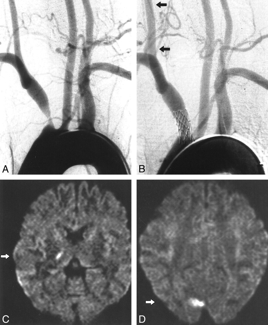
  • fig 2.
    • Download figure
    • Open in new tab
    • Download powerpoint
    fig 2.

    Patient 5.

    A, Aortic arch injection (early phase). Stenosis of the innominate artery with antegrade flow in the right carotid artery. (Retrograde flow in the right VA with subclavian steal in the late phase is not shown).

    B, Result after stent implantation (balloon-expandable stent, 8 mm diameter) with antegrade flow in the right VA (arrows). Post-procedural diffusion-weighted images (6000/103; number of excitations, one) obtained with a diffusion sensitization level of b = 1000 s/mm2.

    C, New ipsilateral lesion: 5 to 10 mm, PCA deep territory, thalamus (arrow).

    D, New ipsilateral lesion: 15 mm, PCA cortical territory, occipital lobe (arrow).

  • fig 3.
    • Download figure
    • Open in new tab
    • Download powerpoint
    fig 3.

    Patient 6.

    A, Stenosis of left VA (occlusion of right VA is not shown).

    B, Result after percutaneous angioplasty (3.5-mm balloon).

    C, Left VA injection. Note the anterior inferior-posterior inferior cerebellar artery complex on the right side (arrow). Post-procedural diffusion-weighted images (6000/103; number of excitations, one) obtained with a diffusion sensitization level of b = 1000 s;clmm2.

    D, New cerebellar lesions: 5 to 10 mm, superior cerebellar artery territory (arrows).

    E, New cerebellar lesion: 20 mm, posterior inferior cerebellar artery territory (arrow).
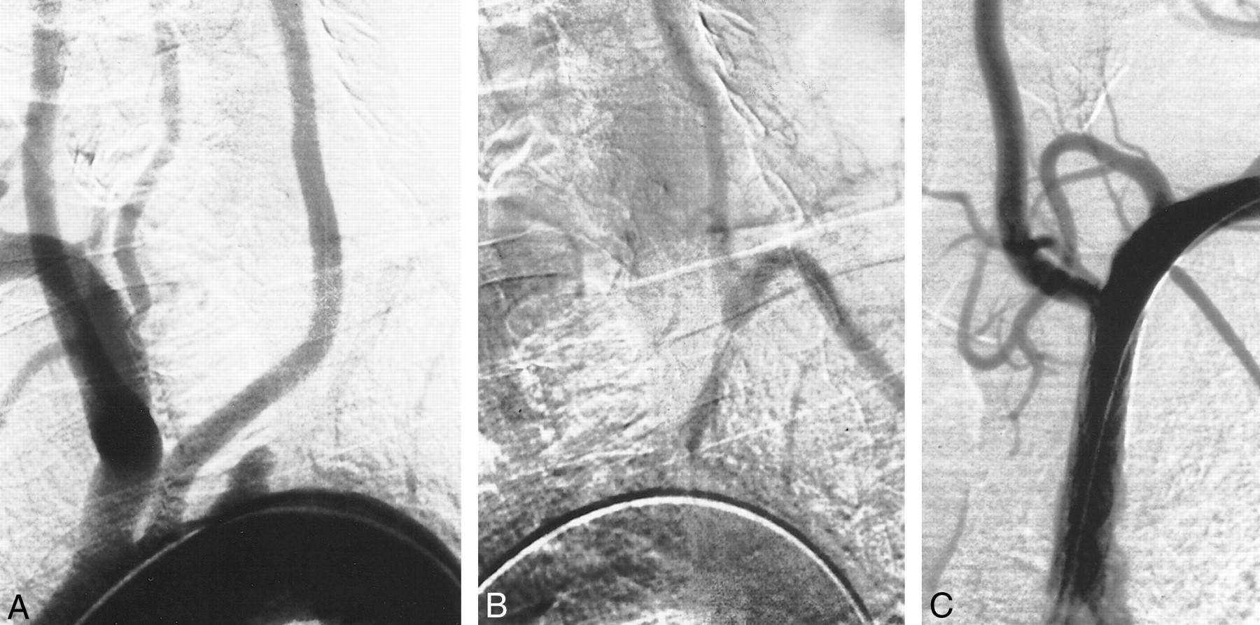
  • fig 4.
    • Download figure
    • Open in new tab
    • Download powerpoint
    fig 4.

    A, Aortic arch injection (early phase). Occlusion of the proximal part of the left SA.

    B, Aortic arch injection (late phase). Retrograde flow in left VA with subclavian steal.

    C, Result after stent implantation (balloon-expandable stent, 7 mm) with antegrade flow in left VA.

Tables

  • Figures
  • TABLE 1:
    • View popup
    • Download powerpoint
    TABLE 1:

    Characteristics of lesions and interventions

  • TABLE 2:
    • View popup
    • Download powerpoint
    TABLE 2:

    Characteristics of new postprocedural lesion in DW-MR imaging

PreviousNext
Back to top

In this issue

American Journal of Neuroradiology: 22 (7)
American Journal of Neuroradiology
Vol. 22, Issue 7
1 Aug 2001
  • Table of Contents
  • Index by author
Advertisement
Print
Download PDF
Email Article

Thank you for your interest in spreading the word on American Journal of Neuroradiology.

NOTE: We only request your email address so that the person you are recommending the page to knows that you wanted them to see it, and that it is not junk mail. We do not capture any email address.

Enter multiple addresses on separate lines or separate them with commas.
Diffusion-weighted MR Imaging After Angioplasty or Angioplasty Plus Stenting of Arteries Supplying the Brain
(Your Name) has sent you a message from American Journal of Neuroradiology
(Your Name) thought you would like to see the American Journal of Neuroradiology web site.
CAPTCHA
This question is for testing whether or not you are a human visitor and to prevent automated spam submissions.
Cite this article
Horst J. Jaeger, Klaus D. Mathias, Robert Drescher, Elke Hauth, Georg Bockisch, Eren Demirel, Hans Martin Gissler
Diffusion-weighted MR Imaging After Angioplasty or Angioplasty Plus Stenting of Arteries Supplying the Brain
American Journal of Neuroradiology Aug 2001, 22 (7) 1251-1259;

Citation Manager Formats

  • BibTeX
  • Bookends
  • EasyBib
  • EndNote (tagged)
  • EndNote 8 (xml)
  • Medlars
  • Mendeley
  • Papers
  • RefWorks Tagged
  • Ref Manager
  • RIS
  • Zotero
0 Responses
Respond to this article
Share
Bookmark this article
Diffusion-weighted MR Imaging After Angioplasty or Angioplasty Plus Stenting of Arteries Supplying the Brain
Horst J. Jaeger, Klaus D. Mathias, Robert Drescher, Elke Hauth, Georg Bockisch, Eren Demirel, Hans Martin Gissler
American Journal of Neuroradiology Aug 2001, 22 (7) 1251-1259;
del.icio.us logo Twitter logo Facebook logo Mendeley logo
  • Tweet Widget
  • Facebook Like
  • Google Plus One

Jump to section

  • Article
    • Abstract
    • Methods
    • Results
    • Discussion
    • Conclusion
    • Acknowledgments
    • Footnotes
    • References
  • Figures & Data
  • Info & Metrics
  • Responses
  • References
  • PDF

Related Articles

  • No related articles found.
  • PubMed
  • Google Scholar

Cited By...

  • New Ischemic Brain Lesions on Diffusion-Weighted MRI after Carotid Artery Stenting with Filter Protection: Frequency and Relationship with Plaque Morphology
  • Reporting standards for angioplasty and stent-assisted angioplasty for intracranial atherosclerosis
  • The Influence of Carotid Artery Catheterization Technique on the Incidence of Thromboembolism during Carotid Artery Stenting
  • Reporting Standards for Angioplasty and Stent-Assisted Angioplasty for Intracranial Atherosclerosis
  • Late Cerebral Embolization After Emboli-Protected Carotid Artery Stenting Assessed by Sequential Diffusion-Weighted Magnetic Resonance Imaging
  • Incidence of New Brain Lesions After Carotid Stenting With and Without Cerebral Protection
  • Silent Cerebral Ischemia Detected With Diffusion-Weighted Imaging in Patients Treated With Protected and Unprotected Carotid Artery Stenting
  • Training, competency, and credentialing standards for diagnostic cervicocerebral angiography, carotid stenting, and cerebrovascular intervention: A Joint Statement from the American Academy of Neurology, the American Association of Neurological Surgeons, the American Society of Interventional and Therapeutic Neuroradiology, the American Society of Neuroradiology, the Congress of Neurological Surgeons, the AANS/CNS Cerebrovascular Section, and the Society of Interventional Radiology
  • Cerebral Ischemia and Infarction From Atheroemboli <100 {micro}m in Size
  • Early Outcome of Carotid Angioplasty and Stenting With and Without Cerebral Protection Devices: A Systematic Review of the Literature
  • Crossref
  • Google Scholar

This article has not yet been cited by articles in journals that are participating in Crossref Cited-by Linking.

More in this TOC Section

  • Evaluating the Effects of White Matter Multiple Sclerosis Lesions on the Volume Estimation of 6 Brain Tissue Segmentation Methods
  • Quiet PROPELLER MRI Techniques Match the Quality of Conventional PROPELLER Brain Imaging Techniques
  • Predictors of Reperfusion in Patients with Acute Ischemic Stroke
Show more Brain

Similar Articles

Advertisement

Indexed Content

  • Current Issue
  • Accepted Manuscripts
  • Article Preview
  • Past Issues
  • Editorials
  • Editor's Choice
  • Fellows' Journal Club
  • Letters to the Editor
  • Video Articles

Cases

  • Case Collection
  • Archive - Case of the Week
  • Archive - Case of the Month
  • Archive - Classic Case

More from AJNR

  • Trainee Corner
  • Imaging Protocols
  • MRI Safety Corner
  • Book Reviews

Multimedia

  • AJNR Podcasts
  • AJNR Scantastics

Resources

  • Turnaround Time
  • Submit a Manuscript
  • Submit a Video Article
  • Submit an eLetter to the Editor/Response
  • Manuscript Submission Guidelines
  • Statistical Tips
  • Fast Publishing of Accepted Manuscripts
  • Graphical Abstract Preparation
  • Imaging Protocol Submission
  • Evidence-Based Medicine Level Guide
  • Publishing Checklists
  • Author Policies
  • Become a Reviewer/Academy of Reviewers
  • News and Updates

About Us

  • About AJNR
  • Editorial Board
  • Editorial Board Alumni
  • Alerts
  • Permissions
  • Not an AJNR Subscriber? Join Now
  • Advertise with Us
  • Librarian Resources
  • Feedback
  • Terms and Conditions
  • AJNR Editorial Board Alumni

American Society of Neuroradiology

  • Not an ASNR Member? Join Now

© 2025 by the American Society of Neuroradiology All rights, including for text and data mining, AI training, and similar technologies, are reserved.
Print ISSN: 0195-6108 Online ISSN: 1936-959X

Powered by HighWire