Advertisement

Main menu

  • Home
  • Content
    • Current Issue
    • Accepted Manuscripts
    • Article Preview
    • Past Issue Archive
    • Video Articles
    • AJNR Case Collection
    • Case of the Week Archive
    • Case of the Month Archive
    • Classic Case Archive
  • Special Collections
    • AJNR Awards
    • Low-Field MRI
    • Alzheimer Disease
    • ASNR Foundation Special Collection
    • Photon-Counting CT
    • View All
  • Multimedia
    • AJNR Podcasts
    • AJNR SCANtastic
    • Trainee Corner
    • MRI Safety Corner
    • Imaging Protocols
  • For Authors
    • Submit a Manuscript
    • Submit a Video Article
    • Submit an eLetter to the Editor/Response
    • Manuscript Submission Guidelines
    • Statistical Tips
    • Fast Publishing of Accepted Manuscripts
    • Graphical Abstract Preparation
    • Imaging Protocol Submission
    • Author Policies
  • About Us
    • About AJNR
    • Editorial Board
    • Editorial Board Alumni
  • More
    • Become a Reviewer/Academy of Reviewers
    • Subscribers
    • Permissions
    • Alerts
    • Feedback
    • Advertisers
    • ASNR Home

User menu

  • Alerts
  • Log in

Search

  • Advanced search
American Journal of Neuroradiology

American Journal of Neuroradiology

ASHNR American Society of Functional Neuroradiology ASHNR American Society of Pediatric Neuroradiology ASSR
  • Alerts
  • Log in

Advanced Search

  • Home
  • Content
    • Current Issue
    • Accepted Manuscripts
    • Article Preview
    • Past Issue Archive
    • Video Articles
    • AJNR Case Collection
    • Case of the Week Archive
    • Case of the Month Archive
    • Classic Case Archive
  • Special Collections
    • AJNR Awards
    • Low-Field MRI
    • Alzheimer Disease
    • ASNR Foundation Special Collection
    • Photon-Counting CT
    • View All
  • Multimedia
    • AJNR Podcasts
    • AJNR SCANtastic
    • Trainee Corner
    • MRI Safety Corner
    • Imaging Protocols
  • For Authors
    • Submit a Manuscript
    • Submit a Video Article
    • Submit an eLetter to the Editor/Response
    • Manuscript Submission Guidelines
    • Statistical Tips
    • Fast Publishing of Accepted Manuscripts
    • Graphical Abstract Preparation
    • Imaging Protocol Submission
    • Author Policies
  • About Us
    • About AJNR
    • Editorial Board
    • Editorial Board Alumni
  • More
    • Become a Reviewer/Academy of Reviewers
    • Subscribers
    • Permissions
    • Alerts
    • Feedback
    • Advertisers
    • ASNR Home
  • Follow AJNR on Twitter
  • Visit AJNR on Facebook
  • Follow AJNR on Instagram
  • Join AJNR on LinkedIn
  • RSS Feeds

AJNR Awards, New Junior Editors, and more. Read the latest AJNR updates

Research ArticleBrain

Quantitative Proton MR Spectroscopic Imaging in Acute Disseminated Encephalomyelitis

Alberto Bizzi, Aziz M. Uluğ, Thomas O. Crawford, Ted Passe, Marianna Bugiani, R. Nick Bryan and Peter B. Barker
American Journal of Neuroradiology June 2001, 22 (6) 1125-1130;
Alberto Bizzi
  • Find this author on Google Scholar
  • Find this author on PubMed
  • Search for this author on this site
Aziz M. Uluğ
  • Find this author on Google Scholar
  • Find this author on PubMed
  • Search for this author on this site
Thomas O. Crawford
  • Find this author on Google Scholar
  • Find this author on PubMed
  • Search for this author on this site
Ted Passe
  • Find this author on Google Scholar
  • Find this author on PubMed
  • Search for this author on this site
Marianna Bugiani
  • Find this author on Google Scholar
  • Find this author on PubMed
  • Search for this author on this site
R. Nick Bryan
  • Find this author on Google Scholar
  • Find this author on PubMed
  • Search for this author on this site
Peter B. Barker
  • Find this author on Google Scholar
  • Find this author on PubMed
  • Search for this author on this site
  • Article
  • Figures & Data
  • Info & Metrics
  • Responses
  • References
  • PDF
Loading

Article Figures & Data

Figures

  • Tables
  • fig 1.
    • Download figure
    • Open in new tab
    • Download powerpoint
    fig 1.

    A–C. T2-weighted MR images at 3000/100/1 (TR/TE/excitation) at the following times after initial symptom (headache) onset. A, At 14 days, bilateral thalamic lesions (left slightly more prominent than right). B, At 21 days, thalamic lesions have increased in size and new lesion has appeared in left cerebral peduncle (lesions in left globus pallidus and forceps major not shown). C, At 32 days, further progression of thalamic lesions to involve right geniculate body, right posterior limb of internal capsule, and complete involvement of left putamen and globus pallidus tracking down to left cerebral peduncle (bilateral lesions in hemispheric white matter and left frontoparietal cortex not shown)
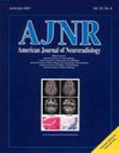
  • fig 1.
    • Download figure
    • Open in new tab
    • Download powerpoint
    fig 1.

    D–F. D, At 8 weeks, pattern of involvement dramatically changed, with resolution of lesions in thalami and left cerebral peduncle, and with new lesions in left caudatum, putamen, and globus pallidus, and in right globus pallidus, anterior thalamus, cerebral peduncle, and posterior limb of internal capsule. (Hyperintensity of subcortical white matter of right temporal lobe not shown.) E, At 17 weeks, nearly all lesions resolved, with only minimal hyperintensity of left caudate nucleus and frontal periventricular white matter (not shown). F, At 25 weeks, all T2 hyperintense lesions completely resolved

  • fig 2.
    • Download figure
    • Open in new tab
    • Download powerpoint
    fig 2.

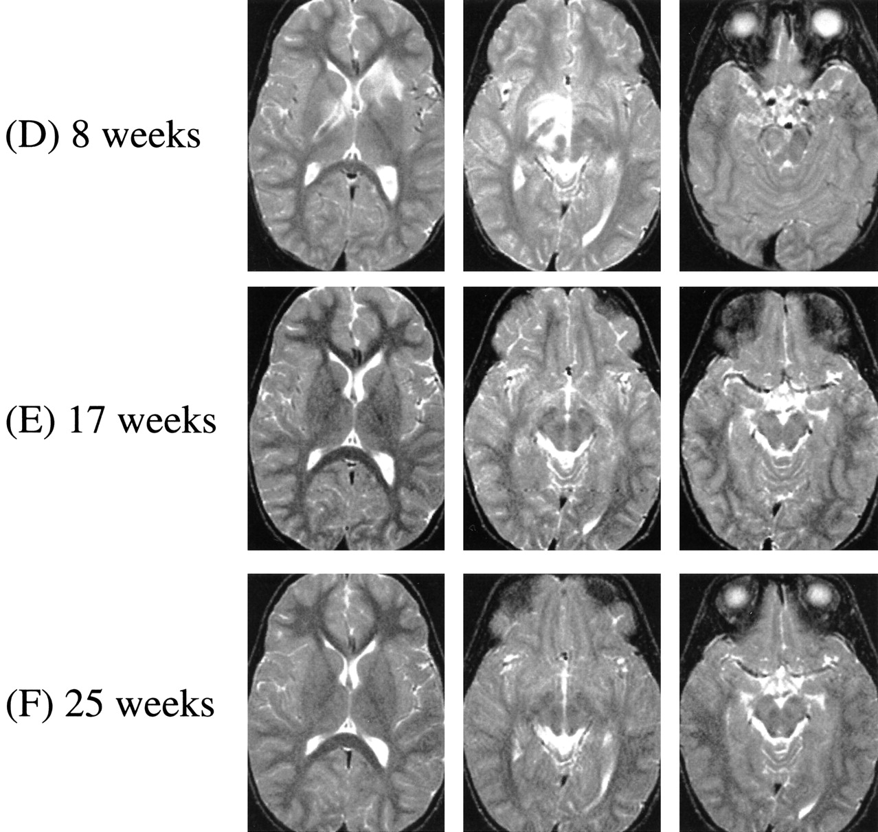
    Proton MRSI at 8 (top) and 25 (bottom) weeks after initial symptom onset. Localizer T1-weighted MR images at 300/13/1 (TR/TE/excitation) and metabolic images at 2300/280/1 (TR/TE/excitation) of choline and NAA from the second of four sections shown. Selected spectrum from left caudatum shown at both time points (voxel location indicated on T1-weighted images). At 8 weeks, NAA image shows decreased levels in left caudatum, lenticular nuclei, and right internal capsule, corresponding to lesions seen in T2-weighted MR images (fig 1D). Choline signals within normal limits; note high choline signal in thalamic region is normal for these regions (Table 2). By 25 weeks, NAA recovered to normal levels in all these regions, whereas creatine and choline levels remain stable (Table 2)

Tables

  • Figures
  • TABLE 1:
    • View popup
    • Download powerpoint
    TABLE 1:

    CSF white and red blood cell counts and protein and glucose levels

  • TABLE 2:
    • View popup
    • Download powerpoint
    TABLE 2:

    Metabolite concentrations (mM) in selected brain structures at 8 and 25 weeks after first onset of symptoms, and ratios of metabolite levels at 25 and 8 weeks (% increase or decrease)

PreviousNext
Back to top

In this issue

American Journal of Neuroradiology: 22 (6)
American Journal of Neuroradiology
Vol. 22, Issue 6
1 Jun 2001
  • Table of Contents
  • Index by author
Advertisement
Print
Download PDF
Email Article

Thank you for your interest in spreading the word on American Journal of Neuroradiology.

NOTE: We only request your email address so that the person you are recommending the page to knows that you wanted them to see it, and that it is not junk mail. We do not capture any email address.

Enter multiple addresses on separate lines or separate them with commas.
Quantitative Proton MR Spectroscopic Imaging in Acute Disseminated Encephalomyelitis
(Your Name) has sent you a message from American Journal of Neuroradiology
(Your Name) thought you would like to see the American Journal of Neuroradiology web site.
CAPTCHA
This question is for testing whether or not you are a human visitor and to prevent automated spam submissions.
Cite this article
Alberto Bizzi, Aziz M. Uluğ, Thomas O. Crawford, Ted Passe, Marianna Bugiani, R. Nick Bryan, Peter B. Barker
Quantitative Proton MR Spectroscopic Imaging in Acute Disseminated Encephalomyelitis
American Journal of Neuroradiology Jun 2001, 22 (6) 1125-1130;

Citation Manager Formats

  • BibTeX
  • Bookends
  • EasyBib
  • EndNote (tagged)
  • EndNote 8 (xml)
  • Medlars
  • Mendeley
  • Papers
  • RefWorks Tagged
  • Ref Manager
  • RIS
  • Zotero
0 Responses
Respond to this article
Share
Bookmark this article
Quantitative Proton MR Spectroscopic Imaging in Acute Disseminated Encephalomyelitis
Alberto Bizzi, Aziz M. Uluğ, Thomas O. Crawford, Ted Passe, Marianna Bugiani, R. Nick Bryan, Peter B. Barker
American Journal of Neuroradiology Jun 2001, 22 (6) 1125-1130;
del.icio.us logo Twitter logo Facebook logo Mendeley logo
  • Tweet Widget
  • Facebook Like
  • Google Plus One

Jump to section

  • Article
    • Abstract
    • Case Report
    • Discussion
    • Acknowledgments
    • Footnotes
    • References
  • Figures & Data
  • Info & Metrics
  • Responses
  • References
  • PDF

Related Articles

  • No related articles found.
  • PubMed
  • Google Scholar

Cited By...

  • What is MR spectroscopy?
  • Acute disseminated encephalomyelitis: Updates on an inflammatory CNS syndrome
  • The groom who could not say "I do"
  • Acute disseminated encephalomyelitis
  • Acute disseminated encephalomyelitis
  • Crossref
  • Google Scholar

This article has not yet been cited by articles in journals that are participating in Crossref Cited-by Linking.

More in this TOC Section

  • Multimodal CT Provides Improved Performance for Lacunar Infarct Detection
  • Optimal MRI Sequence for Identifying Occlusion Location in Acute Stroke: Which Value of Time-Resolved Contrast-Enhanced MRA?
  • Evaluating the Effects of White Matter Multiple Sclerosis Lesions on the Volume Estimation of 6 Brain Tissue Segmentation Methods
Show more Brain

Similar Articles

Advertisement

Indexed Content

  • Current Issue
  • Accepted Manuscripts
  • Article Preview
  • Past Issues
  • Editorials
  • Editor's Choice
  • Fellows' Journal Club
  • Letters to the Editor
  • Video Articles

Cases

  • Case Collection
  • Archive - Case of the Week
  • Archive - Case of the Month
  • Archive - Classic Case

More from AJNR

  • Trainee Corner
  • Imaging Protocols
  • MRI Safety Corner
  • Book Reviews

Multimedia

  • AJNR Podcasts
  • AJNR Scantastics

Resources

  • Turnaround Time
  • Submit a Manuscript
  • Submit a Video Article
  • Submit an eLetter to the Editor/Response
  • Manuscript Submission Guidelines
  • Statistical Tips
  • Fast Publishing of Accepted Manuscripts
  • Graphical Abstract Preparation
  • Imaging Protocol Submission
  • Evidence-Based Medicine Level Guide
  • Publishing Checklists
  • Author Policies
  • Become a Reviewer/Academy of Reviewers
  • News and Updates

About Us

  • About AJNR
  • Editorial Board
  • Editorial Board Alumni
  • Alerts
  • Permissions
  • Not an AJNR Subscriber? Join Now
  • Advertise with Us
  • Librarian Resources
  • Feedback
  • Terms and Conditions
  • AJNR Editorial Board Alumni

American Society of Neuroradiology

  • Not an ASNR Member? Join Now

© 2025 by the American Society of Neuroradiology All rights, including for text and data mining, AI training, and similar technologies, are reserved.
Print ISSN: 0195-6108 Online ISSN: 1936-959X

Powered by HighWire