
Article Figures & Data
Figures
- fig 1.
Images from the case of a 34-year-old man with a large grade II astrocytoma in the inferior frontal and parietal area. Differentiation between edema and tumor is unclear on all sequences.
A, Fast T2-weighted spin-echo image.
B, Contrast-enhanced T1-weighted spin-echo image. The stereotactic biopsy was performed in the area of faint contrast material uptake (arrow).
C, Diffusion-weighted echo-planar image acquired with x sensitizing gradient.
D, Diffusion-weighted echo-planar image acquired with y sensitizing gradient.
E, Diffusion-weighted echo-planar image acquired with z sensitizing gradient.
F, Tensor ADC map.
G, Lattice index map. The red/yellow areas represent high/medium anisotropy, and the blue/dark blue areas represent lower anisotropy
- fig 2.
Images from the case of a 52-year-old man with a large glioblastoma.
A, Invasion of corpus callosum and of occipitotemporal white matter is recognized on fast T2-weighted spin-echo image.
B, Invasion of corpus callosum and of occipitotemporal white matter is recognized on contrast-enhanced T1-weighted spin-echo image.
C, Invasion of subinsular white matter is clearly recognized only on the perfusion-weighted echo-planar image (arrow).
D, On the diffusion-weighted image, only cystic components of tumor are clearly shown. There is no clear difference between contrast-enhancing tumor (arrowhead) and edema (arrow).
E, On the ADC map, only cystic components of tumor are clearly shown. There is no clear difference between contrast-enhancing tumor (arrowhead) and edema (arrow).
F, Histologic examination (hematoxylin and eosin; original magnification, ×250) shows a medium sized extracellular space and moderate cellularity
- fig 2.
fig 3. Images from the case of a 72-year-old woman with a cerebral lymphoma. A, Fast T2-weighted spin-echo image shows lesion to be moderately hyperintense. B, T1-weighted spin-echo image shows strong enhancement of lesion after the administration of contrast material. C, Diffusion-weighted image shows the lymphoma also to be hyperintense. D, ADC values are in the range of 0.55 to 0.6, similar to the values described in association with acute infarction. E, Histologic examination (hematoxylin and eosin; original magnification, ×250) shows high cellularity and small extracellular space, which was significantly associated with reduced ADC values
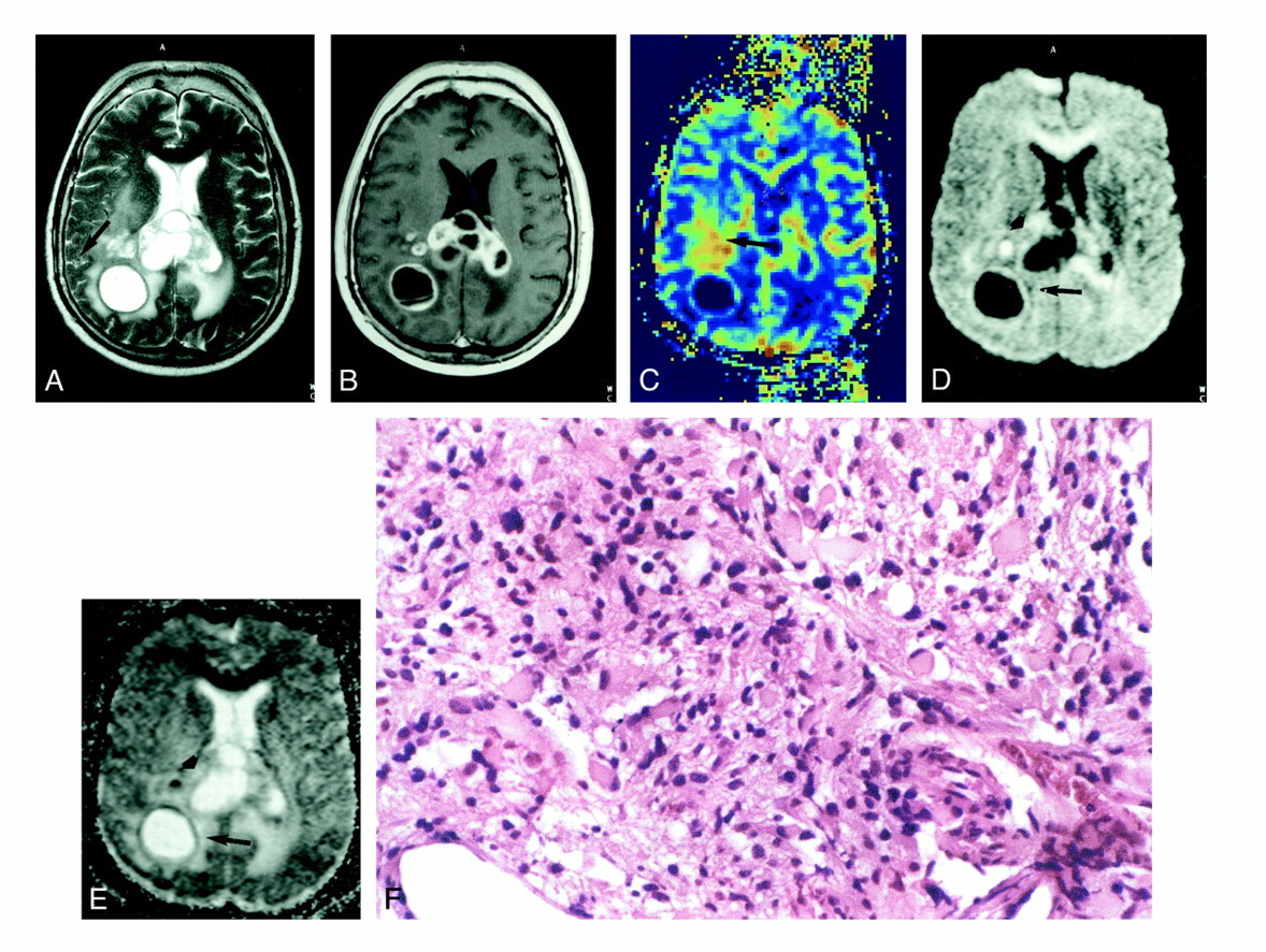
- fig 4.
Images from the case of a 70-year-old man with a streptococcus abscess, recent history of acute dizziness, and focal seizures.
A, T2-weighted spin-echo image is consistent with the diagnosis of glioblastoma or necrotic metastasis.
B, Contrast-enhanced T1-weighted spin-echo image is consistent with the diagnosis of glioblastoma or necrotic metastasis.
C, On the diffusion-weighted echo-planar image, however, the “necrotic” area shows high signal intensity (arrow).
D, On the ADC map, very low ADC values (0.29 e−3 mm2/s) are found (arrow). Such behavior was never present in the necrotic parts of gliomas (compare with fig. 3D and E) or metastases and may be highly specific for abscess formation


Tables

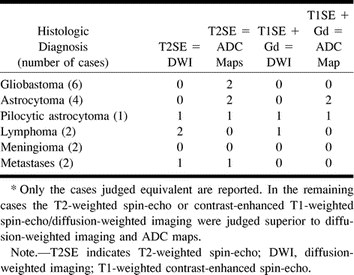
- TABLE 2:
Comparison of fast T2-weighted spin-echo and contrast-enhanced T1-weighted spin-echo with diffusion-weighted echo-planar imaging and ADC maps in delineation of cerebral tumor boundaries (n = 17)*
- TABLE 3:
Tumor/white matter contrast values on diffusion-weighted images acquired in “z” direction for solid components
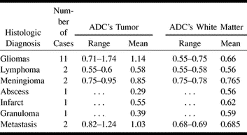
- TABLE 4:
ADC's values for solid tumor components and for white matter acquired in “z” direction


In this issue


Jump to section
Related Articles
Cited By...
- A Multiparametric Model for Mapping Cellularity in Glioblastoma Using Radiographically Localized Biopsies
- MR Imaging Features of Acute Mastoiditis and Their Clinical Relevance
- Diffusion-Weighted Imaging in Cancer: Physical Foundations and Applications of Restriction Spectrum Imaging
- "Dazed and diffused": making sense of diffusion abnormalities in neurologic pathologies
- Longitudinal Restriction Spectrum Imaging Is Resistant to Pseudoresponse in Patients with High-Grade Gliomas Treated with Bevacizumab
- Correlation of MRI-Derived Apparent Diffusion Coefficients in Newly Diagnosed Gliomas with [18F]-Fluoro-L-Dopa PET: What Are We Really Measuring with Minimum ADC?
- Diffusion-weighted imaging characteristics of biopsy-proven demyelinating brain lesions
- The Added Value of Apparent Diffusion Coefficient to Cerebral Blood Volume in the Preoperative Grading of Diffuse Gliomas
- Methodology of diffusion-weighted, diffusion tensor and magnetisation transfer imaging
- Central Nervous System Lymphoma: Characteristic Findings on Traditional and Advanced Imaging
- Apparent Diffusion Coefficient of Glial Neoplasms: Correlation with Fluorodeoxyglucose-Positron-Emission Tomography and Gadolinium-Enhanced MR Imaging
- Diffusion-Weighted MR Imaging Derived Apparent Diffusion Coefficient Is Predictive of Clinical Outcome in Primary Central Nervous System Lymphoma
- Diffusion Tensor Imaging in Glioblastoma Multiforme and Brain Metastases: The Role of p, q, L, and Fractional Anisotropy
- MR Imaging of Orbital Inflammatory Syndrome, Orbital Cellulitis, and Orbital Lymphoid Lesions: The Role of Diffusion-Weighted Imaging
- Glioblastoma multiforme with atypical diffusion-weighted MR findings
- MR diffusion-weighted imaging in a case of West Nile virus encephalitis
- Differentiation of Toxoplasmosis and Lymphoma in AIDS Patients by Using Apparent Diffusion Coefficients