Advertisement

Main menu

  • Home
  • Content
    • Current Issue
    • Accepted Manuscripts
    • Article Preview
    • Past Issue Archive
    • Video Articles
    • AJNR Case Collection
    • Case of the Week Archive
    • Case of the Month Archive
    • Classic Case Archive
  • Special Collections
    • AJNR Awards
    • Low-Field MRI
    • Alzheimer Disease
    • ASNR Foundation Special Collection
    • Photon-Counting CT
    • View All
  • Multimedia
    • AJNR Podcasts
    • AJNR SCANtastic
    • Trainee Corner
    • MRI Safety Corner
    • Imaging Protocols
  • For Authors
    • Submit a Manuscript
    • Submit a Video Article
    • Submit an eLetter to the Editor/Response
    • Manuscript Submission Guidelines
    • Statistical Tips
    • Fast Publishing of Accepted Manuscripts
    • Graphical Abstract Preparation
    • Imaging Protocol Submission
    • Author Policies
  • About Us
    • About AJNR
    • Editorial Board
    • Editorial Board Alumni
  • More
    • Become a Reviewer/Academy of Reviewers
    • Subscribers
    • Permissions
    • Alerts
    • Feedback
    • Advertisers
    • ASNR Home

User menu

  • Alerts
  • Log in

Search

  • Advanced search
American Journal of Neuroradiology

American Journal of Neuroradiology

ASHNR American Society of Functional Neuroradiology ASHNR American Society of Pediatric Neuroradiology ASSR
  • Alerts
  • Log in

Advanced Search

  • Home
  • Content
    • Current Issue
    • Accepted Manuscripts
    • Article Preview
    • Past Issue Archive
    • Video Articles
    • AJNR Case Collection
    • Case of the Week Archive
    • Case of the Month Archive
    • Classic Case Archive
  • Special Collections
    • AJNR Awards
    • Low-Field MRI
    • Alzheimer Disease
    • ASNR Foundation Special Collection
    • Photon-Counting CT
    • View All
  • Multimedia
    • AJNR Podcasts
    • AJNR SCANtastic
    • Trainee Corner
    • MRI Safety Corner
    • Imaging Protocols
  • For Authors
    • Submit a Manuscript
    • Submit a Video Article
    • Submit an eLetter to the Editor/Response
    • Manuscript Submission Guidelines
    • Statistical Tips
    • Fast Publishing of Accepted Manuscripts
    • Graphical Abstract Preparation
    • Imaging Protocol Submission
    • Author Policies
  • About Us
    • About AJNR
    • Editorial Board
    • Editorial Board Alumni
  • More
    • Become a Reviewer/Academy of Reviewers
    • Subscribers
    • Permissions
    • Alerts
    • Feedback
    • Advertisers
    • ASNR Home
  • Follow AJNR on Twitter
  • Visit AJNR on Facebook
  • Follow AJNR on Instagram
  • Join AJNR on LinkedIn
  • RSS Feeds

AJNR Awards, New Junior Editors, and more. Read the latest AJNR updates

Research ArticleBrain

Correlation of Cerebrovascular Reserve as Measured by Acetazolamide-challenged SPECT with Angiographic Flow Patterns and Intra- or Extracranial Arterial Stenosis

Hasan T. Ozgur, T. Kent Walsh, Anthony Masaryk, Joachim F. Seeger, Walter Williams, Elizabeth Krupinski, Miguel Melgar and Enrique Labadie
American Journal of Neuroradiology May 2001, 22 (5) 928-936;
Hasan T. Ozgur
  • Find this author on Google Scholar
  • Find this author on PubMed
  • Search for this author on this site
T. Kent Walsh
  • Find this author on Google Scholar
  • Find this author on PubMed
  • Search for this author on this site
Anthony Masaryk
  • Find this author on Google Scholar
  • Find this author on PubMed
  • Search for this author on this site
Joachim F. Seeger
  • Find this author on Google Scholar
  • Find this author on PubMed
  • Search for this author on this site
Walter Williams
  • Find this author on Google Scholar
  • Find this author on PubMed
  • Search for this author on this site
Elizabeth Krupinski
  • Find this author on Google Scholar
  • Find this author on PubMed
  • Search for this author on this site
Miguel Melgar
  • Find this author on Google Scholar
  • Find this author on PubMed
  • Search for this author on this site
Enrique Labadie
  • Find this author on Google Scholar
  • Find this author on PubMed
  • Search for this author on this site
  • Article
  • Figures & Data
  • Info & Metrics
  • Responses
  • References
  • PDF
Loading

Article Figures & Data

Figures

  • Tables
  • fig 1.
    • Download figure
    • Open in new tab
    • Download powerpoint
    fig 1.

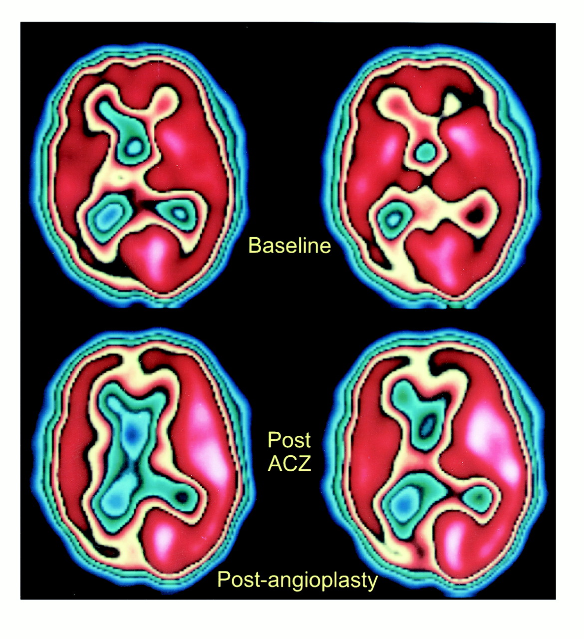
    58-year-old man with neurosyphilis and progressive neurologic symptoms. SPECT scans before right MCA angioplasty show relatively normal baseline perfusion (top row) but significantly decreased perfusion in the right ACA, MCA, and PCA territories after acetazolamide administration (bottom row), consistent with decreased rCVR. (The printed color scale representing brain perfusion is different from the linear 10-level color scale used for interpreting nuclear medicine studies; however, it allows easier comparison with fig 3.)

  • fig 2.
    • Download figure
    • Open in new tab
    • Download powerpoint
    fig 2.

    A, Anteroposterior view of right ICA angiogram shows occlusion of the right ACA and high-grade stenosis of the right MCA (arrow).

    B and C, Anteroposterior views of early (B) and late (C) arterial phases of left CCA angiogram show complete occlusion of the proximal left ACA and reconstitution of both pericallosal arteries (arrows) and the anterior communicating artery from leptomeningeal collaterals (arrowheads).

    D and E, Lateral views of early (D) and late (E) arterial phases of left CCA angiogram show reconstitution of the pericallosal artery (arrows) in the late arterial phase by leptomeningeal collaterals

  • fig 2.
    • Download figure
    • Open in new tab
    • Download powerpoint
    fig 2.

    F and G, Lateral views of early arterial (F) and capillary (G) phases of left vertebral angiogram show high-grade stenosis of the mid-basilar artery (arrow, F) and retrograde filling of the pericallosal artery (arrows, G) via PCA collaterals.

    H, Anteroposterior view of left vertebral angiogram shows an occluded right PCA.

    I, Anteroposterior view of right ICA angiogram after right MCA angioplasty shows almost complete resolution of the right MCA stenosis

  • fig 3.
    • Download figure
    • Open in new tab
    • Download powerpoint
    fig 3.

    SPECT scans after right MCA angioplasty show improved CBF in right ACA and MCA territories on postacetazolamide study (bottom row), consistent with improved rCVR. The rCVR in the right PCA territory appears to be worse as compared with baseline study (top row)

Tables

  • Figures
  • TABLE 1:
    • View popup
    • Download powerpoint
    TABLE 1:

    Cerebral vascular reserve and angiographic findings in 27 patients

  • TABLE 2:
    • View popup
    • Download powerpoint
    TABLE 2:

    Comparison of angiographic cerebral flow patterns in the anterior, middle, and posterior cerebral artery territories with rCVR

  • TABLE 3:
    • View popup
    • Download powerpoint
    TABLE 3:

    Comparison of angiographic anterior circulation arterial stenosis or occlusion with rCVR

PreviousNext
Back to top

In this issue

American Journal of Neuroradiology
Vol. 22, Issue 5
1 May 2001
  • Table of Contents
  • Index by author
Advertisement
Print
Download PDF
Email Article

Thank you for your interest in spreading the word on American Journal of Neuroradiology.

NOTE: We only request your email address so that the person you are recommending the page to knows that you wanted them to see it, and that it is not junk mail. We do not capture any email address.

Enter multiple addresses on separate lines or separate them with commas.
Correlation of Cerebrovascular Reserve as Measured by Acetazolamide-challenged SPECT with Angiographic Flow Patterns and Intra- or Extracranial Arterial Stenosis
(Your Name) has sent you a message from American Journal of Neuroradiology
(Your Name) thought you would like to see the American Journal of Neuroradiology web site.
CAPTCHA
This question is for testing whether or not you are a human visitor and to prevent automated spam submissions.
Cite this article
Hasan T. Ozgur, T. Kent Walsh, Anthony Masaryk, Joachim F. Seeger, Walter Williams, Elizabeth Krupinski, Miguel Melgar, Enrique Labadie
Correlation of Cerebrovascular Reserve as Measured by Acetazolamide-challenged SPECT with Angiographic Flow Patterns and Intra- or Extracranial Arterial Stenosis
American Journal of Neuroradiology May 2001, 22 (5) 928-936;

Citation Manager Formats

  • BibTeX
  • Bookends
  • EasyBib
  • EndNote (tagged)
  • EndNote 8 (xml)
  • Medlars
  • Mendeley
  • Papers
  • RefWorks Tagged
  • Ref Manager
  • RIS
  • Zotero
0 Responses
Respond to this article
Share
Bookmark this article
Correlation of Cerebrovascular Reserve as Measured by Acetazolamide-challenged SPECT with Angiographic Flow Patterns and Intra- or Extracranial Arterial Stenosis
Hasan T. Ozgur, T. Kent Walsh, Anthony Masaryk, Joachim F. Seeger, Walter Williams, Elizabeth Krupinski, Miguel Melgar, Enrique Labadie
American Journal of Neuroradiology May 2001, 22 (5) 928-936;
del.icio.us logo Twitter logo Facebook logo Mendeley logo
  • Tweet Widget
  • Facebook Like
  • Google Plus One

Jump to section

  • Article
    • Abstract
    • Methods
    • Results
    • Discussion
    • Conclusion
    • Acknowledgments
    • Footnotes
    • References
  • Figures & Data
  • Info & Metrics
  • Responses
  • References
  • PDF

Related Articles

  • No related articles found.
  • PubMed
  • Google Scholar

Cited By...

  • Angiographic Correlates of Cerebral Hemodynamic Changes With Diamox Challenge Assessed by Quantitative Magnetic Resonance Angiography
  • History of the Letzte Wiese/Last Meadow Concept of Brain Ischemia
  • Expression of cellular retinoic acid-binding protein-I (CRABP-I) in the cerebrospinal fluid of adult onset moyamoya disease and its association with clinical presentation and postoperative haemodynamic change
  • Standard of practice: endovascular treatment of intracranial atherosclerosis
  • Systematic Review of Methods for Assessing Leptomeningeal Collateral Flow
  • Vasodilatory Capacity of the Cerebral Vasculature in Patients with Carotid Artery Stenosis
  • Reporting standards for angioplasty and stent-assisted angioplasty for intracranial atherosclerosis
  • Recommendations for Imaging of Acute Ischemic Stroke: A Scientific Statement From the American Heart Association
  • Reporting Standards for Angioplasty and Stent-Assisted Angioplasty for Intracranial Atherosclerosis
  • Efficacy Assessment of Cerebral Arterial Bypass Surgery Using Statistical Parametric Mapping and Probabilistic Brain Atlas on Basal/Acetazolamide Brain Perfusion SPECT
  • Prominent Laterality of the Posterior Cerebral Artery at Three-Dimensional Time-of-Flight MR Angiography in M1-Segment Middle Cerebral Artery Occlusion
  • Qualitative versus Quantitative Assessment of Cerebrovascular Reactivity to Acetazolamide Using iodine-123-N-Isopropyl-p-Iodoamphetamine SPECT in Patients with Unilateral Major Cerebral Artery Occlusive Disease
  • Collateral flow and ischemic brain lesions in patients with unilateral carotid artery occlusion
  • Quantitative Measurement of Regional Cerebrovascular Reactivity to Acetazolamide Using 123I-N-Isopropyl-p-Iodoamphetamine Autoradiography with SPECT: Validation Study Using H215O with PET
  • Guidelines and Recommendations for Perfusion Imaging in Cerebral Ischemia: A Scientific Statement for Healthcare Professionals by the Writing Group on Perfusion Imaging, From the Council on Cardiovascular Radiology of the American Heart Association
  • Crossref
  • Google Scholar

This article has not yet been cited by articles in journals that are participating in Crossref Cited-by Linking.

More in this TOC Section

  • Multimodal CT Provides Improved Performance for Lacunar Infarct Detection
  • Optimal MRI Sequence for Identifying Occlusion Location in Acute Stroke: Which Value of Time-Resolved Contrast-Enhanced MRA?
  • Evaluating the Effects of White Matter Multiple Sclerosis Lesions on the Volume Estimation of 6 Brain Tissue Segmentation Methods
Show more Brain

Similar Articles

Advertisement

Indexed Content

  • Current Issue
  • Accepted Manuscripts
  • Article Preview
  • Past Issues
  • Editorials
  • Editor's Choice
  • Fellows' Journal Club
  • Letters to the Editor
  • Video Articles

Cases

  • Case Collection
  • Archive - Case of the Week
  • Archive - Case of the Month
  • Archive - Classic Case

More from AJNR

  • Trainee Corner
  • Imaging Protocols
  • MRI Safety Corner
  • Book Reviews

Multimedia

  • AJNR Podcasts
  • AJNR Scantastics

Resources

  • Turnaround Time
  • Submit a Manuscript
  • Submit a Video Article
  • Submit an eLetter to the Editor/Response
  • Manuscript Submission Guidelines
  • Statistical Tips
  • Fast Publishing of Accepted Manuscripts
  • Graphical Abstract Preparation
  • Imaging Protocol Submission
  • Evidence-Based Medicine Level Guide
  • Publishing Checklists
  • Author Policies
  • Become a Reviewer/Academy of Reviewers
  • News and Updates

About Us

  • About AJNR
  • Editorial Board
  • Editorial Board Alumni
  • Alerts
  • Permissions
  • Not an AJNR Subscriber? Join Now
  • Advertise with Us
  • Librarian Resources
  • Feedback
  • Terms and Conditions
  • AJNR Editorial Board Alumni

American Society of Neuroradiology

  • Not an ASNR Member? Join Now

© 2025 by the American Society of Neuroradiology All rights, including for text and data mining, AI training, and similar technologies, are reserved.
Print ISSN: 0195-6108 Online ISSN: 1936-959X

Powered by HighWire