Advertisement

Main menu

  • Home
  • Content
    • Current Issue
    • Accepted Manuscripts
    • Article Preview
    • Past Issue Archive
    • Video Articles
    • AJNR Case Collection
    • Case of the Week Archive
    • Case of the Month Archive
    • Classic Case Archive
  • Special Collections
    • AJNR Awards
    • Low-Field MRI
    • Alzheimer Disease
    • ASNR Foundation Special Collection
    • Photon-Counting CT
    • View All
  • Multimedia
    • AJNR Podcasts
    • AJNR SCANtastic
    • Trainee Corner
    • MRI Safety Corner
    • Imaging Protocols
  • For Authors
    • Submit a Manuscript
    • Submit a Video Article
    • Submit an eLetter to the Editor/Response
    • Manuscript Submission Guidelines
    • Statistical Tips
    • Fast Publishing of Accepted Manuscripts
    • Graphical Abstract Preparation
    • Imaging Protocol Submission
    • Author Policies
  • About Us
    • About AJNR
    • Editorial Board
    • Editorial Board Alumni
  • More
    • Become a Reviewer/Academy of Reviewers
    • Subscribers
    • Permissions
    • Alerts
    • Feedback
    • Advertisers
    • ASNR Home

User menu

  • Alerts
  • Log in

Search

  • Advanced search
American Journal of Neuroradiology

American Journal of Neuroradiology

ASHNR American Society of Functional Neuroradiology ASHNR American Society of Pediatric Neuroradiology ASSR
  • Alerts
  • Log in

Advanced Search

  • Home
  • Content
    • Current Issue
    • Accepted Manuscripts
    • Article Preview
    • Past Issue Archive
    • Video Articles
    • AJNR Case Collection
    • Case of the Week Archive
    • Case of the Month Archive
    • Classic Case Archive
  • Special Collections
    • AJNR Awards
    • Low-Field MRI
    • Alzheimer Disease
    • ASNR Foundation Special Collection
    • Photon-Counting CT
    • View All
  • Multimedia
    • AJNR Podcasts
    • AJNR SCANtastic
    • Trainee Corner
    • MRI Safety Corner
    • Imaging Protocols
  • For Authors
    • Submit a Manuscript
    • Submit a Video Article
    • Submit an eLetter to the Editor/Response
    • Manuscript Submission Guidelines
    • Statistical Tips
    • Fast Publishing of Accepted Manuscripts
    • Graphical Abstract Preparation
    • Imaging Protocol Submission
    • Author Policies
  • About Us
    • About AJNR
    • Editorial Board
    • Editorial Board Alumni
  • More
    • Become a Reviewer/Academy of Reviewers
    • Subscribers
    • Permissions
    • Alerts
    • Feedback
    • Advertisers
    • ASNR Home
  • Follow AJNR on Twitter
  • Visit AJNR on Facebook
  • Follow AJNR on Instagram
  • Join AJNR on LinkedIn
  • RSS Feeds

AJNR Awards, New Junior Editors, and more. Read the latest AJNR updates

Research ArticleHead and Neck Imaging

Tumefactive Fibroinflammatory Lesion of the Neck with Progressive Invasion of the Meninges, Skull Base, Orbit, and Brain

Andrei I. Holodny, Claudia F. Kirsch, Meera Hameed and Gary Sclar
American Journal of Neuroradiology May 2001, 22 (5) 876-879;
Andrei I. Holodny
  • Find this author on Google Scholar
  • Find this author on PubMed
  • Search for this author on this site
Claudia F. Kirsch
  • Find this author on Google Scholar
  • Find this author on PubMed
  • Search for this author on this site
Meera Hameed
  • Find this author on Google Scholar
  • Find this author on PubMed
  • Search for this author on this site
Gary Sclar
  • Find this author on Google Scholar
  • Find this author on PubMed
  • Search for this author on this site
  • Article
  • Figures & Data
  • Info & Metrics
  • Responses
  • References
  • PDF
Loading

Article Figures & Data

Figures

  • fig 1.
    • Download figure
    • Open in new tab
    • Download powerpoint
    fig 1.

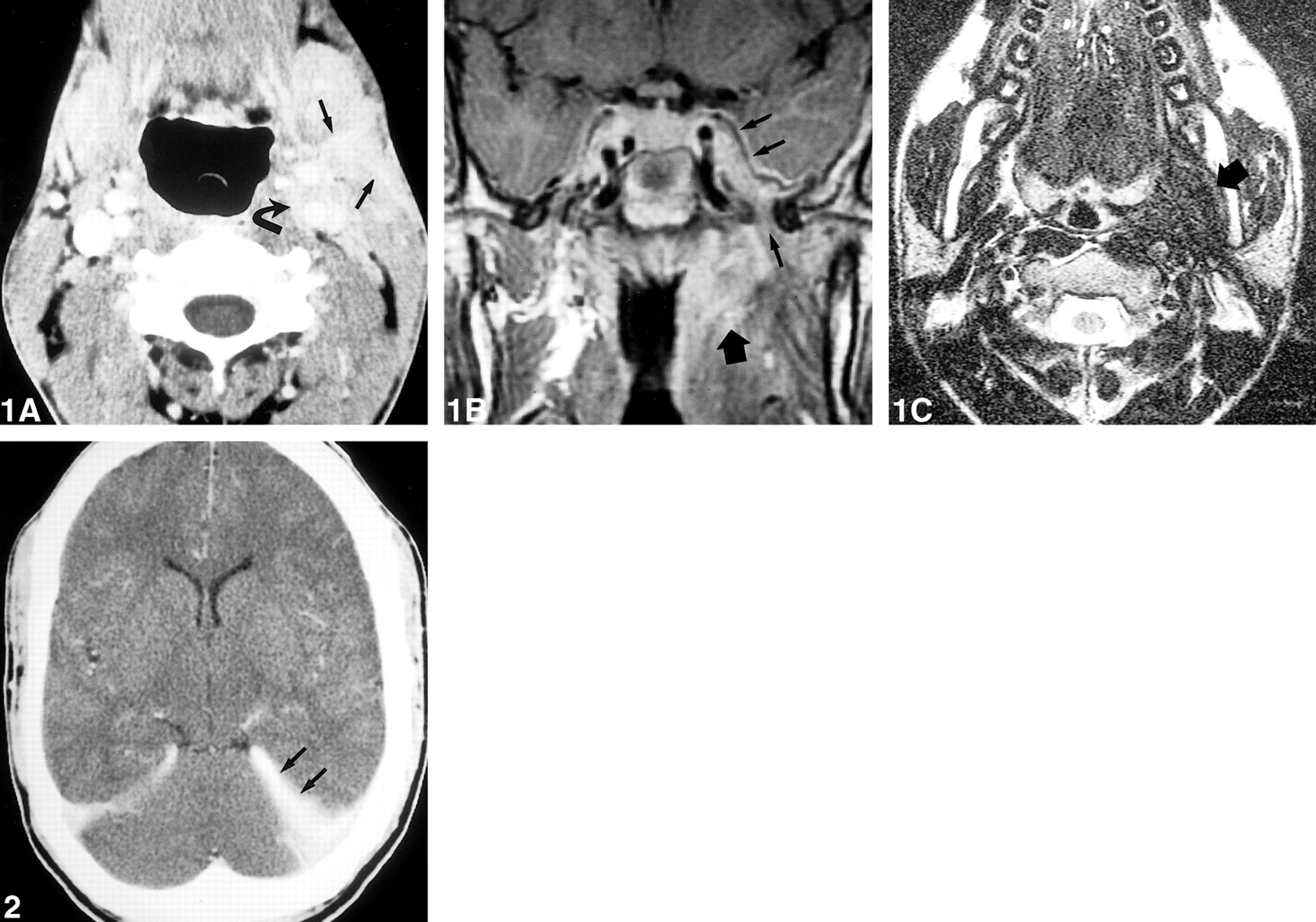
    23-year-old man with 2-year history of trismus and dysphagia. A–C, Imaging studies at the time of the first admission. A, Axial contrast-enhanced CT scan through the neck at the level of the tongue and the submandibular triangle. The mildly enhancing lesion has infiltrated into the left carotid space (curved arrow) and the left submandibular triangle (straight arrows). B and C, Coronal contrast-enhanced T1-weighted (600/14/1) (B) and axial T2-weighted (4000/105/1) (C) MR images show infiltration by the lesion of the left nasopharynx (wide arrow). It invades into the left middle cranial fossa and the left cavernous sinus (double arrows) through the foramen ovale (single arrow). It is mildly enhancing on the T1-weighted image and is hypointense on the T2-weighted image. The lesion replaces adipose tissue in the left parapharyngeal space. fig 2. Contrast-enhanced CT study at the time of the second admission shows thickening and enhancement of the tentorium cerebelli, greater on the left (arrows). This finding was not seen previously

  • fig 3.
    • Download figure
    • Open in new tab
    • Download powerpoint
    fig 3.

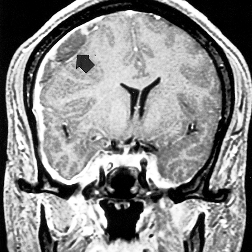
    A–E, Imaging studies at the time of the third admission. A and B, Coronal T1-weighted (600/23/1) (A) and coronal contrast-enhanced T1-weighted (500/14/1) (B) MR images show that the lesion has progressed to involve the contralateral cavernous sinus and the meninges of the right cranial fossa (double arrows, B). The lesion significantly narrows the right cavernous carotid artery and has invaded the right masticator space from the right middle cranial fossa through the foramen ovale (single arrow, B). The unenhanced image shows that the lesion has replaced the normally seen adipose tissue in the right infratemporal fossa. C–E, Coronal T2-weighted (4000/102/1) MR image (C) shows abnormal signal intensity in theright temporal lobe, which involves both the gray and white matter. Axial fat-suppressed, contrast-enhanced T1-weighted (600/20/1) MR images (D and E) show marked thickening and enhancement of the meninges of the right middle cranial fossa extending into the sulci as well as wisps of enhancement of the temporal lobe itself. There is marked narrowing of the cavernous carotid artery. The right lateral rectus muscle is enlarged and enhancing more than the other extraocular muscles, indicating probable involvement by the lesion

  • fig 4.
    • Download figure
    • Open in new tab
    • Download powerpoint
    fig 4.

    Coronal contrast-enhanced T1-weighted (500/5.7/1) MR image at the time of the fourth admission shows a large right convexity subdural hematoma (arrow), leading to a large midline shift

PreviousNext
Back to top

In this issue

American Journal of Neuroradiology
Vol. 22, Issue 5
1 May 2001
  • Table of Contents
  • Index by author
Advertisement
Print
Download PDF
Email Article

Thank you for your interest in spreading the word on American Journal of Neuroradiology.

NOTE: We only request your email address so that the person you are recommending the page to knows that you wanted them to see it, and that it is not junk mail. We do not capture any email address.

Enter multiple addresses on separate lines or separate them with commas.
Tumefactive Fibroinflammatory Lesion of the Neck with Progressive Invasion of the Meninges, Skull Base, Orbit, and Brain
(Your Name) has sent you a message from American Journal of Neuroradiology
(Your Name) thought you would like to see the American Journal of Neuroradiology web site.
CAPTCHA
This question is for testing whether or not you are a human visitor and to prevent automated spam submissions.
Cite this article
Andrei I. Holodny, Claudia F. Kirsch, Meera Hameed, Gary Sclar
Tumefactive Fibroinflammatory Lesion of the Neck with Progressive Invasion of the Meninges, Skull Base, Orbit, and Brain
American Journal of Neuroradiology May 2001, 22 (5) 876-879;

Citation Manager Formats

  • BibTeX
  • Bookends
  • EasyBib
  • EndNote (tagged)
  • EndNote 8 (xml)
  • Medlars
  • Mendeley
  • Papers
  • RefWorks Tagged
  • Ref Manager
  • RIS
  • Zotero
0 Responses
Respond to this article
Share
Bookmark this article
Tumefactive Fibroinflammatory Lesion of the Neck with Progressive Invasion of the Meninges, Skull Base, Orbit, and Brain
Andrei I. Holodny, Claudia F. Kirsch, Meera Hameed, Gary Sclar
American Journal of Neuroradiology May 2001, 22 (5) 876-879;
del.icio.us logo Twitter logo Facebook logo Mendeley logo
  • Tweet Widget
  • Facebook Like
  • Google Plus One

Jump to section

  • Article
    • Abstract
    • Case Report
    • Discussion
    • Footnotes
    • References
  • Figures & Data
  • Info & Metrics
  • Responses
  • References
  • PDF

Related Articles

  • No related articles found.
  • PubMed
  • Google Scholar

Cited By...

  • Tumefactive fibroinflammatory lesion: a rare aetiology for a neck mass in an old Iranian man
  • Crossref
  • Google Scholar

This article has not yet been cited by articles in journals that are participating in Crossref Cited-by Linking.

More in this TOC Section

  • MRI of the Parasellar Ligaments
  • ASL Sensitivity for Head and Neck Paraganglioma
  • Post SRS Peritumoral Hyperintense Signal of VSs
Show more Head and Neck Imaging

Similar Articles

Advertisement

Indexed Content

  • Current Issue
  • Accepted Manuscripts
  • Article Preview
  • Past Issues
  • Editorials
  • Editor's Choice
  • Fellows' Journal Club
  • Letters to the Editor
  • Video Articles

Cases

  • Case Collection
  • Archive - Case of the Week
  • Archive - Case of the Month
  • Archive - Classic Case

More from AJNR

  • Trainee Corner
  • Imaging Protocols
  • MRI Safety Corner
  • Book Reviews

Multimedia

  • AJNR Podcasts
  • AJNR Scantastics

Resources

  • Turnaround Time
  • Submit a Manuscript
  • Submit a Video Article
  • Submit an eLetter to the Editor/Response
  • Manuscript Submission Guidelines
  • Statistical Tips
  • Fast Publishing of Accepted Manuscripts
  • Graphical Abstract Preparation
  • Imaging Protocol Submission
  • Evidence-Based Medicine Level Guide
  • Publishing Checklists
  • Author Policies
  • Become a Reviewer/Academy of Reviewers
  • News and Updates

About Us

  • About AJNR
  • Editorial Board
  • Editorial Board Alumni
  • Alerts
  • Permissions
  • Not an AJNR Subscriber? Join Now
  • Advertise with Us
  • Librarian Resources
  • Feedback
  • Terms and Conditions
  • AJNR Editorial Board Alumni

American Society of Neuroradiology

  • Not an ASNR Member? Join Now

© 2025 by the American Society of Neuroradiology All rights, including for text and data mining, AI training, and similar technologies, are reserved.
Print ISSN: 0195-6108 Online ISSN: 1936-959X

Powered by HighWire