
Article Figures & Data
Figures
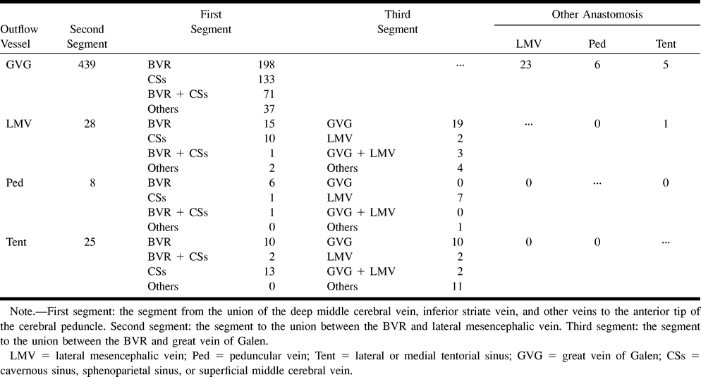
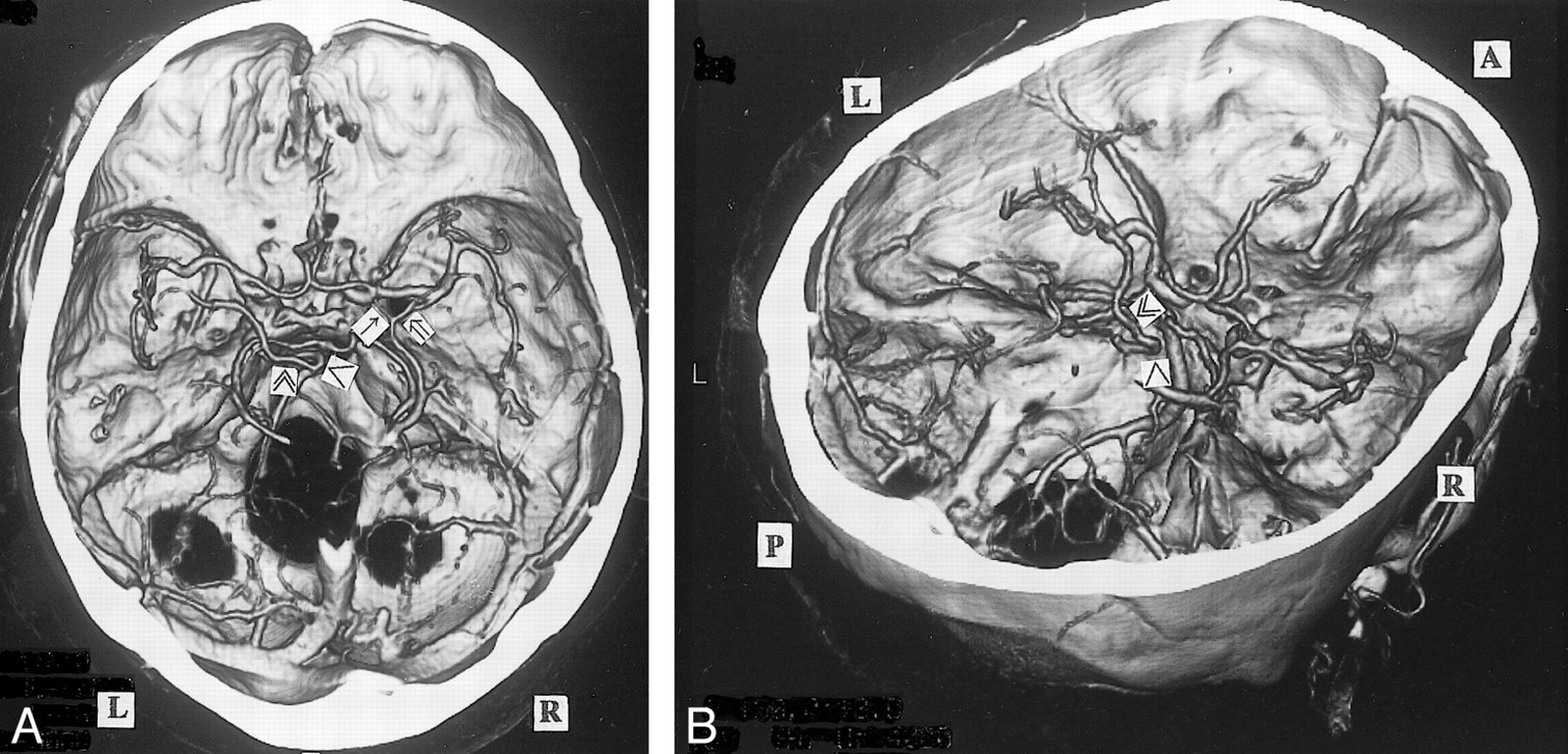
- fig 1.
A, 3D CT angiogram (axial view) and B, MIP image. The left BVR (▴) typically courses posteriorly after receiving the deep middle cerebral vein. The anterior cerebral vein cannot be identified (A). The right BVR (▵) is not depicted by 3D CT angiography (A), but is seen by MIP imaging (B)
- fig 2.
A, 3D CT angiogram (axial view) and B, MIP image. The left BVR (▴) enters the great vein of Galen, but communicates via the uncal vein () with the superficial middle cerebral vein (). The right deep middle cerebral vein (⇒) joins the superficial middle cerebral vein () and enters the cavernous sinus without forming the anastomosis between the first and second segment. This anastomosis is not visible on the MIP image
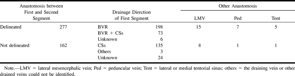
- fig 3.
A, 3D CT angiogram (axial view) and B, posteroanterior stereoscopic views. The right anterior cerebral vein (↑) and the deep middle cerebral vein (⇒) enter the sphenoparietal sinus via the uncal vein () without forming the anastomosis between the first and second segment, and also communicate through the peduncular vein (≫) with the contralateral BVR and the anterior pontomesencephalic vein (>). The left BVR communicates via the uncal vein () with the cavernous sinus, via the deep middle cerebral vein with the insular vein () and the superficial middle cerebral vein (), via the peduncular vein (≫) with the anterior pontomesencephalic vein (>), and the great vein of Galen posteriorly
- fig 4.
3D CT angiogram (axial view). The right BVR flows through the lateral mesencephalic vein () to the superior petrosal sinus (SuPS) and enters the transverse sinus via the lateral or medial tentorial sinus (). The left BVR enters the great vein of Galen () and also flows into the lateral or medial tentorial sinus ()
- fig 5.
A, 3D CT angiogram (axial view) and B, posteroanterior stereoscopic views. The left BVR communicates with the great vein of Galen and lateral mesencephalic vein (). The inferior ventricular veins (∀) originating from the choroid plexuses () are clearly seen bilaterally
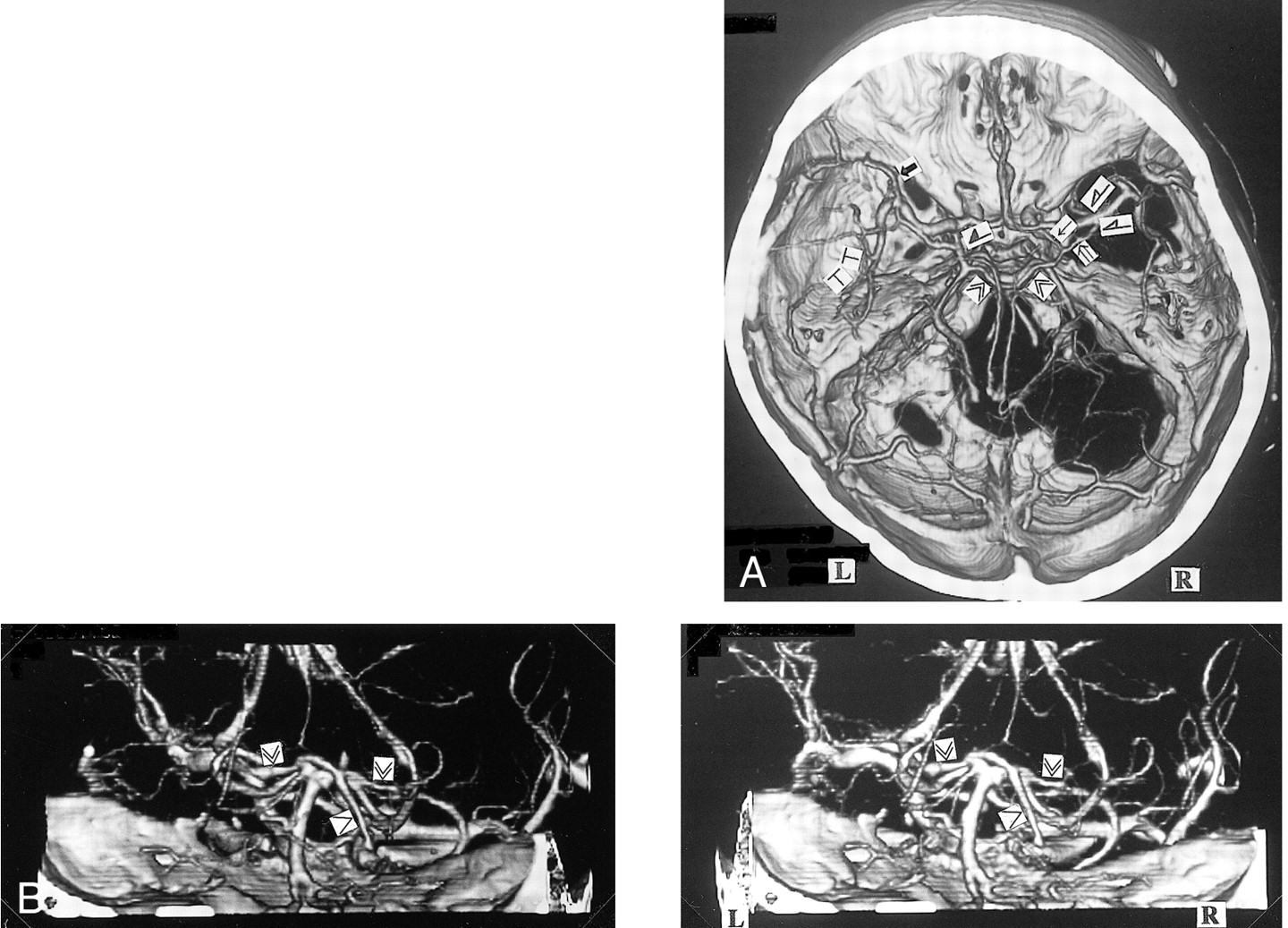
- fig 6.
A, 3D CT angiogram (axial, stereoscopic views) and B, posteroanterior stereoscopic views. The bilateral BVRs drain via the lateral mesencephalic vein () into the superior petrosal sinus. In the presence of a hypoplastic third segment of the BVR, the right inferior ventricular vein (∀) enters the lateral mesencephalic vein. The left third segment enters the great vein of Galen.
- fig 7.
A, 3D CT angiogram (axial view) and B, right posterosuperior oblique view. The left BVR enters the superior petrosal sinus via the peduncular vein (≫) and anterior pontomesencephalic vein (>). The right first segment is formed from the union of the deep middle cerebral vein (⇒) and the anterior cerebral vein (↑)
- fig 8.
3D CT angiogram (axial view). The bilateral BVRs course along the medial edge of the tentorium () and enter the straight sinus. The first segment of the right BVR and the third segment of the left BVR cannot be confirmed
- fig 9.
Four components constituting the BVR and the drainage pathways (1–5). The components develop as the telencephalic vein, diencephalic vein, and mesencephalic vein in the 14- to 18-mm embryonic stage and differentiate to the deep telencephalic vein (A), the ventral diencephalic vein (B), and the dorsal diencephalic vein (C). The BVR is formed by the anastomoses between these components and the mesencephalic vein (D) during the 60- to 80- mm embryonic stage (Padget). Each primitive vein (four vessels) has one or more drainage pathways (five routes). The extent of the anastomoses and the drainage pathways result in a huge number of variations.fig 10. Longitudinal anastomoses of the primitive veins and the drainage routes of the BVR. Five drainage pathways to be considered when the BVR is evaluated. 1: to great vein of Galen, 2: to cavernous sinus or sphenoparietal sinus, 3: to superior petrosal sinus via lateral mesencephalic vein, 4: to superior petrosal sinus via the peduncular vein, 5: to transverse sinus or straight sinus via the tentorium


Tables
 TABLE 1:
Summary of drainage pathways from the basal vein of Rosenthal (BVR)
- TABLE 2:
Summary of the basal vein of Rosenthal variations flowing into great vein of Galen
In this issue


Jump to section
Related Articles
Cited By...
- Endovascular management of anterior falcotentorial dural arteriovenous fistulas: importance of functionality of deep venous system and existence of accompanying choroidal arteriovenous malformation
- Republished: Primary intraventricular haemorrhage due to rupture of giant varix of the basal vein of Rosenthal in a patient with long-standing direct CCF: angiographic features and treatment considerations
- Primary intraventricular haemorrhage due to rupture of giant varix of the basal vein of Rosenthal in a patient with long-standing direct CCF: angiographic features and treatment considerations
- Relationship between cerebral arterial inflow and venous outflow during dynamic supine exercise
- Use of Phase-Contrast MRA to Assess Intracranial Venous Sinus Resistance to Drainage in Healthy Individuals
- Diagnosis and Management of Cerebral Venous Thrombosis: A Statement for Healthcare Professionals From the American Heart Association/American Stroke Association