
Article Figures & Data
Figures
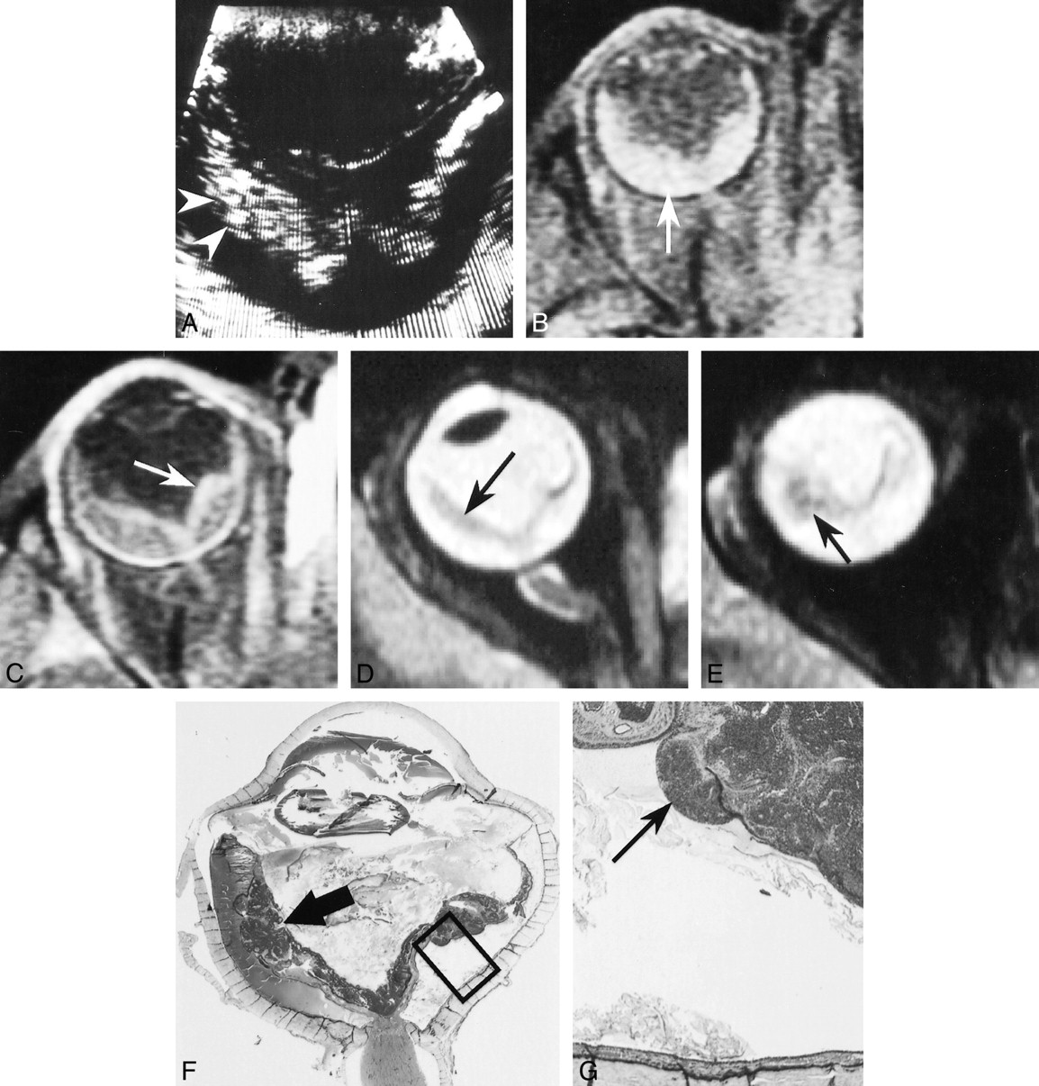
- fig 1.
Patient 1: 3½-year-old boy with leukokoria, posterior synechiae, and total retinal detachment.
A, Sonogram shows detachment and thickening of the retina (arrow).
B, Contrast-enhanced CT scan shows crescent-shaped hyperdensity of the posterior pole due to retinal detachment (arrow), with no calcification.
C and D, Unenhanced (C) and enhanced (D) T1-weighted MR images show retinal detachment containing material with a high signal intensity (arrow, C) and moderate enhancement of the zone of detachment.
E, T2-weighted MR image shows an irregularly thickened, detached retina (arrow).
F and G, Low- (F) and high-power (G) histologic sections show retinoblastoma with an infiltrating component (arrows, G) destroying the retinal architecture (arrowheads, G) and an exophytic component (arrow, F) in the temporal zone of detachment (hematein-eosin-saffron stain, original magnifications ×2 and ×25, respectively). (Box in F denotes magnified area in G.)
- fig 2.
Patient 2: 3-year-old girl with leukokoria, red and painful eye, vitreous opacities, total retinal detachment, and neovascularization of the iris.
A, Sonogram shows detachment and thickening of the retina with two hyperreflective micronodules (arrowheads).
B and C, Unenhanced (B) and enhanced (C) T1-weighted MR images show retinal detachment with frankly hyperintense subretinal space as compared with the vitreous (arrow, B) and locally nodular enhancement (arrow, C) of detached retinal leaflets.
D and E, T2-weighted MR images show detached retina with localized thickening and relatively low signal intensity (arrows).
F and G, Low- (F) and high-power (G) histologic sections show retinoblastoma with infiltrating (arrow, G) and endophytic (arrow, F) components predominantly involving the temporal territory (hematein-eosin-saffron stain, original magnifications ×2 and ×25, respectively). (Box in F denotes magnified area in G.)
- fig 3.
Coats disease in a 2½-year-old boy who presented with strabismus and red eye. Retinal detachment and a whitish mass behind the lens were found on examination of the ocular fundus.
A, Contrast-enhanced CT scan shows large hyperdense detachment (arrow).
B and C, Unenhanced (B) and enhanced (C) T1-weighted MR images show retinal detachment containing material with high signal intensity (arrow, B) and enhancement of the detached retina, forming a mass in the nasal territory (arrow, C).
D, T2-weighted MR image shows the mass has relatively low signal intensity (arrow).
E and F, Low- (F) and high-power (G) histologic sections show Coats disease with retinal detachment, reactive gliosis, and preretinal fibrosis locally constituting a fibrotic block (arrows) containing neovessels (hematein-eosin-saffron stain, original magnifications ×2 and ×25, respectively). (Box in F denotes magnified area in G.)


In this issue


Jump to section
Related Articles
Cited By...
- Diffuse infiltrating retinoblastoma: a multicentre, international, data-sharing study
- Retinoblastoma: What the Neuroradiologist Needs to Know
- Teaching NeuroImages: A diffuse infiltrating retinoblastoma
- MRI of retinoblastoma
- Is CT Still Useful in the Study Protocol of Retinoblastoma?
- Anterior-Segment Retinoblastoma Mimicking Pseudoinflammatory Angle-Closure Glaucoma: Review of the Literature and the Important Role of Imaging