Advertisement

Main menu

  • Home
  • Content
    • Current Issue
    • Accepted Manuscripts
    • Article Preview
    • Past Issue Archive
    • Video Articles
    • AJNR Case Collection
    • Case of the Week Archive
    • Case of the Month Archive
    • Classic Case Archive
  • Special Collections
    • AJNR Awards
    • Low-Field MRI
    • Alzheimer Disease
    • ASNR Foundation Special Collection
    • Photon-Counting CT
    • View All
  • Multimedia
    • AJNR Podcasts
    • AJNR SCANtastic
    • Trainee Corner
    • MRI Safety Corner
    • Imaging Protocols
  • For Authors
    • Submit a Manuscript
    • Submit a Video Article
    • Submit an eLetter to the Editor/Response
    • Manuscript Submission Guidelines
    • Statistical Tips
    • Fast Publishing of Accepted Manuscripts
    • Graphical Abstract Preparation
    • Imaging Protocol Submission
    • Author Policies
  • About Us
    • About AJNR
    • Editorial Board
    • Editorial Board Alumni
  • More
    • Become a Reviewer/Academy of Reviewers
    • Subscribers
    • Permissions
    • Alerts
    • Feedback
    • Advertisers
    • ASNR Home

User menu

  • Alerts
  • Log in

Search

  • Advanced search
American Journal of Neuroradiology

American Journal of Neuroradiology

ASHNR American Society of Functional Neuroradiology ASHNR American Society of Pediatric Neuroradiology ASSR
  • Alerts
  • Log in

Advanced Search

  • Home
  • Content
    • Current Issue
    • Accepted Manuscripts
    • Article Preview
    • Past Issue Archive
    • Video Articles
    • AJNR Case Collection
    • Case of the Week Archive
    • Case of the Month Archive
    • Classic Case Archive
  • Special Collections
    • AJNR Awards
    • Low-Field MRI
    • Alzheimer Disease
    • ASNR Foundation Special Collection
    • Photon-Counting CT
    • View All
  • Multimedia
    • AJNR Podcasts
    • AJNR SCANtastic
    • Trainee Corner
    • MRI Safety Corner
    • Imaging Protocols
  • For Authors
    • Submit a Manuscript
    • Submit a Video Article
    • Submit an eLetter to the Editor/Response
    • Manuscript Submission Guidelines
    • Statistical Tips
    • Fast Publishing of Accepted Manuscripts
    • Graphical Abstract Preparation
    • Imaging Protocol Submission
    • Author Policies
  • About Us
    • About AJNR
    • Editorial Board
    • Editorial Board Alumni
  • More
    • Become a Reviewer/Academy of Reviewers
    • Subscribers
    • Permissions
    • Alerts
    • Feedback
    • Advertisers
    • ASNR Home
  • Follow AJNR on Twitter
  • Visit AJNR on Facebook
  • Follow AJNR on Instagram
  • Join AJNR on LinkedIn
  • RSS Feeds

AJNR Awards, New Junior Editors, and more. Read the latest AJNR updates

Research ArticleNeurointervention

Endovascular Treatment of Experimental Aneurysms by Use of Biologically Modified Embolic Devices: Coil-mediated Intraaneurysmal Delivery of Fibroblast Tissue Allografts

William F. Marx, Harry J. Cloft, Gregory A. Helm, John G. Short, Huy M. Do, Mary E. Jensen and David F. Kallmes
American Journal of Neuroradiology February 2001, 22 (2) 323-333;
William F. Marx
  • Find this author on Google Scholar
  • Find this author on PubMed
  • Search for this author on this site
Harry J. Cloft
  • Find this author on Google Scholar
  • Find this author on PubMed
  • Search for this author on this site
Gregory A. Helm
  • Find this author on Google Scholar
  • Find this author on PubMed
  • Search for this author on this site
John G. Short
  • Find this author on Google Scholar
  • Find this author on PubMed
  • Search for this author on this site
Huy M. Do
  • Find this author on Google Scholar
  • Find this author on PubMed
  • Search for this author on this site
Mary E. Jensen
  • Find this author on Google Scholar
  • Find this author on PubMed
  • Search for this author on this site
David F. Kallmes
  • Find this author on Google Scholar
  • Find this author on PubMed
  • Search for this author on this site
  • Article
  • Figures & Data
  • Info & Metrics
  • Responses
  • References
  • PDF
Loading

Article Figures & Data

Figures

  • Tables
  • fig 1.
    • Download figure
    • Open in new tab
    • Download powerpoint
    fig 1.

    Electron microscopy image of GDC coils co-cultured with fibroblasts. Note fibroblasts growing on surface (A) and within the central lumen (B) of the coils

  • fig 2.
    • Download figure
    • Open in new tab
    • Download powerpoint
    fig 2.

    A, Experimental aneurysm accessed with microcatheter.

    B, Coils deployed under roadmap fluoroscopy.

    C, Control angiogram shows complete occlusion of the aneurysm.

  • fig 3.
    • Download figure
    • Open in new tab
    • Download powerpoint
    fig 3.

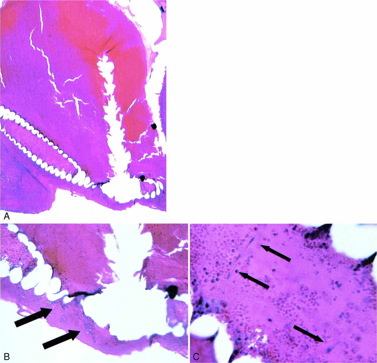
    A, 14-day H&E-stained control aneurysm filled with unorganized thrombus.

    B, 100× magnification shows coil wind surrounded only by RBCs.

  • fig 4.
    • Download figure
    • Open in new tab
    • Download powerpoint
    fig 4.

    A, 40× H&E-stained 3-day sample showing coil bridging aneurysm neck.

    B, 100× H&E-stained slide demonstrating nucleated cells within coil interstices (arrows) adjacent to endothelialized aneurysm neck.

    C, 400× magnification shows fibroblasts (arrows) present within central lumen of coil.

  • fig 5.
    • Download figure
    • Open in new tab
    • Download powerpoint
    fig 5.

    A, 100× H&E-stained 7-day sample. The arrow marks tissue that was present within the central lumen of the implanted coil. The basophilic, nucleated cells in the central region (arrow) are transplanted fibroblasts.

    B, 400× magnification demonstrates fibroblasts (arrows) present within central lumen of coil.

  • fig 6.
    • Download figure
    • Open in new tab
    • Download powerpoint
    fig 6.

    A, 40× H&E-stained view of 14-day speciment embolized with fibroblast-bearing GDC coil. The coil bridged the aneurysm neck, which developed neoendothelial lining.

    B, 100× view of same specimen illustrates fibroblasts (arrow) present within the coil wind interstices and extending into the adjacent thrombus and aneurysm neck.

  • fig 7.
    • Download figure
    • Open in new tab
    • Download powerpoint
    fig 7.

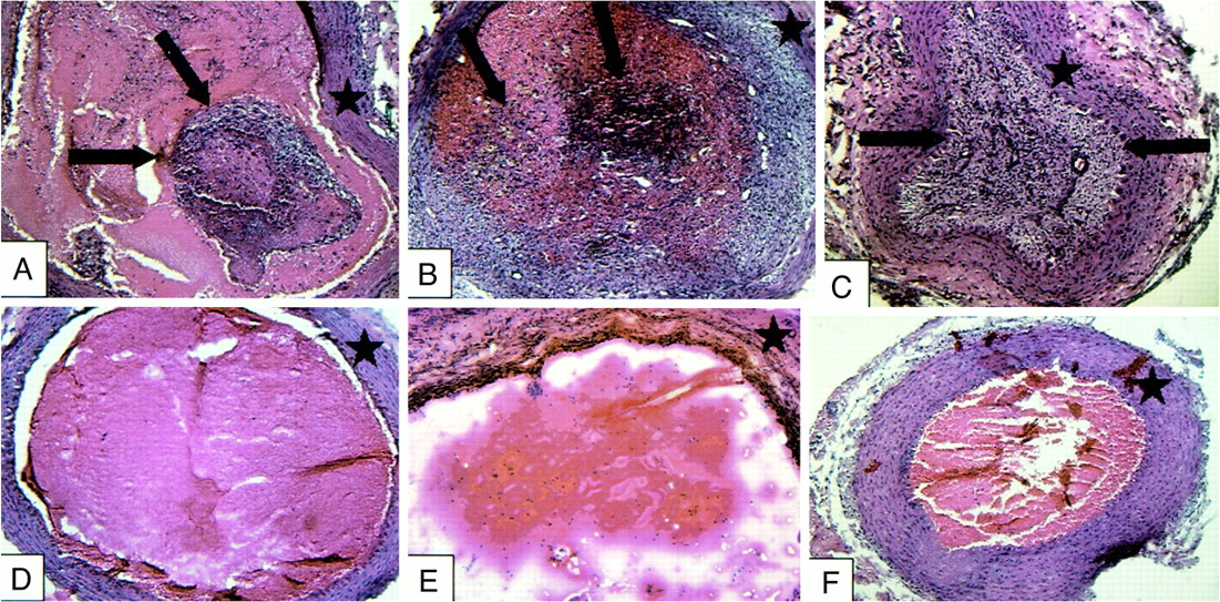
    Treatment vessels containing implanted fibroblasts (A–C) and control vessels (D–F) at 3-day (A and D), 7-day (B and E), and 14-day (C and F) intervals. Treatment vessels show progressive intraluminal cellular proliferation and fibrosis (arrows). Control vessels are filled with unorganized thrombus with scant cellular infiltration. Star indicates vessel wall

  • fig 8.
    • Download figure
    • Open in new tab
    • Download powerpoint
    fig 8.

    H&E (A) and fluorescent micrograph (B) of artery segment implanted 2 weeks previously with fluorescently labeled cells. The lumen of the arterial segment is nearly completely occluded, with a dense cellular infiltrate. The fluorescent images confirm the presence of labeled cells that are most intensely labeled in the central lumen of the vessel. Weak fluorescent labeling of the cells peripherally is likely secondary to the dilution effect on the membrane dye owing to cell division

  • fig 9.
    • Download figure
    • Open in new tab
    • Download powerpoint
    fig 9.

    Estimated percent fibrosis of the treatment and control vessels at each time interval. There was no organized fibrosis in the control vessels at any time point. The treatment vessels had fibroblast proliferation at 3 days, progressing to complete fibrosis of the vessel lumen at 14 days

  • fig 10.
    • Download figure
    • Open in new tab
    • Download powerpoint
    fig 10.

    Segments of GDC coils co-cultured with fluorescence-labeled fibroblasts.

    A, Fluorescent cells in culture adherent to the surface of the coil and extending into interstices of the coil lumen.

    B, Immediately after withdrawal of coil into housing, the cells on the surface are stripped. Cells remain within coil lumen. Coils exposed to systemic arterial flow for 5 minutes (C), 10 minutes (D), 20 minutes (E), and 40 minutes (F). Numerous cells remain at all time intervals. The density of cells is diminished at the 20- and 40-minute time points relative to earlier samples.

  • fig 11.
    • Download figure
    • Open in new tab
    • Download powerpoint
    fig 11.

    Histologic grade immediately adjacent to the implanted coils on the basis of the presence and amount of nucleated fibroblasts. Fibroblasts were seen adjacent to or within the central lumen of coils in eight (88%) of nine anuerysms treated with cell-bearing GDCs. Nucleated cells were present in none of the nine control coil subjects

Tables

  • Figures
  • Table1

    TABLE: Pericoil Histologic Grading

PreviousNext
Back to top

In this issue

American Journal of Neuroradiology
Vol. 22, Issue 2
1 Feb 2001
  • Table of Contents
  • Index by author
Advertisement
Print
Download PDF
Email Article

Thank you for your interest in spreading the word on American Journal of Neuroradiology.

NOTE: We only request your email address so that the person you are recommending the page to knows that you wanted them to see it, and that it is not junk mail. We do not capture any email address.

Enter multiple addresses on separate lines or separate them with commas.
Endovascular Treatment of Experimental Aneurysms by Use of Biologically Modified Embolic Devices: Coil-mediated Intraaneurysmal Delivery of Fibroblast Tissue Allografts
(Your Name) has sent you a message from American Journal of Neuroradiology
(Your Name) thought you would like to see the American Journal of Neuroradiology web site.
CAPTCHA
This question is for testing whether or not you are a human visitor and to prevent automated spam submissions.
Cite this article
William F. Marx, Harry J. Cloft, Gregory A. Helm, John G. Short, Huy M. Do, Mary E. Jensen, David F. Kallmes
Endovascular Treatment of Experimental Aneurysms by Use of Biologically Modified Embolic Devices: Coil-mediated Intraaneurysmal Delivery of Fibroblast Tissue Allografts
American Journal of Neuroradiology Feb 2001, 22 (2) 323-333;

Citation Manager Formats

  • BibTeX
  • Bookends
  • EasyBib
  • EndNote (tagged)
  • EndNote 8 (xml)
  • Medlars
  • Mendeley
  • Papers
  • RefWorks Tagged
  • Ref Manager
  • RIS
  • Zotero
0 Responses
Respond to this article
Share
Bookmark this article
Endovascular Treatment of Experimental Aneurysms by Use of Biologically Modified Embolic Devices: Coil-mediated Intraaneurysmal Delivery of Fibroblast Tissue Allografts
William F. Marx, Harry J. Cloft, Gregory A. Helm, John G. Short, Huy M. Do, Mary E. Jensen, David F. Kallmes
American Journal of Neuroradiology Feb 2001, 22 (2) 323-333;
del.icio.us logo Twitter logo Facebook logo Mendeley logo
  • Tweet Widget
  • Facebook Like
  • Google Plus One

Jump to section

  • Article
    • Abstract
    • Methods
    • Results
    • Part 2
    • Discussion
    • Conclusion
    • Footnotes
    • References
  • Figures & Data
  • Info & Metrics
  • Responses
  • References
  • PDF

Related Articles

  • No related articles found.
  • PubMed
  • Google Scholar

Cited By...

  • Autologous adipose-derived mesenchymal stem cells improve healing of coiled experimental saccular aneurysms: an angiographic and histopathological study
  • Mechanisms of Healing in Coiled Intracranial Aneurysms: A Review of the Literature
  • 1-Hexyl n-cyanoacrylate compound (Neucrylate™ AN), a new treatment for berry aneurysm. III: Initial clinical results
  • Embolization of intracranial aneurysms with second-generation Matrix-2 detachable coils: mid-term and long-term results
  • Bioactivity and bioinactivity: two sides of the same coin
  • Cerecyte versus Platinum Coils in the Treatment of Intracranial Aneurysms: Packing Attenuation and Clinical and Angiographic Midterm Results
  • Endovascular Histologic Effects of Ultrathin Gold- or Vitronectin-Coated Platinum Aneurysm Coils in a Rodent Arterial Occlusion Model: A Preliminary Investigation
  • Morbidity and Mortality Associated with Creation of Elastase-Induced Saccular Aneurysms in a Rabbit Model
  • Control of Aneurysm Volume by Adjusting the Position of Ligation During Creation of Elastase-Induced Aneurysms: A Prospective Study
  • Brain Aneurysms and Arteriovenous Malformations: Advancements and Emerging Treatments in Endovascular Embolization
  • Endovascular Treatment of Experimental Aneurysms by Use of Fibroblast-Coated Platinum Coils: An Angiographic and Histopathologic Study
  • Polyglycolide/Polylactide-Coated Platinum Coils for Patients With Ruptured and Unruptured Cerebral Aneurysms: A Single-Center Experience
  • Matrix and Bioabsorbable Polymeric Coils Accelerate Healing of Intracranial Aneurysms: Long-Term Experimental Study
  • Crossref
  • Google Scholar

This article has not yet been cited by articles in journals that are participating in Crossref Cited-by Linking.

More in this TOC Section

  • Rescue Reentry in Carotid Near-Occlusion
  • Contour Neurovascular System: Five Year Follow Up
  • Effect of SARS-CoV2 on Endovascular Thrombectomy
Show more Neurointervention

Similar Articles

Advertisement

Indexed Content

  • Current Issue
  • Accepted Manuscripts
  • Article Preview
  • Past Issues
  • Editorials
  • Editor's Choice
  • Fellows' Journal Club
  • Letters to the Editor
  • Video Articles

Cases

  • Case Collection
  • Archive - Case of the Week
  • Archive - Case of the Month
  • Archive - Classic Case

More from AJNR

  • Trainee Corner
  • Imaging Protocols
  • MRI Safety Corner
  • Book Reviews

Multimedia

  • AJNR Podcasts
  • AJNR Scantastics

Resources

  • Turnaround Time
  • Submit a Manuscript
  • Submit a Video Article
  • Submit an eLetter to the Editor/Response
  • Manuscript Submission Guidelines
  • Statistical Tips
  • Fast Publishing of Accepted Manuscripts
  • Graphical Abstract Preparation
  • Imaging Protocol Submission
  • Evidence-Based Medicine Level Guide
  • Publishing Checklists
  • Author Policies
  • Become a Reviewer/Academy of Reviewers
  • News and Updates

About Us

  • About AJNR
  • Editorial Board
  • Editorial Board Alumni
  • Alerts
  • Permissions
  • Not an AJNR Subscriber? Join Now
  • Advertise with Us
  • Librarian Resources
  • Feedback
  • Terms and Conditions
  • AJNR Editorial Board Alumni

American Society of Neuroradiology

  • Not an ASNR Member? Join Now

© 2025 by the American Society of Neuroradiology All rights, including for text and data mining, AI training, and similar technologies, are reserved.
Print ISSN: 0195-6108 Online ISSN: 1936-959X

Powered by HighWire