
Article Figures & Data
Figures
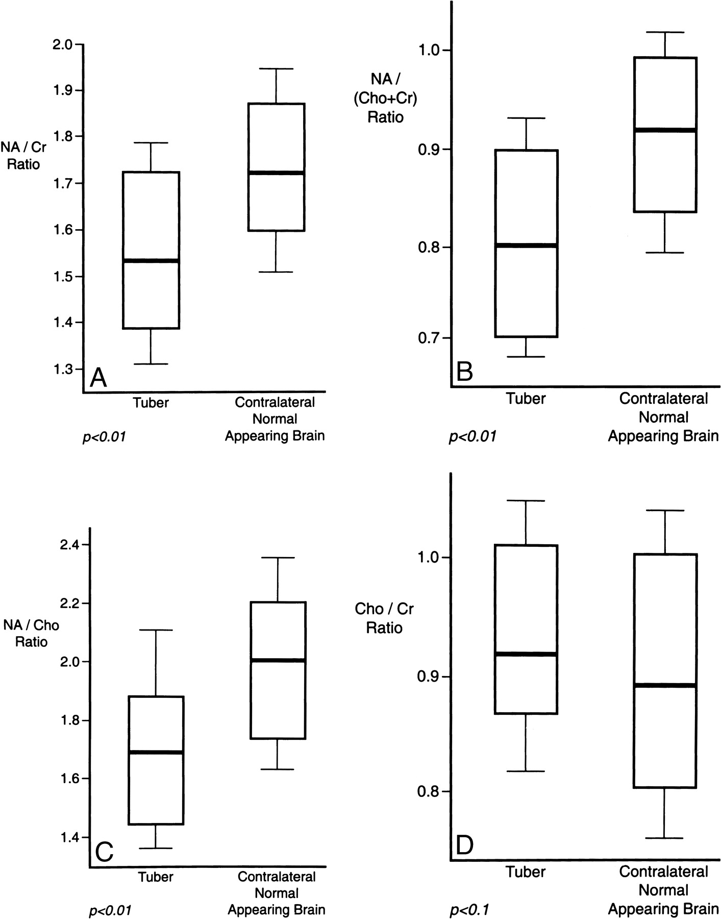
- fig 1.
A–D, Comparisons of mean NA/Cr (A), NA/(Cho+Cr) (B), NA/Cho (C), and Cho/Cr (D) ratios in cortical tubers and contralateral normal-appearing brain. The inferior and superior margins of each box represent the first and third quartiles, the central line is the mean, and the bars at either end represent the standard deviations
- fig 2.
Typical normal spectrum from patient with TSC.
A, Axial fluid-attenuated inversion recovery (FLAIR) image shows the sampled region of interest over an area of normal-appearing brain in the cerebral hemisphere contralateral to the cortical tuber.
B, MR spectrum shows that the Cho and Cr peaks are approximately equal in height while the NA peak is approximately double the height of the Cr peak.
C, Axial FLAIR image shows a region of interest for spectroscopic analysis centered over a typical cortical tuber.
D, MR spectrum from the cortical tuber shows that the Cho and Cr peaks are identical in height to those of normal-appearing brain. There is, however, a marked reduction in the height of the NA peak.
- fig 3.
9-year-old girl thought to have TSC by the referring pediatrician. A calcified lesion was found in the anterior left temporal lobe on CT (not shown).
A, Axial FLAIR MR image shows a high-signal-intensity abnormality in the left temporal lobe that corresponds to the area of calcification on CT scan.
B, Coronal contrast-enhanced T1-weighted image shows no enhancement.
C, Proton MR spectrum of this lesion shows an increase in Cho and a marked reduction in the NA peak, in keeping with a neoplasm rather than a cortical tuber. Review by pediatric neurologists and dermatologists confirmed that TSC was not present clinically.
D and E, Follow-up MR study 6 months later shows the lesion has increased in size on FLAIR image (D), but still does not enhance after contrast administration (E). A diagnosis of low-grade astrocytoma was made on the basis of biopsy findings.


