Advertisement

Main menu

  • Home
  • Content
    • Current Issue
    • Accepted Manuscripts
    • Article Preview
    • Past Issue Archive
    • Video Articles
    • AJNR Case Collection
    • Case of the Week Archive
    • Case of the Month Archive
    • Classic Case Archive
  • Special Collections
    • AJNR Awards
    • Low-Field MRI
    • Alzheimer Disease
    • ASNR Foundation Special Collection
    • Photon-Counting CT
    • View All
  • Multimedia
    • AJNR Podcasts
    • AJNR SCANtastic
    • Trainee Corner
    • MRI Safety Corner
    • Imaging Protocols
  • For Authors
    • Submit a Manuscript
    • Submit a Video Article
    • Submit an eLetter to the Editor/Response
    • Manuscript Submission Guidelines
    • Statistical Tips
    • Fast Publishing of Accepted Manuscripts
    • Graphical Abstract Preparation
    • Imaging Protocol Submission
    • Author Policies
  • About Us
    • About AJNR
    • Editorial Board
    • Editorial Board Alumni
  • More
    • Become a Reviewer/Academy of Reviewers
    • Subscribers
    • Permissions
    • Alerts
    • Feedback
    • Advertisers
    • ASNR Home

User menu

  • Alerts
  • Log in

Search

  • Advanced search
American Journal of Neuroradiology

American Journal of Neuroradiology

ASHNR American Society of Functional Neuroradiology ASHNR American Society of Pediatric Neuroradiology ASSR
  • Alerts
  • Log in

Advanced Search

  • Home
  • Content
    • Current Issue
    • Accepted Manuscripts
    • Article Preview
    • Past Issue Archive
    • Video Articles
    • AJNR Case Collection
    • Case of the Week Archive
    • Case of the Month Archive
    • Classic Case Archive
  • Special Collections
    • AJNR Awards
    • Low-Field MRI
    • Alzheimer Disease
    • ASNR Foundation Special Collection
    • Photon-Counting CT
    • View All
  • Multimedia
    • AJNR Podcasts
    • AJNR SCANtastic
    • Trainee Corner
    • MRI Safety Corner
    • Imaging Protocols
  • For Authors
    • Submit a Manuscript
    • Submit a Video Article
    • Submit an eLetter to the Editor/Response
    • Manuscript Submission Guidelines
    • Statistical Tips
    • Fast Publishing of Accepted Manuscripts
    • Graphical Abstract Preparation
    • Imaging Protocol Submission
    • Author Policies
  • About Us
    • About AJNR
    • Editorial Board
    • Editorial Board Alumni
  • More
    • Become a Reviewer/Academy of Reviewers
    • Subscribers
    • Permissions
    • Alerts
    • Feedback
    • Advertisers
    • ASNR Home
  • Follow AJNR on Twitter
  • Visit AJNR on Facebook
  • Follow AJNR on Instagram
  • Join AJNR on LinkedIn
  • RSS Feeds

AJNR Awards, New Junior Editors, and more. Read the latest AJNR updates

Research ArticleINTERVENTIONAL

Aneurysms of the Posterior Cerebral Artery: Classification and Endovascular Treatment

Elisa F. Ciceri, Richard P. Klucznik, Robert G. Grossman, James E. Rose and Michel E. Mawad
American Journal of Neuroradiology January 2001, 22 (1) 27-34;
Elisa F. Ciceri
  • Find this author on Google Scholar
  • Find this author on PubMed
  • Search for this author on this site
Richard P. Klucznik
  • Find this author on Google Scholar
  • Find this author on PubMed
  • Search for this author on this site
Robert G. Grossman
  • Find this author on Google Scholar
  • Find this author on PubMed
  • Search for this author on this site
James E. Rose
  • Find this author on Google Scholar
  • Find this author on PubMed
  • Search for this author on this site
Michel E. Mawad
  • Find this author on Google Scholar
  • Find this author on PubMed
  • Search for this author on this site
  • Article
  • Figures & Data
  • Info & Metrics
  • Responses
  • References
  • PDF
Loading

Article Figures & Data

Figures

  • Tables
  • fig 1.
    • Download figure
    • Open in new tab
    • Download powerpoint
    fig 1.

    Anatomic segments of the PCA and their branches.

    Composite line tracing of all four anatomic segments of the PCA.

  • fig 2.
    • Download figure
    • Open in new tab
    • Download powerpoint
    fig 2.

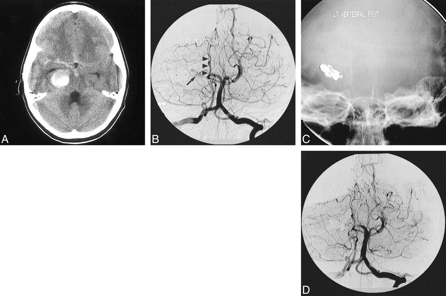
    Giant serpentine aneurysm of the P2A segment of the PCA presenting with intracerebral and subarachnoid hemorrhage in a 12-year-old boy.

    A, Noncontrast CT scan of the head shows the large partially clotted aneurysm in the perimesencephalic cistern, associated with SAH.

    B, Towne's view of the vertebral angiogram shows the residual lumen of the partially clotted aneurysm (arrow), non-filling of the distal branches of the ipsilateral PCA, and avascular mass effect on the brain stem structures (arrowheads).

    C, The lumen of the aneurysm and the parent artery were permanently occluded with GDC. The nonsubtracted Towne's view shows the metallic cast of the coils.

    D, The corresponding subtracted image of the posttreatment vertebral angiogram shows complete obliteration of the aneurysm.

  • fig 3.
    • Download figure
    • Open in new tab
    • Download powerpoint
    fig 3.

    Successful obliteration of a ruptured saccular aneurysm of the PCA with preservation of the parent artery.

    A, The oblique subtracted view of a vertebral angiogram shows an irregular bilobed saccular aneurysm arising from the origin of the P3/P4 segments. The patient is a 52-year-old woman presenting with acute SAH.

    B, The oblique nonsubtracted view of the posttreatment vertebral angiogram shows the cast of the GDC obliterating the aneurysm with preservation of the parent artery.

    C, The oblique subtracted view of the follow-up vertebral angiogram, obtained 2 years after the treatment, shows persistent obliteration of the aneurysm.

  • fig 4.
    • Download figure
    • Open in new tab
    • Download powerpoint
    fig 4.

    Parent artery occlusion of a giant serpentine aneurysm of the PCA complicated by cerebral infarction in the distal arterial territory.

    A, Axial T2-weighted MR image of the head showing a giant serpentine aneurysm of the P2/P3 segments of the left PCA.

    B, Towne's view of a left vertebral angiogram showing the serpentine nature of the giant aneurysm, which involves the P2 and P3 segments of the left PCA.

    C, Subtracted Towne's view of the posttreatment vertebral angiogram shows obliteration of the aneurysm and no antegrade flow in the distal branches of the left PCA. Note the retrograde flow from the cortical branches of the ipsilateral middle cerebral artery (arrows).

    D, Homonymous hemianopsia and contralateral hemiparesis complicated the treatment. The posttreatment axial T2-weighted MR image shows an acute infarction in the distribution of the left PCA territory.

  • fig 5.
    • Download figure
    • Open in new tab
    • Download powerpoint
    fig 5.

    P1 segment aneurysm associated with a distal cortical AVM.

    A, Pretreatment Towne's view of a vertebral angiogram showing a P1 segment aneurysm associated with a temporooccipital AVM. Note the poor ratio of the fundus of the aneurysm to the size of its neck.

    B, Same view of the vertebral angiogram after the endovascular treatment of the aneurysm with GDC

    C, Follow-up vertebral angiogram obtained 42 months after the initial treatment showing complete obliteration of the AVM and questionable minimal residual filling of the aneurysm.

  • fig 6.
    • Download figure
    • Open in new tab
    • Download powerpoint
    fig 6.

    Multiple PCA associated with Moya-Moya disease, successfully treated with GDC.

    A, Frontal view of left internal carotid angiogram showing occlusion of the anterior and a middle cerebral arteries and Moya-Moya collateral circulation. There is increased flow in a dysplastic and enlarged ipsilateral PCA (arrow).

    B, Oblique view of the internal carotid angiogram showing an aneurysm originating from the P1/P2 junction of the left posterior cerebral artery (arrow) and a second aneurysm arising from the P2P segment (arrowhead). Note the presence of at least two flow-related aneurysms arising from the supraclinoid segment of the internal carotid artery (double arrows).

    C, Oblique view of the internal carotid angiogram, obtained 9 months after treatment, shows persistent obliteration of both PCA aneurysms (arrows).

Tables

  • Figures
  • Table1

    TABLE: Summary of 20 patients with 21 PCA aneurysms

PreviousNext
Back to top

In this issue

American Journal of Neuroradiology
Vol. 22, Issue 1
1 Jan 2001
  • Table of Contents
  • Index by author
Advertisement
Print
Download PDF
Email Article

Thank you for your interest in spreading the word on American Journal of Neuroradiology.

NOTE: We only request your email address so that the person you are recommending the page to knows that you wanted them to see it, and that it is not junk mail. We do not capture any email address.

Enter multiple addresses on separate lines or separate them with commas.
Aneurysms of the Posterior Cerebral Artery: Classification and Endovascular Treatment
(Your Name) has sent you a message from American Journal of Neuroradiology
(Your Name) thought you would like to see the American Journal of Neuroradiology web site.
CAPTCHA
This question is for testing whether or not you are a human visitor and to prevent automated spam submissions.
Cite this article
Elisa F. Ciceri, Richard P. Klucznik, Robert G. Grossman, James E. Rose, Michel E. Mawad
Aneurysms of the Posterior Cerebral Artery: Classification and Endovascular Treatment
American Journal of Neuroradiology Jan 2001, 22 (1) 27-34;

Citation Manager Formats

  • BibTeX
  • Bookends
  • EasyBib
  • EndNote (tagged)
  • EndNote 8 (xml)
  • Medlars
  • Mendeley
  • Papers
  • RefWorks Tagged
  • Ref Manager
  • RIS
  • Zotero
0 Responses
Respond to this article
Share
Bookmark this article
Aneurysms of the Posterior Cerebral Artery: Classification and Endovascular Treatment
Elisa F. Ciceri, Richard P. Klucznik, Robert G. Grossman, James E. Rose, Michel E. Mawad
American Journal of Neuroradiology Jan 2001, 22 (1) 27-34;
del.icio.us logo Twitter logo Facebook logo Mendeley logo
  • Tweet Widget
  • Facebook Like
  • Google Plus One

Jump to section

  • Article
    • Abstract
    • Methods
    • Results
    • Discussion
    • Conclusion
    • Footnotes
    • References
  • Figures & Data
  • Info & Metrics
  • Responses
  • References
  • PDF

Related Articles

  • No related articles found.
  • PubMed
  • Google Scholar

Cited By...

  • Micro-WADA and balloon test occlusion for sacrifice of distal P2 aneurysm
  • Saccular posterior cerebral artery aneurysm encased within a lipoma
  • Relationship between cerebral aneurysms and variations in cerebral basal arterial network: a morphometric cross-sectional study in Computed Tomography Angiograms from a neurointerventional unit
  • Radical treatment of ruptured dissecting aneurysm on the P1 segment with monotherapy using multiple LVIS stents
  • Traumatic dissecting pathology of posterior cerebral artery: a report of two cases--aneurysm and pial arteriovenous fistula
  • Endovascular parent artery occlusion of proximal posterior cerebral artery aneurysms: a report of two cases
  • Endoluminal Reconstruction for Nonsaccular Aneurysms of the Proximal Posterior Cerebral Artery with the Pipeline Embolization Device
  • Third-nerve palsy heralding dissecting aneurysm of posterior cerebral artery: digital subtraction angiography and magnetic resonance appearance
  • Crossref
  • Google Scholar

This article has not yet been cited by articles in journals that are participating in Crossref Cited-by Linking.

More in this TOC Section

  • Safety, Efficacy, and Durability of Stent-Assisted Coiling Treatment of M2 (Insular) Segment MCA Aneurysms
  • Endovascular Management of Intracranial Dural AVFs: Transvenous Approach
  • A Meta-analysis of Combined Aspiration Catheter and Stent Retriever versus Stent Retriever Alone for Large-Vessel Occlusion Ischemic Stroke
Show more INTERVENTIONAL

Similar Articles

Advertisement

Indexed Content

  • Current Issue
  • Accepted Manuscripts
  • Article Preview
  • Past Issues
  • Editorials
  • Editor's Choice
  • Fellows' Journal Club
  • Letters to the Editor
  • Video Articles

Cases

  • Case Collection
  • Archive - Case of the Week
  • Archive - Case of the Month
  • Archive - Classic Case

More from AJNR

  • Trainee Corner
  • Imaging Protocols
  • MRI Safety Corner
  • Book Reviews

Multimedia

  • AJNR Podcasts
  • AJNR Scantastics

Resources

  • Turnaround Time
  • Submit a Manuscript
  • Submit a Video Article
  • Submit an eLetter to the Editor/Response
  • Manuscript Submission Guidelines
  • Statistical Tips
  • Fast Publishing of Accepted Manuscripts
  • Graphical Abstract Preparation
  • Imaging Protocol Submission
  • Evidence-Based Medicine Level Guide
  • Publishing Checklists
  • Author Policies
  • Become a Reviewer/Academy of Reviewers
  • News and Updates

About Us

  • About AJNR
  • Editorial Board
  • Editorial Board Alumni
  • Alerts
  • Permissions
  • Not an AJNR Subscriber? Join Now
  • Advertise with Us
  • Librarian Resources
  • Feedback
  • Terms and Conditions
  • AJNR Editorial Board Alumni

American Society of Neuroradiology

  • Not an ASNR Member? Join Now

© 2025 by the American Society of Neuroradiology All rights, including for text and data mining, AI training, and similar technologies, are reserved.
Print ISSN: 0195-6108 Online ISSN: 1936-959X

Powered by HighWire