
Article Figures & Data
Figures
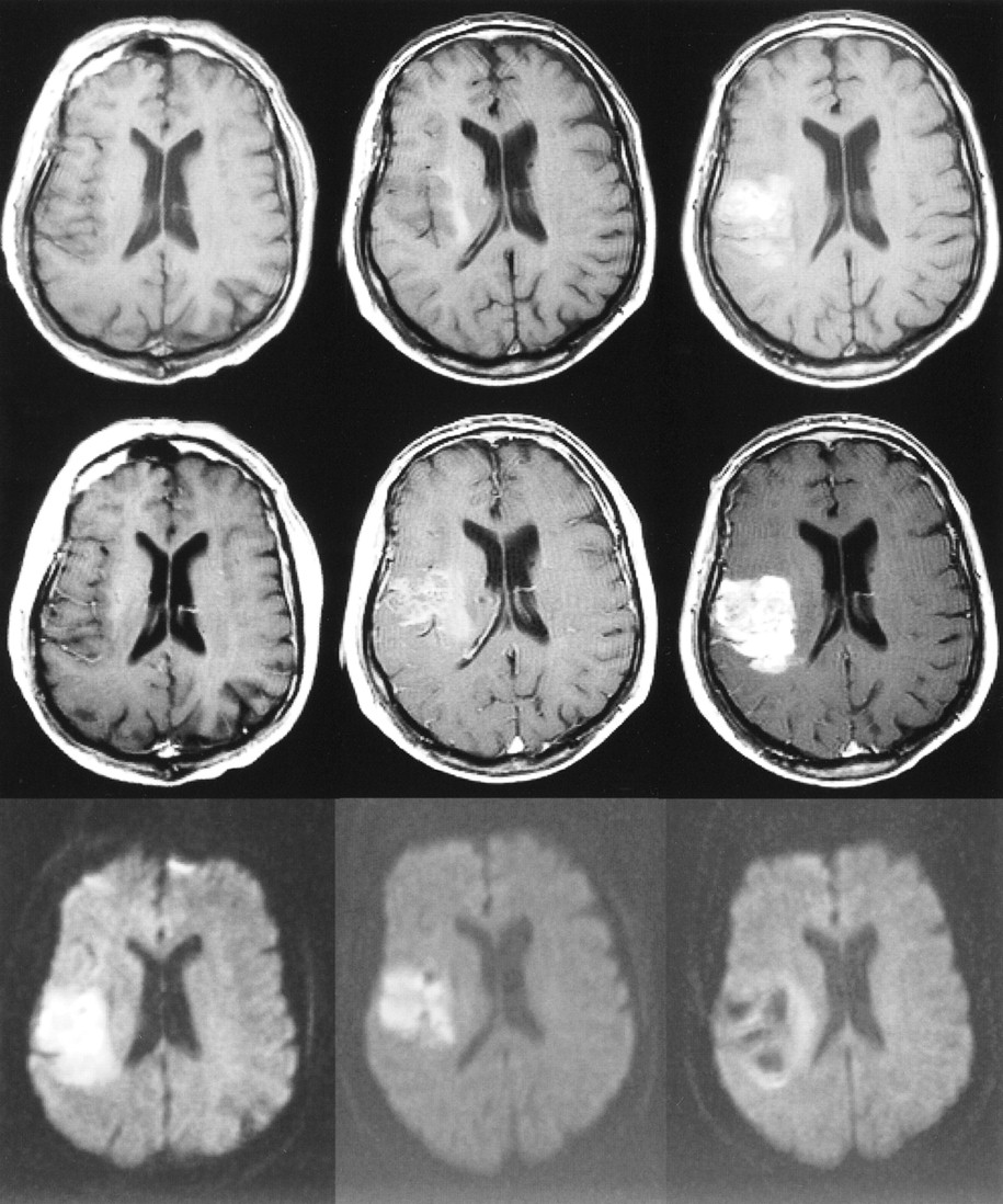
- fig 1.
75-year-old woman with dysphasia and weakness in the right arm. The first MR examination (left column) was performed 8 hours after the onset of symptoms, the second MR examination (middle column) was performed on the second day, and the third MR examination (right column) 1 week after stroke. The slice positions are not exactly the same but this does not change the interpretation.
Top row, Unenhanced T1-weighted images (600/14/1) show progressing low signal mass effect of the infarcted tissue.
Middle row, Contrast-enhanced T1-weighted images (600/14/1) show intravascular enhancement over the infarct on the first and second days and moderate cortical and subcortical enhancement 1 week after stroke.
Bottom row, Diffusion-weighted trace images (4000/103/1, in raw image acquisition) show the extent of infarcted tissue as areas of increased signal intensity (bright) in the territory of the left middle cerebral artery.
- fig 2.
63-year-old woman with right hemiparesis and aphasia. The first MR examination (left column) was performed 6.5 hours after the onset of symptoms, the second MR examination (middle column) was performed on the second day, and the third MR examination (right column) 1 week after stroke.
Top row, Unenhanced T1-weighted images (600/14/1) show progressing low signal mass effect of the infarcted tissue. Hemorrhagic transformation is detected as areas of increased signal intensity on the second day and at 1 week. The gyral pattern of increased signal intensity at 1 week may also represent cortical laminar necrosis.
Middle row, Contrast-enhanced T1-weighted images (600/14/1) show intravascular enhancement over the infarct on the first and second days. Moderate cortical and deep enhancement is detected at 1 week.
Bottom row, Diffusion-weighted trace images (4000/103/1, in raw image acquisition) show the extent of infarcted tissue as areas of increased signal intensity in the territory of the left middle cerebral artery. Hemorrhagic transformation is detected as a dark area due to the susceptibility effect of breakdown products of hemoglobin.
- fig 3.
71-year-old man with left hemiparesis. The first MR examination (left column) was performed 15 hours after the onset of symptoms, the second MR examination (middle column) was performed on the second day, and the third MR examination (right column) 1 week after stroke.
Top row, Unenhanced T1-weighted images (600/14/1) show progressing low signal mass effect of the infarcted tissue. Hemorrhagic transformation is detected as areas of increased signal intensity on the second day and at 1 week.
Middle row, Contrast-enhanced T1-weighted images (600/14/1) show intravascular enhancement over the infarct on the first and second days. Moderate cortical and subcortical enhancement are detected on the second day. Intense parenchymal enhancement is detected in the infarct at 1 week.
Bottom row, Diffusion-weighted trace images (4000/103/1, in raw image acquisition) show the extent of infarcted tissue as areas of increased signal intensity in the territory of the right middle cerebral artery. Hemorrhagic transformation is detected as dark areas due to the susceptibility effect of the breakdown products of hemoglobin.
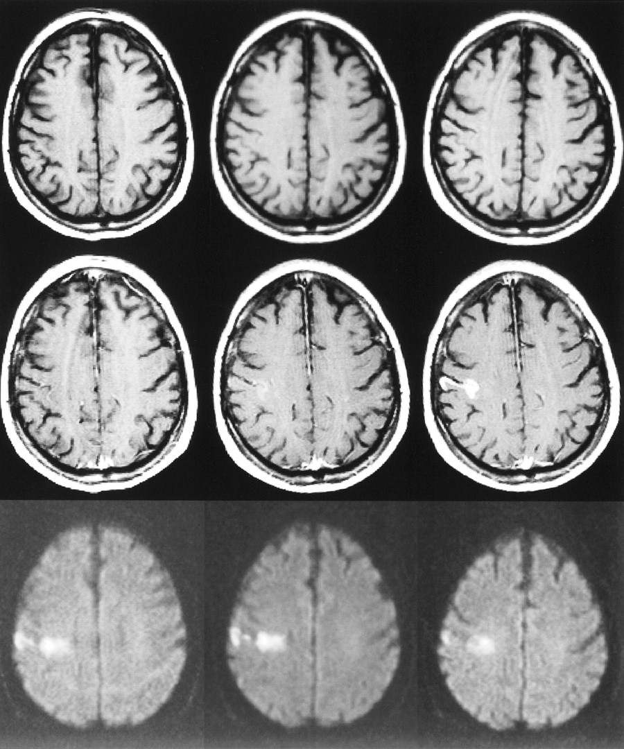
- fig 4.
73-year-old man with left hemiparesis. The first MR examination (left column) was performed 23 hours after the onset of symptoms, the second MR examination (middle column) was performed on the second day, and the third MR examination (right column) 1 week after stroke.
Top row, Unenhanced T1-weighted images (600/14/1) are normal on the first and second days, but at 1 week, slightly increased signal intensity is observed in the cortex due to microhemorrhage or cortical laminar necrosis.
Middle row, Contrast-enhanced T1-weighted images (600/14/1) show intravascular enhancement over the infarct on the first day. Mild cortical enhancement is detected on the second day, progressing to intensive cortical enhancement at 1 week.
Bottom row, Diffusion-weighted trace images (4000/103/1, in raw image acquisition) show the extent of infarcted tissue as areas of increased signal intensity in the territory of the right middle cerebral artery.


Tables
 TABLE 1:
Patterns of contrast enhancement during the first week after stroke onset
- TABLE 2:
Intensities of parenchymal contrast enhancement during the first week after stroke onset


In this issue


Jump to section
Related Articles
Cited By...
- Hyperperfusion and Blood-Brain Barrier Disruption beyond the Diffusion-Restricted Infarct 1 Day after Successful Mechanical Thrombectomy
- Comprehensive Update and Review of Clinical and Imaging Features of SMART Syndrome
- Comprehensive Update and Review of Clinical and Imaging Features of SMART Syndrome
- Clinical biomarkers differentiate myelitis from vascular and other causes of myelopathy
- Delayed enhancing lesions after coil embolization of aneurysms: clinical experience and benchtop analyses
- DCE-MRI blood-brain barrier assessment in acute ischemic stroke
- Effects of Microvascular Permeability Changes on Contrast-Enhanced T1 and Pharmacokinetic MR Imagings After Ischemia
- Restricted diffusion preceding gadolinium enhancement in large or tumefactive demyelinating lesions
- Hemorrhagic Risk of Recent Silent Cerebral Infarct on Prethrombolysis MR Imaging in Acute Stroke