
Article Figures & Data
Figures
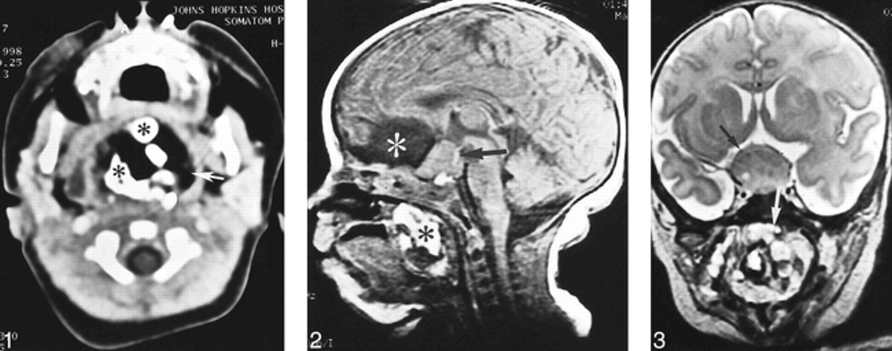
- fig 1.
Contrast-enhanced CT of the neck shows a large, heterogeneous, nasopharyngeal mass with fatty (arrow) and calcified (asterisk) densities compatible with a teratoma.fig 2. Midsagittal T1-weighted (450/12 [TR/TE]) MR image reveals a large well-defined mass in the suprasellar region (arrow) that is isointense to gray matter. An interhemispheric cyst, which is isointense to CSF (white asterisk), is also seen anterior to the suprasellar mass. Another heterogeneous mass with foci of T1 hyperintensities (black asterisk) fills the nasopharynx.fig 3. Coronal fast spin-echo T2-weighted (6000/98 [TR/TEeff]) MR image through the sellar area shows a large mass (black arrow) isointense to gray matter. The optic nerves and chiasm are not recognized because of severe compression and displacement. In the nasopharynx, a mixed signal mass (white arrow) with foci of low and high signal intensity is noted
- fig 4.
High-power microphotograph of the tissue specimen stained with hematoxylin and eosin shows molecular layer (asterisk) and granular cell layer (large arrow) separated by a monolayer of Purkinje cells (small arrow)


