Advertisement

Main menu

  • Home
  • Content
    • Current Issue
    • Accepted Manuscripts
    • Article Preview
    • Past Issue Archive
    • Video Articles
    • AJNR Case Collection
    • Case of the Week Archive
    • Case of the Month Archive
    • Classic Case Archive
  • Special Collections
    • AJNR Awards
    • Low-Field MRI
    • Alzheimer Disease
    • ASNR Foundation Special Collection
    • Photon-Counting CT
    • View All
  • Multimedia
    • AJNR Podcasts
    • AJNR SCANtastic
    • Trainee Corner
    • MRI Safety Corner
    • Imaging Protocols
  • For Authors
    • Submit a Manuscript
    • Submit a Video Article
    • Submit an eLetter to the Editor/Response
    • Manuscript Submission Guidelines
    • Statistical Tips
    • Fast Publishing of Accepted Manuscripts
    • Graphical Abstract Preparation
    • Imaging Protocol Submission
    • Author Policies
  • About Us
    • About AJNR
    • Editorial Board
    • Editorial Board Alumni
  • More
    • Become a Reviewer/Academy of Reviewers
    • Subscribers
    • Permissions
    • Alerts
    • Feedback
    • Advertisers
    • ASNR Home

User menu

  • Alerts
  • Log in

Search

  • Advanced search
American Journal of Neuroradiology

American Journal of Neuroradiology

ASHNR American Society of Functional Neuroradiology ASHNR American Society of Pediatric Neuroradiology ASSR
  • Alerts
  • Log in

Advanced Search

  • Home
  • Content
    • Current Issue
    • Accepted Manuscripts
    • Article Preview
    • Past Issue Archive
    • Video Articles
    • AJNR Case Collection
    • Case of the Week Archive
    • Case of the Month Archive
    • Classic Case Archive
  • Special Collections
    • AJNR Awards
    • Low-Field MRI
    • Alzheimer Disease
    • ASNR Foundation Special Collection
    • Photon-Counting CT
    • View All
  • Multimedia
    • AJNR Podcasts
    • AJNR SCANtastic
    • Trainee Corner
    • MRI Safety Corner
    • Imaging Protocols
  • For Authors
    • Submit a Manuscript
    • Submit a Video Article
    • Submit an eLetter to the Editor/Response
    • Manuscript Submission Guidelines
    • Statistical Tips
    • Fast Publishing of Accepted Manuscripts
    • Graphical Abstract Preparation
    • Imaging Protocol Submission
    • Author Policies
  • About Us
    • About AJNR
    • Editorial Board
    • Editorial Board Alumni
  • More
    • Become a Reviewer/Academy of Reviewers
    • Subscribers
    • Permissions
    • Alerts
    • Feedback
    • Advertisers
    • ASNR Home
  • Follow AJNR on Twitter
  • Visit AJNR on Facebook
  • Follow AJNR on Instagram
  • Join AJNR on LinkedIn
  • RSS Feeds

AJNR Awards, New Junior Editors, and more. Read the latest AJNR updates

Research ArticleBRAIN

Applicability and Advantages of Flow artifact–insensitive Fluid-attenuated Inversion-recovery MR Sequences for Imaging the Posterior Fossa

Norimitsu Tanaka, Toshi Abe, Kazuyuki Kojima, Hiroshi Nishimura and Naofumi Hayabuchi
American Journal of Neuroradiology June 2000, 21 (6) 1095-1098;
Norimitsu Tanaka
  • Find this author on Google Scholar
  • Find this author on PubMed
  • Search for this author on this site
Toshi Abe
  • Find this author on Google Scholar
  • Find this author on PubMed
  • Search for this author on this site
Kazuyuki Kojima
  • Find this author on Google Scholar
  • Find this author on PubMed
  • Search for this author on this site
Hiroshi Nishimura
  • Find this author on Google Scholar
  • Find this author on PubMed
  • Search for this author on this site
Naofumi Hayabuchi
  • Find this author on Google Scholar
  • Find this author on PubMed
  • Search for this author on this site
  • Article
  • Figures & Data
  • Info & Metrics
  • Responses
  • References
  • PDF
Loading

Article Figures & Data

Figures

  • fig 1.
    • Download figure
    • Open in new tab
    • Download powerpoint
    fig 1.

    Conventional FLAIR images showing artifacts in the cerebellopontine angle cistern (A) and in the third ventricle (B). By combining the information from two reverse-ordered sequences, Scan 1 (C, D) and Scan 2 (E, F), the FAIS-FLAIR images result in a conspicuous absence of flow-related FLAIR artifacts (G, H). It is a simple summation of images, which are processed automatically in line with the image-array processor. Note flow-related artifacts lessen on Scan 1 and Scan 2, but image contrast is not constant. On Scan 1, signal intensity is increasing from C to D, whereas on Scan 2, it is decreasing from E to F.
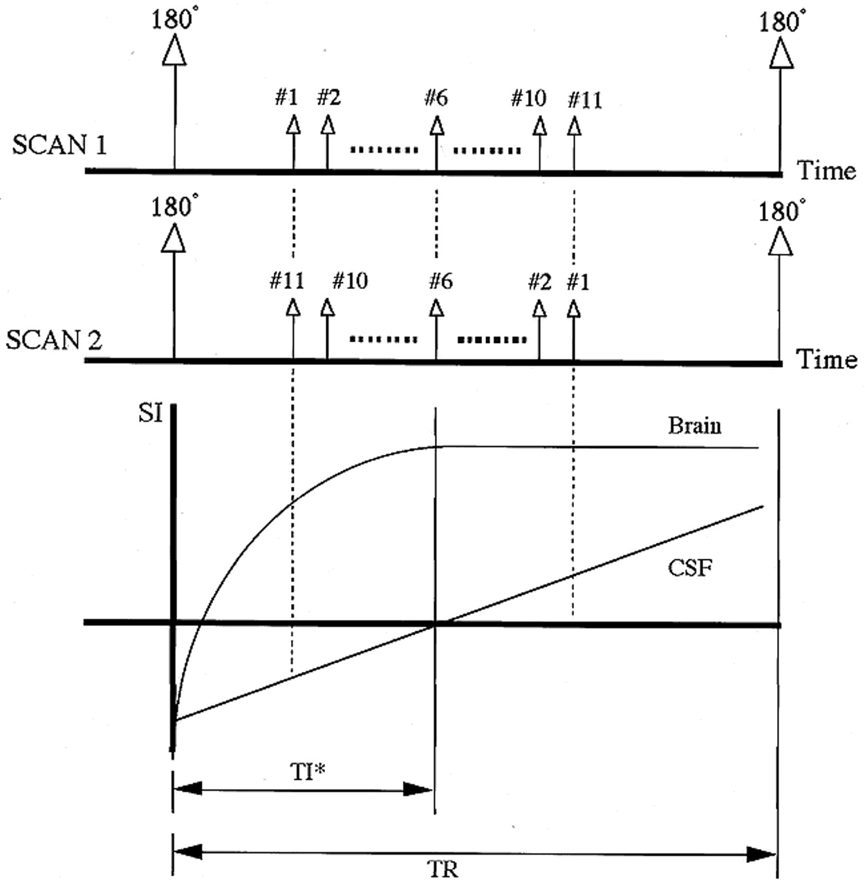
  • fig 2.
    • Download figure
    • Open in new tab
    • Download powerpoint
    fig 2.

    The FAIS-FLAIR sequence consisted of two scans. On the first scan, a 180° inversion prepulse is nonselectively applied to the whole brain, and is then followed by a multislice fast spin-echo sequence. The intent of the prepulse is to null the water-dependent signal by inducing a delay before the center-slice ex

  • fig 3.
    • Download figure
    • Open in new tab
    • Download powerpoint
    fig 3.

    A vestibular schwannoma is demonstrated on multiple images in a 58-year-old woman, including unenhanced conventional FLAIR (A), unenhanced (B) and enhanced (C) FAIS-FLAIR, and T1-weighted (D) images. FAIS-FLAIR shows the tumor in the cerebellopontine cistern more clearly than does T1-weighted imaging. The same lesion is even more visible with postcontrast FAIS-FLAIR imaging. On the FLAIR image, the tumor is depicted, but inflow artifacts in the cerebellopontine and prepontine cistern interfere with interpretation citation. The second scan consists of parameters similar to the first, with the exception that each slice is excited in reverse order. TI* means the interval between 180° inversion prepulse and the excitation pulse of the center slice

  • fig 4.
    • Download figure
    • Open in new tab
    • Download powerpoint
    fig 4.

    The T1 shortening effect with MR contrast agents results in an upward shift of the T1 relaxation curve during FLAIR imaging

PreviousNext
Back to top

In this issue

American Journal of Neuroradiology
Vol. 21, Issue 6
1 Jun 2000
  • Table of Contents
  • Index by author
Advertisement
Print
Download PDF
Email Article

Thank you for your interest in spreading the word on American Journal of Neuroradiology.

NOTE: We only request your email address so that the person you are recommending the page to knows that you wanted them to see it, and that it is not junk mail. We do not capture any email address.

Enter multiple addresses on separate lines or separate them with commas.
Applicability and Advantages of Flow artifact–insensitive Fluid-attenuated Inversion-recovery MR Sequences for Imaging the Posterior Fossa
(Your Name) has sent you a message from American Journal of Neuroradiology
(Your Name) thought you would like to see the American Journal of Neuroradiology web site.
CAPTCHA
This question is for testing whether or not you are a human visitor and to prevent automated spam submissions.
Cite this article
Norimitsu Tanaka, Toshi Abe, Kazuyuki Kojima, Hiroshi Nishimura, Naofumi Hayabuchi
Applicability and Advantages of Flow artifact–insensitive Fluid-attenuated Inversion-recovery MR Sequences for Imaging the Posterior Fossa
American Journal of Neuroradiology Jun 2000, 21 (6) 1095-1098;

Citation Manager Formats

  • BibTeX
  • Bookends
  • EasyBib
  • EndNote (tagged)
  • EndNote 8 (xml)
  • Medlars
  • Mendeley
  • Papers
  • RefWorks Tagged
  • Ref Manager
  • RIS
  • Zotero
0 Responses
Respond to this article
Share
Bookmark this article
Applicability and Advantages of Flow artifact–insensitive Fluid-attenuated Inversion-recovery MR Sequences for Imaging the Posterior Fossa
Norimitsu Tanaka, Toshi Abe, Kazuyuki Kojima, Hiroshi Nishimura, Naofumi Hayabuchi
American Journal of Neuroradiology Jun 2000, 21 (6) 1095-1098;
del.icio.us logo Twitter logo Facebook logo Mendeley logo
  • Tweet Widget
  • Facebook Like
  • Google Plus One

Jump to section

  • Article
    • Abstract
    • Description of Technique and Results
    • Discussion
    • Acknowledgments
    • Footnotes
    • References
  • Figures & Data
  • Info & Metrics
  • Responses
  • References
  • PDF

Related Articles

  • No related articles found.
  • PubMed
  • Google Scholar

Cited By...

  • 3D Fluid-Attenuated Inversion Recovery Imaging: Reduced CSF Artifacts and Enhanced Sensitivity and Specificity for Subarachnoid Hemorrhage
  • Crossref
  • Google Scholar

This article has not yet been cited by articles in journals that are participating in Crossref Cited-by Linking.

More in this TOC Section

  • Quiet PROPELLER MRI Techniques Match the Quality of Conventional PROPELLER Brain Imaging Techniques
  • Predictors of Reperfusion in Patients with Acute Ischemic Stroke
  • Enhanced Axonal Metabolism during Early Natalizumab Treatment in Relapsing-Remitting Multiple Sclerosis
Show more Brain

Similar Articles

Advertisement

Indexed Content

  • Current Issue
  • Accepted Manuscripts
  • Article Preview
  • Past Issues
  • Editorials
  • Editor's Choice
  • Fellows' Journal Club
  • Letters to the Editor
  • Video Articles

Cases

  • Case Collection
  • Archive - Case of the Week
  • Archive - Case of the Month
  • Archive - Classic Case

More from AJNR

  • Trainee Corner
  • Imaging Protocols
  • MRI Safety Corner
  • Book Reviews

Multimedia

  • AJNR Podcasts
  • AJNR Scantastics

Resources

  • Turnaround Time
  • Submit a Manuscript
  • Submit a Video Article
  • Submit an eLetter to the Editor/Response
  • Manuscript Submission Guidelines
  • Statistical Tips
  • Fast Publishing of Accepted Manuscripts
  • Graphical Abstract Preparation
  • Imaging Protocol Submission
  • Evidence-Based Medicine Level Guide
  • Publishing Checklists
  • Author Policies
  • Become a Reviewer/Academy of Reviewers
  • News and Updates

About Us

  • About AJNR
  • Editorial Board
  • Editorial Board Alumni
  • Alerts
  • Permissions
  • Not an AJNR Subscriber? Join Now
  • Advertise with Us
  • Librarian Resources
  • Feedback
  • Terms and Conditions
  • AJNR Editorial Board Alumni

American Society of Neuroradiology

  • Not an ASNR Member? Join Now

© 2025 by the American Society of Neuroradiology All rights, including for text and data mining, AI training, and similar technologies, are reserved.
Print ISSN: 0195-6108 Online ISSN: 1936-959X

Powered by HighWire