
Article Figures & Data
Figures
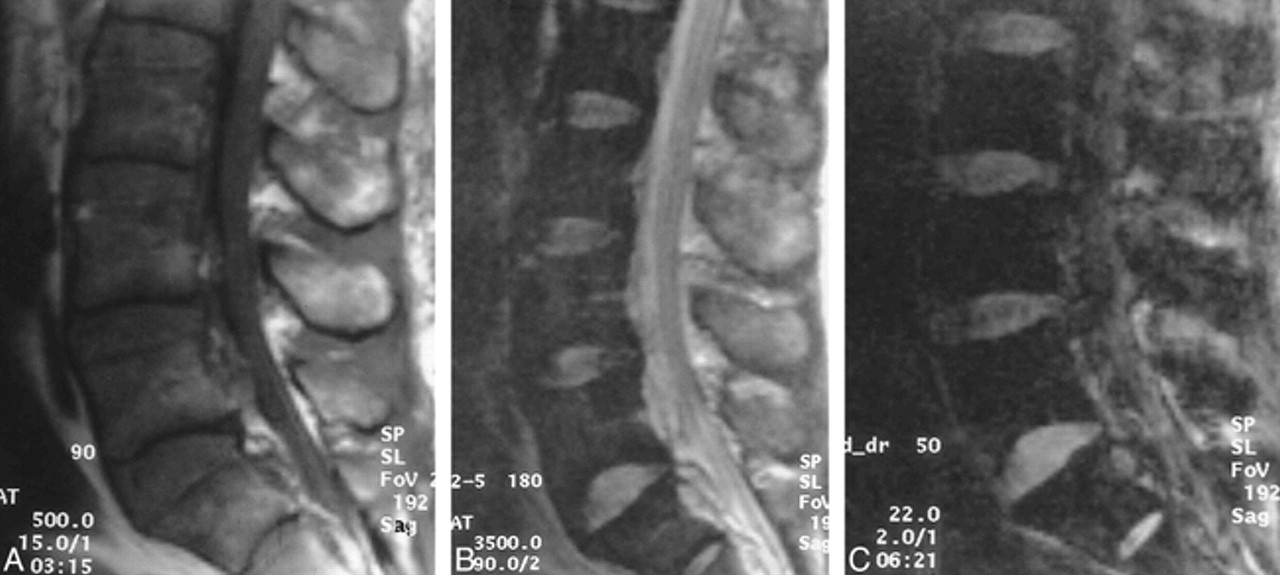
- fig 1.
Involvement of a single vertebra.
A, Midsagittal T1-weighted image in a patient 6 months after spine radiation for breast metastases who presented with new pain and a positive radionuclide bone scan. This image shows hypointensity and partial wedge deformity of T7 (arrow). Note that vertebrae above and below the lesion are hyperintense as a result of prior radiation therapy.
B, Corresponding T2-weighted image shows lesion (arrow) to be minimally hypointense with respect to presumed normal bone marrow above and below.
C, Corresponding diffusion-weighted image shows that T7 (arrow) is hypointense relative to other vertebrae. Although the abnormality is perhaps seen better on diffusion-weighted image than on the T2-weighted image, the diffusion-weighted image is not superior to the T1-weighted image.
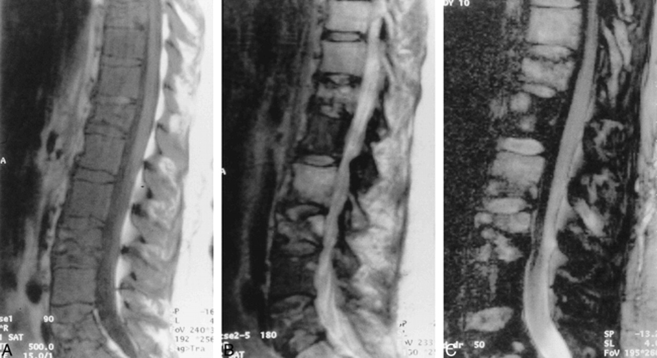
- fig 2.
Patient with diffuse bone metastases from prostate cancer, with hypointensity on all sequences.
A, Midsagittal T1-weighted image shows diffuse and mostly hypointense signal intensity in lower lumbar and sacral vertebrae.
B, Corresponding T2-weighted image shows that the vertebrae are diffusely hypointense.
C, Corresponding diffusion-weighted image shows that the vertebrae involved by metastases are hypointense. This hypointensity on all sequences could be related to sclerosis seen on radiographs of this region. The abnormality is more difficult to perceive on diffusion-weighted image than on T1-weighted image.
- fig 3.
Patient with diffuse involvement from an unknown primary tumor, with hyperintensity on diffusion-weighted images.
A, Sagittal T1-weighted image shows diffuse hypointense metastatic involvement. There are compression fractures involving T12, T9, L5, and L3.
B, Corresponding T2-weighted image shows diffuse hyperintensity without focal lesions throughout all vertebrae visualized.
C, Corresponding diffusion-weighted image shows that the diffuse lesions are mostly hyperintense with minimal patchy appearance. The lesions are better seen on the T1-weighted image than on the T2- and diffusion-weighted sequences.
- fig 4.
Patient with diffuse involvement from lung cancer, with patchy hyperintensity on diffusion-weighted images.
A, Sagittal T1-weighted image shows diffuse hypointensity throughout all visualized vertebral bodies.
B, Corresponding T2-weighted image shows patchy areas of hyperintensity mixed with hypointense regions.
C, Corresponding diffusion-weighted image shows patchy signal intensity throughout the vertebrae. The areas of increased signal intensity correspond to those seen on the T2-weighted image and, although more obvious on the diffusion-weighted image, the diffuse nature of disease is better seen on the T1-weighted image. The areas of hyperintensity on diffusion-weighted images are probably at least partially accounted for by shine-through artifact from the T2-weighted abnormalities. The inhomogeneous appearance of the CSF is due to the inherent sensitivity to macroscopic fluid motion of the type of diffusion-weighted imaging used in this study.


Tables

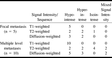
Signal intensity of vertebral metastases
In this issue


Jump to section
Related Articles
Cited By...
- Review of the Imaging Features of Benign Osteoporotic and Malignant Vertebral Compression Fractures
- Predictive Models in Differentiating Vertebral Lesions Using Multiparametric MRI
- Diffusion-Weighted MRI "Claw Sign" Improves Differentiation of Infectious from Degenerative Modic Type 1 Signal Changes of the Spine
- Diffusion-Weighted Imaging of the Spine: Is It Reliable?
- Acute vertebral body compression fractures: discrimination between benign and malignant causes using apparent diffusion coefficients