
Article Figures & Data
Figures
- fig 1.
A, Schematic representation of two capillaries containing contrast agent. The healthy capillary with an intact BBB (left) is not permeable to the contrast agent, which remains intravascular. A diseased capillary with a disrupted BBB may become permeable to the agent, leading to accumulation of contrast agent outside the vessel (right).
B, Because the contrast agent causes relaxation rate enhancement to water in its environment, initial tissue relaxation rate enhancement reflects the fraction of the tissue containing blood vessels (since the contrast agent is, at least initially, confined to the intravascular compartment). The ratio of initial enhancement in tissue to enhancement in a region of 100% blood (eg, the sagittal sinus) will then yield the fBV. Over time, if the contrast agent leaks out of the vessel into the extravascular space of the tissue, the relaxation rate will rise progressively. The rate of increase in relaxation rate is proportional to the permeability of the capillary wall to the contrast agent.
- fig 2.
A, MR image before contrast administration (top left) and six dynamic contrast-enhanced MR images of a grade 2 oligoastrocytoma. Top center, 15 seconds; top right, 45 seconds; middle left, 75 seconds; middle center, 105 seconds; middle right, 135 seconds; and lower left, 165 seconds after contrast administration. From these seven images, spatial maps of fBV and kPS were determined: fBV (lower center) was estimated as 12.8% in the ventral, enhancing part of the tumor and as 9.3% in the more dorsal part. kPS values (lower right) were 0.31 mL/100 cm3 per minute in the tumor (compared with 0.02 mL/100 cm3 per minute in normal brain tissue)
- fig 2.
B, MR image before contrast administration (top left) and six dynamic contrast-enhanced MR images of a grade 4 glioblastoma multiforme. Top center, 15 seconds; top right, 45 seconds; middle left, 75 seconds; middle center, 105 seconds; middle right, 135 seconds; and lower left, 165 seconds after contrast administration. From these seven images, spatial maps of fBV and kPS were determined: fBV (lower center) was estimated as 3.0% in the tumor rim and as 0.01% in the core. kPS values (lower right) were 13.6 mL/100 cm3 per minute in the tumor (compared with 0.5 mL/100 cm3 per minute in the core). Times are defined as the time after contrast that the center of k-space (mid-part of 3D acquisition) was attained.
- fig 3.
A–D, Fitted curves of the ΔSI time course of a typical low-grade (A, B) and a typical high-grade (C, D) tumor. Dynamic postcontrast values for the reference vascular signal (sagittal sinus) are displayed in A and C, respectively, and for tumor curves in B and D, respectively. The equations are displayed on the graphs. The low-grade tumor (patient 8 in Table 1) has an fBV of 3.2%, k1 + k2 is 5.8, and kPS is 5.5; the high-grade tumor (patient 20 in Table 1) has an fBV of 6.7%, k1 + k2 is 13.7, and kPS is 10.8. Times are defined as the time after contrast administration that the center of k-space (mid-part of 3D acquisition) was attained.
- fig 4.
A–C, Correlation between fBV (A), kPS (B), and total permeability, k, (C) and histologically determined tumor grade. While the data points in A are very scattered, reflective of a weak correlation (r = .39) between fBV and tumor grade, the slope is nonetheless positive, suggesting a tendency toward increased fBV in higher-grade tumors. For kPS, the tendency is more pronounced, with reduced scatter (r = .76). In comparison, total permeability, k, yielded a similar correlation (r = .83), with a steeper slope and less overlap between tumor grade groups.


Tables
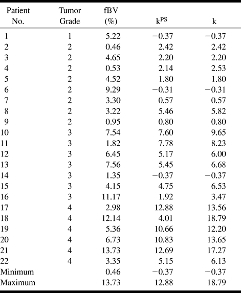
 TABLE 1:
Quantitative estimates of fBV, kPS, and total permeability, k (based on the sum of k1 and k2), for the 22 patients
- TABLE 2:
P values for t-tests for the parameters fBV, kPS, and the “total permeability” k to differentiate among histologically determined tumor gradesa

In this issue


Jump to section
Related Articles
Cited By...
- Tumor-related Perfusion Changes in White Matter Adjacent to Brain Tumors: Pharmacodynamic Analysis of Dynamic 3T Magnetic Resonance Imaging
- MRI Evaluation of Non-Necrotic T2-Hyperintense Foci in Pediatric Diffuse Intrinsic Pontine Glioma
- Improved Leakage Correction for Single-Echo Dynamic Susceptibility Contrast Perfusion MRI Estimates of Relative Cerebral Blood Volume in High-Grade Gliomas by Accounting for Bidirectional Contrast Agent Exchange
- Mitotic Activity in Glioblastoma Correlates with Estimated Extravascular Extracellular Space Derived from Dynamic Contrast-Enhanced MR Imaging
- Improved Brain Tumor Classification by Sodium MR Imaging: Prediction of IDH Mutation Status and Tumor Progression
- Increased Microvascularization and Vessel Permeability Associate With Active Inflammation in Human Atheromata
- Glioma: Application of Histogram Analysis of Pharmacokinetic Parameters from T1-Weighted Dynamic Contrast-Enhanced MR Imaging to Tumor Grading
- Exploratory Evaluation of MR Permeability with 18F-FDG PET Mapping in Pediatric Brain Tumors: A Report from the Pediatric Brain Tumor Consortium
- Magnetic Resonance Imaging Profile of Blood-Brain Barrier Injury in Patients With Acute Intracerebral Hemorrhage
- Semi-automated and automated glioma grading using dynamic susceptibility-weighted contrast-enhanced perfusion MRI relative cerebral blood volume measurements
- Diagnostic Accuracy of Dynamic Contrast-Enhanced MR Imaging Using a Phase-Derived Vascular Input Function in the Preoperative Grading of Gliomas
- MR Imaging of Neoplastic Central Nervous System Lesions: Review and Recommendations for Current Practice
- Perfusion CT Imaging of Brain Tumors: An Overview
- In Vivo Correlation of Tumor Blood Volume and Permeability with Histologic and Molecular Angiogenic Markers in Gliomas
- Increased Blood-Brain Barrier Permeability on Perfusion CT Might Predict Malignant Middle Cerebral Artery Infarction
- Contrast-Enhanced MR Imaging in Acute Ischemic Stroke: T2* Measures of Blood-Brain Barrier Permeability and Their Relationship to T1 Estimates and Hemorrhagic Transformation
- Texture Analysis: A Review of Neurologic MR Imaging Applications
- ZD6474, a Multitargeted Inhibitor for Receptor Tyrosine Kinases, Suppresses Growth of Gliomas Expressing an Epidermal Growth Factor Receptor Mutant, EGFRvIII, in the Brain
- Enhancing Fraction in Glioma and Its Relationship to the Tumoral Vascular Microenvironment: A Dynamic Contrast-Enhanced MR Imaging Study
- Optimized Preload Leakage-Correction Methods to Improve the Diagnostic Accuracy of Dynamic Susceptibility-Weighted Contrast-Enhanced Perfusion MR Imaging in Posttreatment Gliomas
- Recombinant Tissue Plasminogen Activator Increases Blood-Brain Barrier Disruption in Acute Ischemic Stroke: An MR Imaging Permeability Study
- Theoretic Basis and Technical Implementations of CT Perfusion in Acute Ischemic Stroke, Part 1: Theoretic Basis
- Antiangiogenesis Treatment for Glioblastoma Multiforme: Challenges and Opportunities
- Magnetic Resonance Imaging Determination of Tumor Grade and Early Response to Temozolomide in a Genetically Engineered Mouse Model of Glioma
- The Extent and Severity of Vascular Leakage as Evidence of Tumor Aggressiveness in High-Grade Gliomas
- Physiologic and Metabolic Magnetic Resonance Imaging in Gliomas
- Characterizing Extravascular Fluid Transport of Macromolecules in the Tumor Interstitium by Magnetic Resonance Imaging
- The Thioredoxin-1 Inhibitor 1-Methylpropyl 2-Imidazolyl Disulfide (PX-12) Decreases Vascular Permeability in Tumor Xenografts Monitored by Dynamic Contrast Enhanced Magnetic Resonance Imaging
- Dynamic Magnetic Resonance Perfusion Imaging of Brain Tumors
- Perfusion CT: a worthwhile enhancement?
- Quantitative Magnetic Resonance Imaging Analysis of Neovasculature Volume in Carotid Atherosclerotic Plaque
- Blood Volume of Gliomas Determined by Double-Echo Dynamic Perfusion-Weighted MR Imaging: A Preliminary Study