Advertisement

Main menu

  • Home
  • Content
    • Current Issue
    • Accepted Manuscripts
    • Article Preview
    • Past Issue Archive
    • Video Articles
    • AJNR Case Collection
    • Case of the Week Archive
    • Case of the Month Archive
    • Classic Case Archive
  • Special Collections
    • AJNR Awards
    • Low-Field MRI
    • Alzheimer Disease
    • ASNR Foundation Special Collection
    • Photon-Counting CT
    • View All
  • Multimedia
    • AJNR Podcasts
    • AJNR SCANtastic
    • Trainee Corner
    • MRI Safety Corner
    • Imaging Protocols
  • For Authors
    • Submit a Manuscript
    • Submit a Video Article
    • Submit an eLetter to the Editor/Response
    • Manuscript Submission Guidelines
    • Statistical Tips
    • Fast Publishing of Accepted Manuscripts
    • Graphical Abstract Preparation
    • Imaging Protocol Submission
    • Author Policies
  • About Us
    • About AJNR
    • Editorial Board
    • Editorial Board Alumni
  • More
    • Become a Reviewer/Academy of Reviewers
    • Subscribers
    • Permissions
    • Alerts
    • Feedback
    • Advertisers
    • ASNR Home

User menu

  • Alerts
  • Log in

Search

  • Advanced search
American Journal of Neuroradiology

American Journal of Neuroradiology

ASHNR American Society of Functional Neuroradiology ASHNR American Society of Pediatric Neuroradiology ASSR
  • Alerts
  • Log in

Advanced Search

  • Home
  • Content
    • Current Issue
    • Accepted Manuscripts
    • Article Preview
    • Past Issue Archive
    • Video Articles
    • AJNR Case Collection
    • Case of the Week Archive
    • Case of the Month Archive
    • Classic Case Archive
  • Special Collections
    • AJNR Awards
    • Low-Field MRI
    • Alzheimer Disease
    • ASNR Foundation Special Collection
    • Photon-Counting CT
    • View All
  • Multimedia
    • AJNR Podcasts
    • AJNR SCANtastic
    • Trainee Corner
    • MRI Safety Corner
    • Imaging Protocols
  • For Authors
    • Submit a Manuscript
    • Submit a Video Article
    • Submit an eLetter to the Editor/Response
    • Manuscript Submission Guidelines
    • Statistical Tips
    • Fast Publishing of Accepted Manuscripts
    • Graphical Abstract Preparation
    • Imaging Protocol Submission
    • Author Policies
  • About Us
    • About AJNR
    • Editorial Board
    • Editorial Board Alumni
  • More
    • Become a Reviewer/Academy of Reviewers
    • Subscribers
    • Permissions
    • Alerts
    • Feedback
    • Advertisers
    • ASNR Home
  • Follow AJNR on Twitter
  • Visit AJNR on Facebook
  • Follow AJNR on Instagram
  • Join AJNR on LinkedIn
  • RSS Feeds

AJNR Awards, New Junior Editors, and more. Read the latest AJNR updates

Getting new auth cookie, if you see this message a lot, tell someone!
Research ArticleBrain

Demyelinating Plaques in Relapsing-remitting and Secondary-progressive Multiple Sclerosis: Assessment with Diffusion MR Imaging

Alessandro Castriota Scanderbeg, Francesco Tomaiuolo, Umberto Sabatini, Ugo Nocentini, Maria G. Grasso and Carlo Caltagirone
American Journal of Neuroradiology May 2000, 21 (5) 862-868;
Alessandro Castriota Scanderbeg
  • Find this author on Google Scholar
  • Find this author on PubMed
  • Search for this author on this site
Francesco Tomaiuolo
  • Find this author on Google Scholar
  • Find this author on PubMed
  • Search for this author on this site
Umberto Sabatini
  • Find this author on Google Scholar
  • Find this author on PubMed
  • Search for this author on this site
Ugo Nocentini
  • Find this author on Google Scholar
  • Find this author on PubMed
  • Search for this author on this site
Maria G. Grasso
  • Find this author on Google Scholar
  • Find this author on PubMed
  • Search for this author on this site
Carlo Caltagirone
  • Find this author on Google Scholar
  • Find this author on PubMed
  • Search for this author on this site
  • Article
  • Figures & Data
  • Info & Metrics
  • Responses
  • References
  • PDF
Loading

Article Figures & Data

Figures

  • fig 1.
    • Download figure
    • Open in new tab
    • Download powerpoint
    fig 1.

    A–D, Axial SE T1-weighted (A), fast SE T2-weighted (B), and echo-planar T2-weighted (C) MR images, and the orientationally averaged ADC map (D), show the presence of multiple periventricular demyelinating lesions in a patient with secondary-progressive MS. Circle indicates the ROI
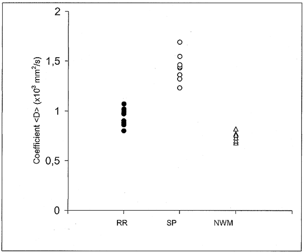
  • fig 2.
    • Download figure
    • Open in new tab
    • Download powerpoint
    fig 2.

    Scatter plot of the coefficient <D> data points for the relapsing-remitting (RR, 10 points), secondary-progressive (SP, 10 points), and control group (NWM, normal white matter, 11 points). Each point corresponds to one subject and represents the mean value of <D> as obtained in each subject from all the ROIs that were drawn on the T2-weighted (b = 0) images. Graph shows there is no overlap between the relapsing-remitting and secondary-progressive groups

  • fig 3.
    • Download figure
    • Open in new tab
    • Download powerpoint
    fig 3.

    Scatter plot of the coefficient <D> vs. EDSS score shows a significant correlation. Each point corresponds to one patient and represents the mean value of <D> as obtained from all the ROIs that were drawn on the T2-weighted (b = 0) images. Closed circles represent the subjects with relapsing-remitting MS; open circles, the subjects with secondary-progressive MS
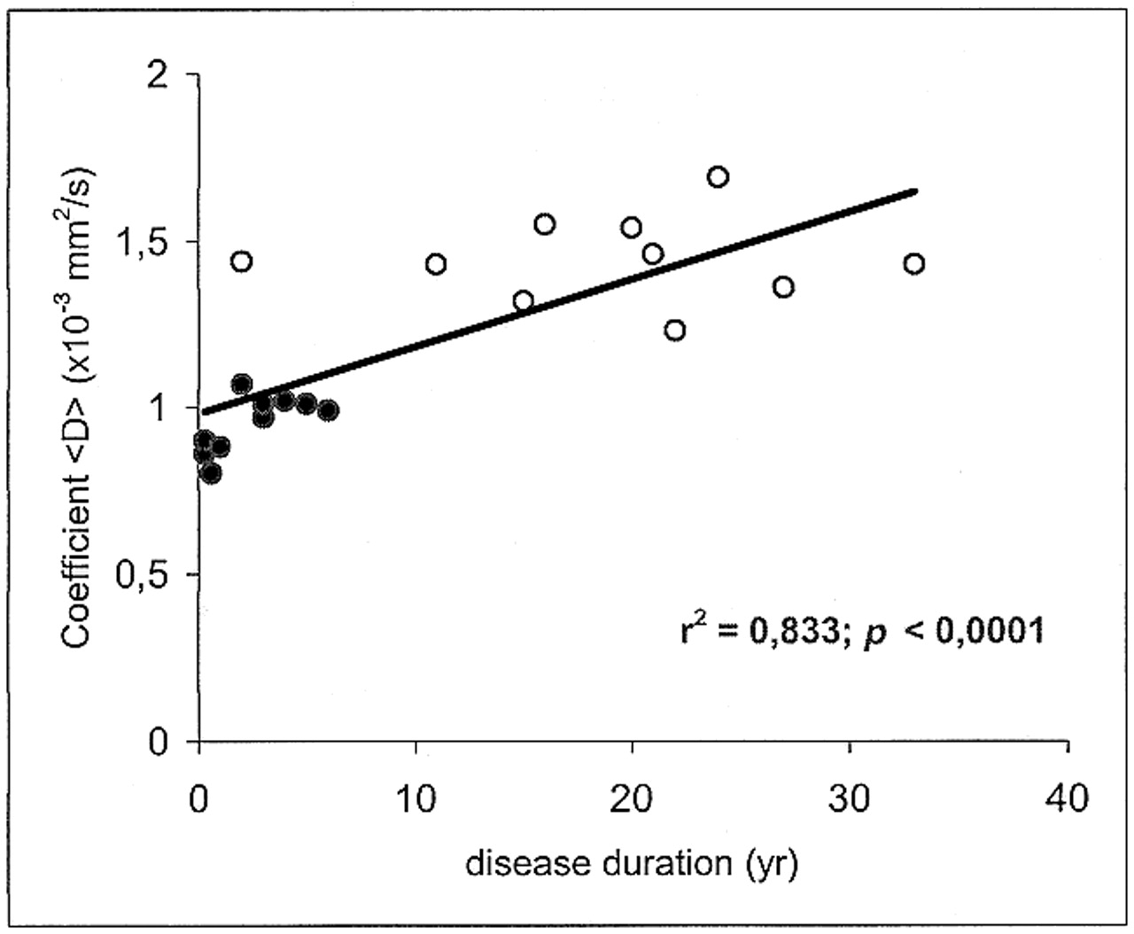
  • fig 4.
    • Download figure
    • Open in new tab
    • Download powerpoint
    fig 4.

    Scatter plot of coefficient <D> vs. disease duration shows a significant correlation. Each point corresponds to one patient and represents the mean value of <D> as obtained from all the ROIs that were drawn on the T2-weighted (b = 0) images. Closed circles represent the patients with relapsing-remitting MS; open circles, the patients with secondary-progressive MS

  • fig 5.
    • Download figure
    • Open in new tab
    • Download powerpoint
    fig 5.

    Scatter plot of coefficient <D> vs. SIlesion/SICSF ratio on SE T1-weighted images for the 41 T1 hypointense lesions studied. There is a significant inverse correlation between <D> and SIlesion/SICSF ratio. Each point corresponds to one lesion. Closed circles represent lesions from the relapsing-remitting group (11 lesions); open circles, lesions from the secondary-progressive group (30 lesions)

PreviousNext
Back to top

In this issue

American Journal of Neuroradiology
Vol. 21, Issue 5
1 May 2000
  • Table of Contents
  • Index by author
Advertisement
Print
Download PDF
Email Article

Thank you for your interest in spreading the word on American Journal of Neuroradiology.

NOTE: We only request your email address so that the person you are recommending the page to knows that you wanted them to see it, and that it is not junk mail. We do not capture any email address.

Enter multiple addresses on separate lines or separate them with commas.
Demyelinating Plaques in Relapsing-remitting and Secondary-progressive Multiple Sclerosis: Assessment with Diffusion MR Imaging
(Your Name) has sent you a message from American Journal of Neuroradiology
(Your Name) thought you would like to see the American Journal of Neuroradiology web site.
CAPTCHA
This question is for testing whether or not you are a human visitor and to prevent automated spam submissions.
Cite this article
Alessandro Castriota Scanderbeg, Francesco Tomaiuolo, Umberto Sabatini, Ugo Nocentini, Maria G. Grasso, Carlo Caltagirone
Demyelinating Plaques in Relapsing-remitting and Secondary-progressive Multiple Sclerosis: Assessment with Diffusion MR Imaging
American Journal of Neuroradiology May 2000, 21 (5) 862-868;

Citation Manager Formats

  • BibTeX
  • Bookends
  • EasyBib
  • EndNote (tagged)
  • EndNote 8 (xml)
  • Medlars
  • Mendeley
  • Papers
  • RefWorks Tagged
  • Ref Manager
  • RIS
  • Zotero
0 Responses
Respond to this article
Share
Bookmark this article
Demyelinating Plaques in Relapsing-remitting and Secondary-progressive Multiple Sclerosis: Assessment with Diffusion MR Imaging
Alessandro Castriota Scanderbeg, Francesco Tomaiuolo, Umberto Sabatini, Ugo Nocentini, Maria G. Grasso, Carlo Caltagirone
American Journal of Neuroradiology May 2000, 21 (5) 862-868;
del.icio.us logo Twitter logo Facebook logo Mendeley logo
  • Tweet Widget
  • Facebook Like
  • Google Plus One

Jump to section

  • Article
    • Abstract
    • Methods
    • Results
    • Discussion
    • Conclusion
    • Acknowledgments
    • Footnotes
    • References
  • Figures & Data
  • Info & Metrics
  • Responses
  • References
  • PDF

Related Articles

  • No related articles found.
  • PubMed
  • Google Scholar

Cited By...

  • Evolution of Chronic Lesion Tissue in RRMS patients: An association with disease progression
  • Modelling axonal loss caused by chronic demyelination and slow-burning inflammation at the rim of chronic lesions in MS patients
  • Short-term surrogate biomarkers of chronic lesion expansion
  • Measuring Brain Tissue Integrity during 4 Years Using Diffusion Tensor Imaging
  • Increased diffusivity in acute multiple sclerosis lesions predicts risk of black hole
  • Diffusion-weighted imaging findings in Balo concentric sclerosis.
  • Diffusion MRI in multiple sclerosis
  • A review of structural magnetic resonance neuroimaging
  • Coefficient Dav Is More Sensitive Than Fractional Anisotropy in Monitoring Progression of Irreversible Tissue Damage in Focal Nonactive Multiple Sclerosis Lesions
  • MRI techniques to monitor MS evolution: The present and the future
  • Analysis of Normal-Appearing White Matter in Multiple Sclerosis: Comparison of Diffusion Tensor MR Imaging and Magnetization Transfer Imaging
  • Optimizing the association between disability and biological markers in MS
  • Crossref
  • Google Scholar

This article has not yet been cited by articles in journals that are participating in Crossref Cited-by Linking.

More in this TOC Section

  • Enhanced Axonal Metabolism during Early Natalizumab Treatment in Relapsing-Remitting Multiple Sclerosis
  • Progression of Microstructural Damage in Spinocerebellar Ataxia Type 2: A Longitudinal DTI Study
  • SWI or T2*: Which MRI Sequence to Use in the Detection of Cerebral Microbleeds? The Karolinska Imaging Dementia Study
Show more Brain

Similar Articles

Advertisement

Indexed Content

  • Current Issue
  • Accepted Manuscripts
  • Article Preview
  • Past Issues
  • Editorials
  • Editor's Choice
  • Fellows' Journal Club
  • Letters to the Editor
  • Video Articles

Cases

  • Case Collection
  • Archive - Case of the Week
  • Archive - Case of the Month
  • Archive - Classic Case

More from AJNR

  • Trainee Corner
  • Imaging Protocols
  • MRI Safety Corner
  • Book Reviews

Multimedia

  • AJNR Podcasts
  • AJNR Scantastics

Resources

  • Turnaround Time
  • Submit a Manuscript
  • Submit a Video Article
  • Submit an eLetter to the Editor/Response
  • Manuscript Submission Guidelines
  • Statistical Tips
  • Fast Publishing of Accepted Manuscripts
  • Graphical Abstract Preparation
  • Imaging Protocol Submission
  • Evidence-Based Medicine Level Guide
  • Publishing Checklists
  • Author Policies
  • Become a Reviewer/Academy of Reviewers
  • News and Updates

About Us

  • About AJNR
  • Editorial Board
  • Editorial Board Alumni
  • Alerts
  • Permissions
  • Not an AJNR Subscriber? Join Now
  • Advertise with Us
  • Librarian Resources
  • Feedback
  • Terms and Conditions
  • AJNR Editorial Board Alumni

American Society of Neuroradiology

  • Not an ASNR Member? Join Now

© 2025 by the American Society of Neuroradiology All rights, including for text and data mining, AI training, and similar technologies, are reserved.
Print ISSN: 0195-6108 Online ISSN: 1936-959X

Powered by HighWire