
Article Figures & Data
Figures
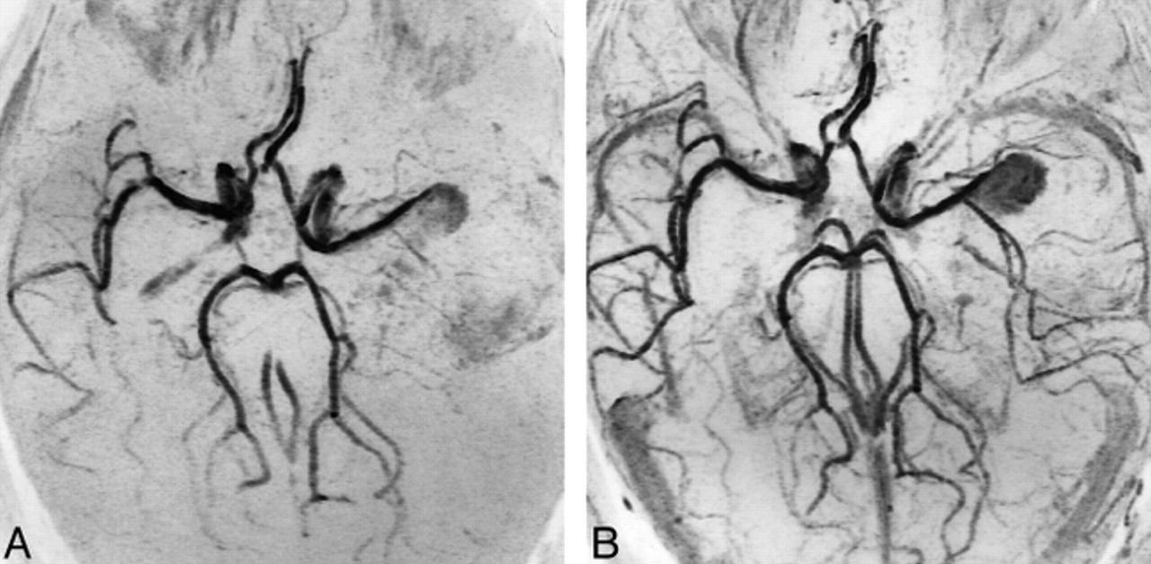
- fig 1.
Small anterior communicating artery aneurysm treated with GDCs. The presence of an artifact does not permit evaluation of the aneurysm neck.
A, 3D-TOF MR angiographic source image (39/6.2/1) shows a region of absence of signal related to the presence of coils in the site of the treated aneurysm. The A2 segment of both anterior cerebral arteries is not visible.
B, Corresponding targeted MIP reconstruction shows the gap in vascular signal at the site of the treated aneurysm and at the level of the origin of both A2 segments.
C, Targeted MIP reconstruction from the ultrashort-TE MR angiographic sequence (5/2.1/1) shows the origin of both A2 segments, although the anterior communicating artery is still not visible.
- fig 2.
Small basilar tip aneurysm found to be incompletely occluded at MR angiography and completely occluded at DSA follow-up after treatment with GDCs.
A, DSA study before treatment shows the presence of a small basilar tip aneurysm.
B, The aneurysm was judged to be completely occluded at the first follow-up DSA study.
C, On the first follow-up 3D-TOF MR angiogram (39/6.2/1), a small residual patency was detected.
D, Residual patency of the aneurysm was confirmed at the second DSA follow-up (6 months after the first follow-up study).
- fig 3.
Small basilar trunk aneurysm incompletely occluded after treatment with GDCs, as demonstrated at the first follow-up examination. The patient was retreated, with complete occlusion of the aneurysm noted at 1-year follow-up study.
A, DSA shows the presence of residual patency of the treated aneurysm near the neck.
B, 3D-TOF MR angiogram (43/8/1) shows residual patency with the same extent as seen at DSA, despite the presence of a vascular signal gap, related to the coils, at the P1 segment of the left posterior cerebral artery.
- fig 4.
Giant middle cerebral artery bifurcation aneurysm partially treated with incomplete occlusion verified at the 6-month follow-up examination.
A, MIP reconstruction from basal 3D-TOF MR angiogram (43/8/1) shows flow within the portion of the aneurysm seen previously; the slow flow is responsible for low signal of both the aneurysm and the middle cerebral artery branches.
B, Enhanced 3D-TOF MR angiogram (43/8/1) better depicts the partially occluded aneurysm and the patency of the middle cerebral artery branches.
- fig 5.
Small aneurysm at the origin of the left PICA, which was completely occluded after treatment with GDCs.
A, 3D-TOF MR angiogram (39/6.2/1) targeted MIP reconstruction shows the aneurysm before treatment.
B, 3D-TOF MR angiogram (39/6.2/1) targeted MIP reconstruction 6 months after treatment shows complete occlusion of the aneurysm and reperfusion of the left PICA.


Tables

- TABLE 3:
Results of group 1 (MR angiography without contrast enhancement)


In this issue


Jump to section
Related Articles
Cited By...
- MRA versus DSA for the follow-up imaging of intracranial aneurysms treated using endovascular techniques: a meta-analysis
- MRA Versus DSA for Follow-Up of Coiled Intracranial Aneurysms: A Meta-Analysis
- Temporal Evolution of Susceptibility Artifacts from Coiled Aneurysms on MR Angiography: An In Vivo Canine Study
- Outcomes of Endovascular Treatments of Aneurysms: Observer Variability and Implications for Interpreting Case Series and Planning Randomized Trials
- Residual Flow After Cerebral Aneurysm Coil Occlusion: Diagnostic Accuracy of MR Angiography
- MR Angiographic Follow-Up of Intracranial Aneurysms Treated with Detachable Coils: Evaluation of a Blood-Pool Contrast Medium
- Intracranial Aneurysms Treated With Guglielmi Detachable Coils: Imaging Follow-Up With Contrast-Enhanced MR Angiography
- Intracranial Aneurysms Treated With Endovascular Coils: Detection of Recurrences Using Unenhanced and Contrast-Enhanced Transcranial Color-Coded Duplex Sonography
- Matrix Detachable Coils for the Endovascular Treatment of Intracranial Aneurysms: Analysis of Early Angiographic and Clinical Outcomes
- CT and MR Imaging Findings and Their Implications in the Follow-up of Patients with Intracranial Aneurysms Treated with Endosaccular Occlusion with Onyx
- Current theory in imaging of intracranial vascular disease