
Article Figures & Data
Figures
- fig 1.
Case of a 61-year-old man with squamous cell carcinoma of the hard palate.
A, Gray-scale sonogram of a reactive cervical lymph node shows homogeneous and low echogenicity of the parenchyma and normal hilar echoes (arrows).
B, Power Doppler sonogram of the same node as that shown in panel A shows a normal reactive hilar blood flow.
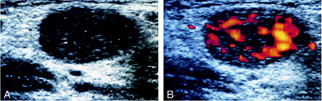
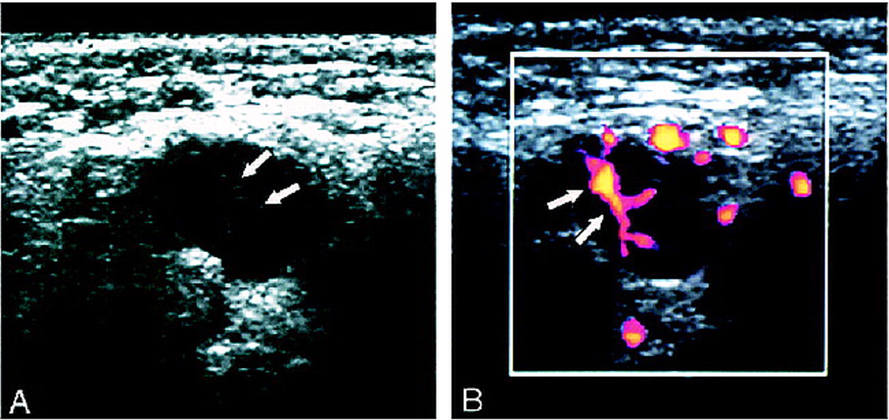
- fig 2.
Case of a 53-year-old man with squamous cell carcinoma of the hypopharynx.
A, Gray-scale sonogram of a metastatic cervical lymph node shows abnormal, heterogeneous echogenicity of the parenchyma.
B, Power Doppler sonogram of the same node as that shown in panel A shows abnormal parenchymal blood flow, characteristic of metastasis.
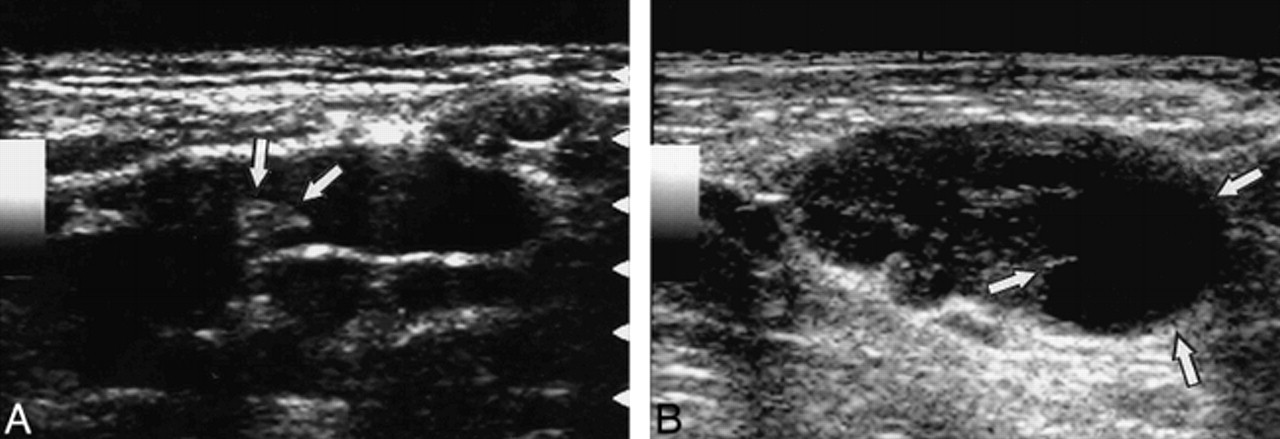
- fig 3.
Sonograms of a reactive node from a 54-year-old man with squamous cell carcinoma of the larynx (A) and a metastatic node from 64-year-old man with squamous cell carcinoma of the hypopharynx (B) show a preferential increase in short axis diameter of the metastatic node.
A, Gray-scale sonogram shows reactive cervical lymph node with relatively flat shape. Arrows indicate hilum.
B, Gray-scale sonogram shows metastatic cervical lymph node. Note that the lymph nodes in panels A and B exhibit similar longitudinal axis lengths, but that the metastatic node in B has a greater short axis length. The metastatic node also shows a focal area of necrosis (arrows).
- fig 4.
Case of a 66-year-old man with squamous cell carcinoma of the upper gingiva. This case shows the value of the presence of hilar echoes over that of increased short axis diameter. The presence of normal hilar flow as detected by Doppler sonography is helpful for confirming the presence of normal hilar echoes.
A, Gray-scale sonogram of a reactive cervical lymph. The short axis length is increased, suggestive of metastatic node; however, the normal hilar echo is present (arrows).
B, Power Doppler sonogram of the same reactive node as that shown in panel A shows a normal hilar flow (arrows).


Tables
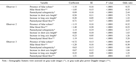
 TABLE 1:
Estimation by three observers of the presence or absence of sonographic criteria in 133 nodes
- TABLE 2:
Univariate logistic regression analysis of lymph node sonographic features
- TABLE 3:
Multivariate logistic regression analysis of sonographic features of the lymph nodes
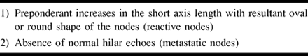
- TABLE 4:
A summary of the most important criteria for detecting cervical lymph node metastasis on sonography


In this issue


Jump to section
Related Articles
Cited By...
- Quantitative evaluation of vascularity within cervical lymph nodes using Doppler ultrasound in patients with oral cancer: relation to lymph node size
- Small Atypical Cervical Nodes Detected on Sonography in Patients With Squamous Cell Carcinoma of the Head and Neck: Probability of Metastasis
- Evaluation of the efficacy of colour Doppler ultrasound in diagnosis of cervical lymphadenopathy
- Sonography as a replacement for sialography for the diagnosis of salivary glands affected by Sjogren's syndrome
- Contribution of Doppler Sonography Blood Flow Information to the Diagnosis of Metastatic Cervical Nodes in Patients with Head and Neck Cancer: Assessment in Relation to Anatomic Levels of the Neck
- Detection of Lymph Node Metastases in the Neck: Radiologic Criteria