Advertisement

Main menu

  • Home
  • Content
    • Current Issue
    • Accepted Manuscripts
    • Article Preview
    • Past Issue Archive
    • Video Articles
    • AJNR Case Collection
    • Case of the Week Archive
    • Case of the Month Archive
    • Classic Case Archive
  • Special Collections
    • AJNR Awards
    • Low-Field MRI
    • Alzheimer Disease
    • ASNR Foundation Special Collection
    • Photon-Counting CT
    • View All
  • Multimedia
    • AJNR Podcasts
    • AJNR SCANtastic
    • Trainee Corner
    • MRI Safety Corner
    • Imaging Protocols
  • For Authors
    • Submit a Manuscript
    • Submit a Video Article
    • Submit an eLetter to the Editor/Response
    • Manuscript Submission Guidelines
    • Statistical Tips
    • Fast Publishing of Accepted Manuscripts
    • Graphical Abstract Preparation
    • Imaging Protocol Submission
    • Author Policies
  • About Us
    • About AJNR
    • Editorial Board
    • Editorial Board Alumni
  • More
    • Become a Reviewer/Academy of Reviewers
    • Subscribers
    • Permissions
    • Alerts
    • Feedback
    • Advertisers
    • ASNR Home

User menu

  • Alerts
  • Log in

Search

  • Advanced search
American Journal of Neuroradiology

American Journal of Neuroradiology

ASHNR American Society of Functional Neuroradiology ASHNR American Society of Pediatric Neuroradiology ASSR
  • Alerts
  • Log in

Advanced Search

  • Home
  • Content
    • Current Issue
    • Accepted Manuscripts
    • Article Preview
    • Past Issue Archive
    • Video Articles
    • AJNR Case Collection
    • Case of the Week Archive
    • Case of the Month Archive
    • Classic Case Archive
  • Special Collections
    • AJNR Awards
    • Low-Field MRI
    • Alzheimer Disease
    • ASNR Foundation Special Collection
    • Photon-Counting CT
    • View All
  • Multimedia
    • AJNR Podcasts
    • AJNR SCANtastic
    • Trainee Corner
    • MRI Safety Corner
    • Imaging Protocols
  • For Authors
    • Submit a Manuscript
    • Submit a Video Article
    • Submit an eLetter to the Editor/Response
    • Manuscript Submission Guidelines
    • Statistical Tips
    • Fast Publishing of Accepted Manuscripts
    • Graphical Abstract Preparation
    • Imaging Protocol Submission
    • Author Policies
  • About Us
    • About AJNR
    • Editorial Board
    • Editorial Board Alumni
  • More
    • Become a Reviewer/Academy of Reviewers
    • Subscribers
    • Permissions
    • Alerts
    • Feedback
    • Advertisers
    • ASNR Home
  • Follow AJNR on Twitter
  • Visit AJNR on Facebook
  • Follow AJNR on Instagram
  • Join AJNR on LinkedIn
  • RSS Feeds

AJNR Awards, New Junior Editors, and more. Read the latest AJNR updates

Research ArticleHead and Neck Imaging

Midline Destructive Lesions of the Sinonasal Tract: Simplified Terminology Based onHistopathologic Criteria

Alexandra Borges, James Fink, Pablo Villablanca, Roy Eversole and Robert Lufkin
American Journal of Neuroradiology February 2000, 21 (2) 331-336;
Alexandra Borges
aFrom the Departments of Radiological Sciences (A.B., J.F., P.V., R.L.) and Oral Pathology (R.E.), University of California, Los Angeles.
  • Find this author on Google Scholar
  • Find this author on PubMed
  • Search for this author on this site
James Fink
aFrom the Departments of Radiological Sciences (A.B., J.F., P.V., R.L.) and Oral Pathology (R.E.), University of California, Los Angeles.
  • Find this author on Google Scholar
  • Find this author on PubMed
  • Search for this author on this site
Pablo Villablanca
aFrom the Departments of Radiological Sciences (A.B., J.F., P.V., R.L.) and Oral Pathology (R.E.), University of California, Los Angeles.
  • Find this author on Google Scholar
  • Find this author on PubMed
  • Search for this author on this site
Roy Eversole
aFrom the Departments of Radiological Sciences (A.B., J.F., P.V., R.L.) and Oral Pathology (R.E.), University of California, Los Angeles.
  • Find this author on Google Scholar
  • Find this author on PubMed
  • Search for this author on this site
Robert Lufkin
aFrom the Departments of Radiological Sciences (A.B., J.F., P.V., R.L.) and Oral Pathology (R.E.), University of California, Los Angeles.
  • Find this author on Google Scholar
  • Find this author on PubMed
  • Search for this author on this site
  • Article
  • Figures & Data
  • Info & Metrics
  • Responses
  • References
  • PDF
Loading

Article Figures & Data

Figures

  • Tables
  • fig 1.
    • Download figure
    • Open in new tab
    • Download powerpoint
    fig 1.

    Patient 5. Initial diagnosis: nonhealing midline granuloma (Stewart's syndrome); final diagnosis: non-Hodgkin's T-cell lymphoma.

    A, CT scan of the paranasal sinuses shows soft-tissue opacification of the anterior ethmoidal air cells bilaterally, soft-tissue density in the region of the right ostiomeatal unit, and mild mucosal thickening in the inferior aspect of the right maxillary sinus. The left maxillary sinus is hypoplastic and the remainder of the sinus cavity is filled with soft tissue. Bony destructive changes are noted with a nasoseptal perforation and destruction of the inferomedial wall of the right orbit. However, the abnormal soft tissue does not appear to infiltrate the extraconal space. The bony defect in the inferior aspect of the left maxillary sinus is postsurgical in nature, due to a prior Caldwell-Luc antrostomy for chronic sinus disease. Some soft-tissue thickening is noted in the left premaxillary region.

    B, Histologic slide of a sinonasal biopsy specimen shows lymphocytic invasion of a blood vessel reflecting the angioinvasive nature of the disease. Also noted is a heterogeneous and polymorphic inflammatory infiltrate (hematoxylin-eosin, original magnification ×400).

    C, Histologic slide of a sinonasal biopsy specimen shows invasion of trabecular bone in the right upper corner of the image (hematoxylin-eosin, original magnification ×100).

    D, Histologic slide of a sinonasal biopsy specimen shows positive staining with CD45RO T-cell marker (original magnification ×200).

    E, Histologic slide of a sinonasal biopsy specimen shows negative staining with CD20, a B-cell marker (original magnification ×200).

  • fig 2.
    • Download figure
    • Open in new tab
    • Download powerpoint
    fig 2.

    Patient 4. Initial diagnosis: malignant midline granuloma; final diagnosis: Wegener's granulomatosis.

    A and B, Axial (A) and coronal (B) CT scans of the paranasal sinuses (bone window) show complete soft-tissue filling of the nasal cavity and nasal vault extending posteriorly to the choana and high nasopharynx. The anterior aspect of the bony nasal septum has been destroyed. The left maxillary sinus is totally opacified and slightly expanded with mild bowing of the medial wall of the sinus. The bony walls of the left maxillary sinus are intact. No abnormal soft tissue is seen in the retroantral fat or pterygopalatine fossa.

    C, Histologic slide of a sinonasal biopsy specimen shows an inflammatory infiltrate composed mainly of histiocytes and eosinophils. Note also an area of fibrinoid necrosis in the center of the slide (hematoxylin-eosin, original magnification ×200).

    D, Histologic slide of a sinonasal biopsy specimen shows typical multinucleated giant cells. A final diagnosis of Wegener's granulomatosis was based on the histologic findings and on necrotizing vasculitis (hematoxylin-eosin, original magnification ×400).

Tables

  • Figures
  • TABLE 1:
    • View popup
    • Download powerpoint
    TABLE 1:

    Diagnosis before and after review of the pathologic specimens and immunohistochemical analysis in seven patients with destructive lesions of the sinonasal tract
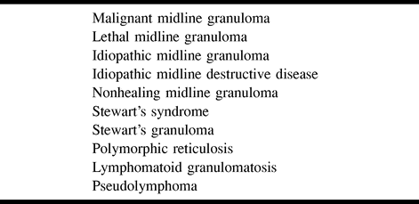
  • TABLE 2:
    • View popup
    • Download powerpoint
    TABLE 2:

    Terms to be abolished from the radiologic lexicon

  • TABLE 3:
    • View popup
    • Download powerpoint
    TABLE 3:

    Differential diagnosis of destructive lesions of the sinonasal tract

PreviousNext
Back to top

In this issue

American Journal of Neuroradiology
Vol. 21, Issue 2
1 Feb 2000
  • Table of Contents
  • Index by author
Advertisement
Print
Download PDF
Email Article

Thank you for your interest in spreading the word on American Journal of Neuroradiology.

NOTE: We only request your email address so that the person you are recommending the page to knows that you wanted them to see it, and that it is not junk mail. We do not capture any email address.

Enter multiple addresses on separate lines or separate them with commas.
Midline Destructive Lesions of the Sinonasal Tract: Simplified Terminology Based onHistopathologic Criteria
(Your Name) has sent you a message from American Journal of Neuroradiology
(Your Name) thought you would like to see the American Journal of Neuroradiology web site.
CAPTCHA
This question is for testing whether or not you are a human visitor and to prevent automated spam submissions.
Cite this article
Alexandra Borges, James Fink, Pablo Villablanca, Roy Eversole, Robert Lufkin
Midline Destructive Lesions of the Sinonasal Tract: Simplified Terminology Based onHistopathologic Criteria
American Journal of Neuroradiology Feb 2000, 21 (2) 331-336;

Citation Manager Formats

  • BibTeX
  • Bookends
  • EasyBib
  • EndNote (tagged)
  • EndNote 8 (xml)
  • Medlars
  • Mendeley
  • Papers
  • RefWorks Tagged
  • Ref Manager
  • RIS
  • Zotero
0 Responses
Respond to this article
Share
Bookmark this article
Midline Destructive Lesions of the Sinonasal Tract: Simplified Terminology Based onHistopathologic Criteria
Alexandra Borges, James Fink, Pablo Villablanca, Roy Eversole, Robert Lufkin
American Journal of Neuroradiology Feb 2000, 21 (2) 331-336;
del.icio.us logo Twitter logo Facebook logo Mendeley logo
  • Tweet Widget
  • Facebook Like
  • Google Plus One

Jump to section

  • Article
    • Abstract
    • Methods
    • Results
    • Discussion
    • Conclusion
    • Footnotes
    • References
  • Figures & Data
  • Info & Metrics
  • Responses
  • References
  • PDF

Related Articles

  • No related articles found.
  • PubMed
  • Google Scholar

Cited By...

  • IgG4-related midline destructive lesion
  • Crossref
  • Google Scholar

This article has not yet been cited by articles in journals that are participating in Crossref Cited-by Linking.

More in this TOC Section

  • MRI of the Parasellar Ligaments
  • ASL Sensitivity for Head and Neck Paraganglioma
  • Post SRS Peritumoral Hyperintense Signal of VSs
Show more Head and Neck Imaging

Similar Articles

Advertisement

Indexed Content

  • Current Issue
  • Accepted Manuscripts
  • Article Preview
  • Past Issues
  • Editorials
  • Editor's Choice
  • Fellows' Journal Club
  • Letters to the Editor
  • Video Articles

Cases

  • Case Collection
  • Archive - Case of the Week
  • Archive - Case of the Month
  • Archive - Classic Case

More from AJNR

  • Trainee Corner
  • Imaging Protocols
  • MRI Safety Corner
  • Book Reviews

Multimedia

  • AJNR Podcasts
  • AJNR Scantastics

Resources

  • Turnaround Time
  • Submit a Manuscript
  • Submit a Video Article
  • Submit an eLetter to the Editor/Response
  • Manuscript Submission Guidelines
  • Statistical Tips
  • Fast Publishing of Accepted Manuscripts
  • Graphical Abstract Preparation
  • Imaging Protocol Submission
  • Evidence-Based Medicine Level Guide
  • Publishing Checklists
  • Author Policies
  • Become a Reviewer/Academy of Reviewers
  • News and Updates

About Us

  • About AJNR
  • Editorial Board
  • Editorial Board Alumni
  • Alerts
  • Permissions
  • Not an AJNR Subscriber? Join Now
  • Advertise with Us
  • Librarian Resources
  • Feedback
  • Terms and Conditions
  • AJNR Editorial Board Alumni

American Society of Neuroradiology

  • Not an ASNR Member? Join Now

© 2025 by the American Society of Neuroradiology All rights, including for text and data mining, AI training, and similar technologies, are reserved.
Print ISSN: 0195-6108 Online ISSN: 1936-959X

Powered by HighWire